Articles
- Page Path
- HOME > J Pathol Transl Med > Volume 50(3); 2016 > Article
-
Review
Pathologic Evaluation of Breast Cancer after Neoadjuvant Therapy - Cheol Keun Park, Woo-Hee Jung, Ja Seung Koo,
-
Journal of Pathology and Translational Medicine 2016;50(3):173-180.
DOI: https://doi.org/10.4132/jptm.2016.02.02
Published online: April 11, 2016
Department of Pathology, Yonsei University College of Medicine, Seoul, Korea
- Corresponding Author: Ja Seung Koo, MD Department of Pathology, Severance Hospital, Yonsei University College of Medicine, 50-1 Yonsei-ro, Seodaemun-gu, Seoul 03722, Korea Tel: +82-2-2228-1772 Fax: +82-2-362-0860 E-mail: kjs1976@yuhs.ac
© 2016 The Korean Society of Pathologists/The Korean Society for Cytopathology
This is an Open Access article distributed under the terms of the Creative Commons Attribution Non-Commercial License (http://creativecommons.org/licenses/by-nc/3.0/) which permits unrestricted non-commercial use, distribution, and reproduction in any medium, provided the original work is properly cited.
Abstract
- Breast cancer, one of the most common cancers in women, has various treatment modalities. Neoadjuvant therapy (NAT) has been used in many clinical trials because it is easy to evaluate the treatment response to therapeutic agents in a short time period; consequently, NAT is currently a standard treatment modality for large-sized and locally advanced breast cancers, and its use in early-stage breast cancer is becoming more common. Thus, chances to encounter breast tissue from patients treated with NAT is increasing. However, systems for handling and evaluating such specimens have not been established. Several evaluation systems emphasize a multidisciplinary approach to increase the accuracy of breast cancer assessment. Thus, detailed and systematic evaluation of clinical, radiologic, and pathologic findings is important. In this review, we compare the major problems of each evaluation system and discuss important points for handling and evaluating NAT-treated breast specimens.
- Specimen handling
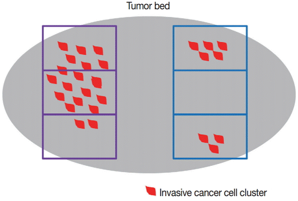
- Identification of the tumor bed is important for the handling of breast specimens after NAT. Close examination of fresh specimens cut into 5-mm sections or smaller is required for identification of the tumor bed. However, some cases require extensive sampling because of uncertainty in the gross identification of tumor bed. There have been attempts to insert metallic clips while conducting breast core biopsy for easy recognition of the tumor bed [15-18]. However, this method cannot locate the tumor bed accurately because the inserted metallic clip shifts over time [19]. Some guidelines suggest that small specimens (<5 cm at the widest diameter or <30 g) should be thinly sectioned and submitted in their entirety so that the specimens can be reintegrated upon histologic evaluation. However, these methods have limitations in that samples for research use cannot be secured [20]. It is crucial to select representative sections when dealing with large specimens, such as those from a large lumpectomy or mastectomy. The important goal in specimen selection is to identify the area that correlates best with clinical and radiologic findings. This area, which is known as the pretreatment area, should comprise grossly identifiable tumor bed, a metallic clip, and peritumoral tissue [20]. After slicing surgical specimens into ≤ 5-mm sections, the cross-section that includes the largest pretreatment area should be selected for sampling. The extent of tissue sampling varies according to guidelines: one or two tissue blocks from every 1 cm of pretreatment tumor [13,20] or 10 blocks at least from an entire specimen [4]. Because histologic patterns of residual post-NAT breast cancer tumors are diverse, different sampling methods can yield different evaluation results (Fig. 1), potentially resulting in sampling error. Even so, submission of large surgical samples in their entirety is not recommended because it is inefficient and offers little information despite the intensive sampling effort required [21]. Thus, the extent of tissue sampling should be optimized and determined on a case-by-case basis considering clinical, macroscopic, and radiologic features. However, it is important, when creating sample specimens, to provide annotations and photographs of each tissue block to clarify the origin of tissue sections; this enables the pathologists conducting evaluations to identify correlations between macroscopic and histologic features [20,21]. Also, exact descriptions, including the size of any grossly visible tumor beds and distances from resection margins, should be recorded.
- Microscopic pathologic report
- Pathologic variables that describe surgical breast cancer specimens that were not exposed to NAT are also important for post-NAT specimen. However, several factors should be taken into account, due to the diversity of evaluation systems for post-NAT breast cancer, including differences in major variables of each evaluation system and histologic factors causing post-NAT changes (Table 1). A summary of the pathologic reports for breast cancer after NAT is provided in Table 2.
- Histologic tumor subtype and grade
- In principle, the method to evaluate histologic subtype and tumor grade in breast cancer patients who received NAT is the same as that used for patients with non-neoadjuvant cancer. However, it is necessary to consider that NAT can affect histologic architecture, nuclear features, and tumor mitosis [5,22-24]. Thus, some cases require comparison with pretreatment biopsy findings.
- Tumor size and extent
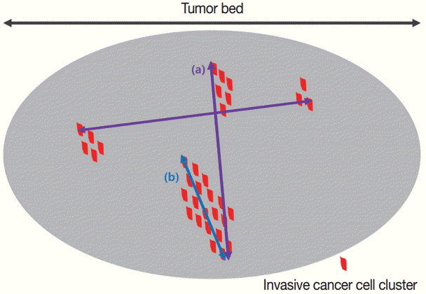
- There are many potential variables that can be used for assessing tumor size/extent in breast cancer patients who received NAT. Variable relevance depends on which tumor-response evaluation system is being used, because each system offers a different definition of significant tumor size. For example, the largest contiguous focus of the invasive cancer is the most important factor in determining ypT stage in ypTNM system [10]. Contrarily, the two dimensions of the largest residual area of remaining invasive cancer are most important according to the Residual Cancer Burden (RCB) system [14]. For the RCB system, however, the residual invasive cancer does not need to be contiguous, leading to a discrepancy in perceived tumor size between the two systems [14]. The largest discrepancies in tumor size/extent due to the differences in measurement methods were observed when the tissue response pattern after NAT manifested as a scattered pattern (Fig. 2).
- Tumor cellularity
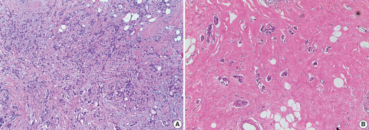
- Though NAT can affect several parameters of breast cancer, tumor cellularity is one of the most representative factors [25]. Tumor cellularity is not always recorded in pathologic reports because it is important in some tumor-response systems [11,13,14] but not in others [10,12,26]. There are several factors that should always be considered when evaluating tumor cellularity. The first factor is the comparison of cellularity in pre- and post-treatment specimens (Fig. 3). Differences between pre- and post-treatment cellularity are important for some tumor-response systems [11]; however, pretreatment cellularity is not considered in the RCB system [14]. The second factor is tumor heterogeneity. Because residual tumor cellularity can appear heterogeneous after NAT, extensive tissue sampling should be performed. However, the majority of systems do not specifically include the cellularity of residual heterogeneous tumors, except for the RCB system, which recommends mentioning the average tumor cellularity [14].
- Lymphovascular invasion
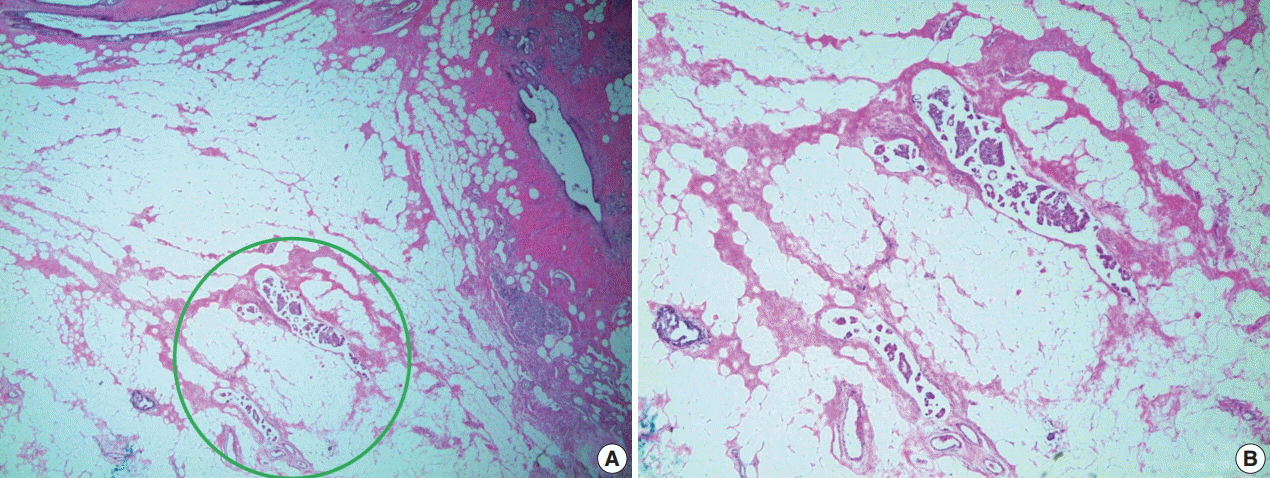
- Lymphovascular invasion (LVI) is documented in most pathologic reports because it is a significant prognostic factor in non-neoadjuvant breast cancer [27,28]. Though there are insufficient data on whether LVI is independently significant in neoadjuvant specimens, it should still be mentioned in pathologic reports [20]. Ductal carcinoma in situ (DCIS) and LVI are defined as resistant breast cancer components after NAT [22]. Therefore, in some situations, the only residual after NAT is tumor emboli in lymphovascular space, with no residual tumor in the breast parenchyma (Fig. 4) [29]. According to these guidelines, researchers have recommended that such cases not be regarded as pathologic complete response (pCR) [20]. Consequently, several LVI measurement methods have been suggested, including measurement according to size [20] or using semi-quantitative methods (focal or extensive) [30].
- Surgical margins
- Evaluation of resection margins is identical to that for non-neoadjuvant breast cancer specimens. Careful examination is required for evaluation of resection margins in neoadjuvant specimens because grossly invisible residual tumors or multiple scattered microscopic tumor foci are common. Furthermore, when the resection margin involves the tumor bed, it should be documented in the pathological report.
- Evaluation of the axillary lymph node after NAT
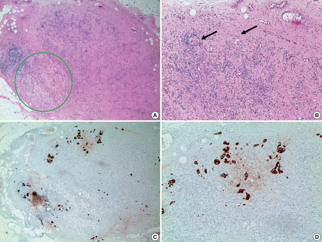
- The evaluation method for axillary lymph nodes is the same as that for non-neoadjuvant cases. Generally, all lymph nodes are sectioned into 2-mm intervals and sampled in their entirety for microscopic evaluation. Sometimes lymph nodes with complete treatment response are observed under microscopic evaluation for characteristic features, such as fibrous scarring, lymphocytic depletion, or histiocytic aggregation, without any identifiable tumor cell clusters (Fig. 5). If lymph nodes with these features are identified during microscopic evaluation, the total number observed should be noted in the pathologic report [31]. When metastatic deposits are observed, the size of the largest metastatic tumor and presence/absence of extranodal extension should be recorded. It is difficult to measure the size of the largest metastatic tumor when the treatment response is accompanied by metastasis. In cases with multiple singly scattered tumor cells that involve the entire lymph node and when the treatment response is not accompanied by fibrosis, the size of the metastatic tumor is determined by measuring the size of the largest cell cluster. Some guidelines recommend measuring the sizes of the tumor cells and intervening stroma—not the largest cell cluster—when accompanied by a tumor response; consensus for these measurements has not been established among the various evaluation systems [20]. Thus, when metastatic deposits are observed during microscopic evaluation, conditions such as macrometastasis, micrometastasis, and isolated tumor cells can be altered by changes in the sizes of metastatic deposits according to applied systems. However, residual disease in the lymph nodes are not considered pCR in most systems [10,20].
- Pathologic complete response
- Though each system that evaluates treatment responses to NAT has a unique definition of pCR, all systems record whether the patient has invasive carcinoma and whether it is identified in the breast parenchyma [9-14]. Significant differences among these evaluation systems are based on the inclusion or exclusion of DCIS and axillary lymph node status. Thus, description of DCIS and axillary lymph node status should always be included in pathologic reports because the treatment response evaluation systems differ across institutions.
- Re-evaluation of biomarkers in breast cancer after NAT
- Estrogen receptor (ER), progesterone receptor (PR), and human epidermal growth factor receptor-2 (HER-2), which are representative biomarkers of breast cancer, should be used for evaluating invasive breast cancer; however, there is no consensus on whether ER, PR, and HER-2 status should be re-evaluated in breast cancer patients who received NAT. Different guidelines suggest different processes based on core biopsy results, because ER, PR, and HER-2 statuses after NAT are evaluated based on the biomarker status of pretreatment core biopsy. If ER, PR, and HER-2 statuses from pre-treatment core biopsy are all positive, there will be no changes in status for most patients; thus, reevaluation is generally not recommended. However, re-evaluation is considered necessary in the following circumstances: (1) negative or equivocal results in core biopsy, (2) only DCIS or insufficient invasive carcinoma in core biopsy, (3) core biopsy performed at another institute, and (4) no treatment response [20,21]. Additionally, re-evaluation should be performed when the patient is enrolled in a clinical trial protocol or when ER, PR, or HER-2 status is unknown.
EVALUATION OF BREAST CANCER SPECIMENS AFTER NEOADJUVANT THERAPY
- The number of existing post-NAT breast cancer specimens has recently increased because NAT is now established as an effective treatment approach for patients with large or locally advanced breast cancer and for cases of early-stage breast cancer. However, guidelines for pathologic evaluation of breast cancer after NAT have not been established; instead, there are several evaluation systems, each with different major main-effect variables. Moreover, from macroscopic examination to microscopic evaluation, there are several obstacles to pathologic evaluation of neoadjuvant breast cancer because there is a diverse range of histologic responses to NAT. Pathologic evaluation of residual disease is the most essential component of post-NAT breast cancer evaluation. Thus, the evaluation should be conducted based on close comparisons and correlations between clinical, radiologic, and pathologic findings.
CONCLUSIONS
Acknowledgments
| System | Included variable | Definition of pCR | Category status | Reference |
|---|---|---|---|---|
| AJCC (y) | Size of invasive carcinoma | No invasive carcinoma in breast and lymph node | Stage 0 | Boughey et al. [8] |
| Lymph nose status (the number of metastatic lymph node and size of metastatic deposit) | Stage 1 | |||
| Stage 2 | ||||
| Stage 3 | ||||
| B-18 | Treatment effect in invasive carcinoma | No invasive carcinoma in breast and lymph node | No pathologic response | Diaz et al. [24] |
| Lymph nose status (the number of metastatic lymph node and size of metastatic deposit) | Pathologic partial response | |||
| Pathologic complete response | ||||
| Miller-Payne | Presence of invasive carcinoma | No invasive carcinoma in breast | Grade 1: no change or some minor alteration in individual malignant cells, but no reduction in overall cellularity | Mamounas et al. [9] |
| Tumor cellularity | Grade 2: a minor loss of tumor cells, but overall high cellularity; up to 30% reduction of cellularity | |||
| Grade 3: between an estimated 30% and 90% reduction in tumor cellularity | ||||
| Grade 4: a marked disappearance of more than 90% of tumor cells such that only small clusters or widely dispersed individual cells remain | ||||
| Grade 5: no invasive malignant cells identifiable in sections from the site of the tumor | ||||
| MNPI | Size of invasive carcinoma | No invasive carcinoma in breast and lymph node | MNPI=0.2×tumor size+lymph node stage+MSBR grade | Carey et al. [10] |
| Tumor grade | Lymph node state: 1, node negative; 2, 1-3 positive; 3, ≥4 positive | |||
| Lymph nose status (the number of metastatic lymph node) | ||||
| Pinder | Tumor proportion (%) in remaining breast | No invasive carcinoma in breast and lymph node | Complete pathologic response | Ogston et al. [11] |
| Lymph nose status (presence of evidence of response) | Partial response to therapy | |||
| < 10% of tumor remaining | ||||
| 10%-50% of tumor remaining | ||||
| > 50% of tumor remaining | ||||
| No evidence of response | ||||
| RCB | Size of tumor bed in two dimension | No invasive carcinoma in breast and lymph node | RCB 0: no residual disease | Abrial et al. [12] |
| Tumor cellularity | RCB 1: minimal residual disease | |||
| Lymph node status (the number of metastatic lymph node and size of metastatic deposit) | RCB 2: moderate residual disease | |||
| RCB 3: extensive residual disease |
- 1. Thompson AM, Moulder-Thompson SL. Neoadjuvant treatment of breast cancer. Ann Oncol 2012; 23 Suppl 10: x231-6. Article
- 2. Moreno-Aspitia A. Neoadjuvant therapy in early-stage breast cancer. Crit Rev Oncol Hematol 2012; 82: 187-99. ArticlePubMed
- 3. Pierga JY, Mouret E, Laurence V, et al. Prognostic factors for survival after neoadjuvant chemotherapy in operable breast cancer: the role of clinical response. Eur J Cancer 2003; 39: 1089-96. ArticlePubMed
- 4. Kuerer HM, Newman LA, Smith TL, et al. Clinical course of breast cancer patients with complete pathologic primary tumor and axillary lymph node response to doxorubicin-based neoadjuvant chemotherapy. J Clin Oncol 1999; 17: 460-9. ArticlePubMed
- 5. Fisher B, Bryant J, Wolmark N, et al. Effect of preoperative chemotherapy on the outcome of women with operable breast cancer. J Clin Oncol 1998; 16: 2672-85. ArticlePubMed
- 6. Esserman LJ, Woodcock J. Accelerating identification and regulatory approval of investigational cancer drugs. JAMA 2011; 306: 2608-9. ArticlePubMed
- 7. Bonadonna G, Veronesi U, Brambilla C, et al. Primary chemotherapy to avoid mastectomy in tumors with diameters of three centimeters or more. J Natl Cancer Inst 1990; 82: 1539-45. ArticlePubMed
- 8. Boughey JC, Peintinger F, Meric-Bernstam F, et al. Impact of preoperative versus postoperative chemotherapy on the extent and number of surgical procedures in patients treated in randomized clinical trials for breast cancer. Ann Surg 2006; 244: 464-70. ArticlePubMedPMC
- 9. Mamounas EP, Anderson SJ, Dignam JJ, et al. Predictors of locoregional recurrence after neoadjuvant chemotherapy: results from combined analysis of National Surgical Adjuvant Breast and Bowel Project B-18 and B-27. J Clin Oncol 2012; 30: 3960-6. ArticlePubMedPMC
- 10. Carey LA, Metzger R, Dees EC, et al. American Joint Committee on Cancer tumor-node-metastasis stage after neoadjuvant chemotherapy and breast cancer outcome. J Natl Cancer Inst 2005; 97: 1137-42. ArticlePubMed
- 11. Ogston KN, Miller ID, Payne S, et al. A new histological grading system to assess response of breast cancers to primary chemotherapy: prognostic significance and survival. Breast 2003; 12: 320-7. ArticlePubMed
- 12. Abrial SC, Penault-Llorca F, Delva R, et al. High prognostic significance of residual disease after neoadjuvant chemotherapy: a retrospective study in 710 patients with operable breast cancer. Breast Cancer Res Treat 2005; 94: 255-63. ArticlePubMed
- 13. Pinder SE, Provenzano E, Earl H, Ellis IO. Laboratory handling and histology reporting of breast specimens from patients who have received neoadjuvant chemotherapy. Histopathology 2007; 50: 409-17. ArticlePubMed
- 14. Symmans WF, Peintinger F, Hatzis C, et al. Measurement of residual breast cancer burden to predict survival after neoadjuvant chemotherapy. J Clin Oncol 2007; 25: 4414-22. ArticlePubMed
- 15. Dash N, Chafin SH, Johnson RR, Contractor FM. Usefulness of tissue marker clips in patients undergoing neoadjuvant chemotherapy for breast cancer. AJR Am J Roentgenol 1999; 173: 911-7. ArticlePubMed
- 16. Oh JL, Nguyen G, Whitman GJ, et al. Placement of radiopaque clips for tumor localization in patients undergoing neoadjuvant chemotherapy and breast conservation therapy. Cancer 2007; 110: 2420-7. ArticlePubMedPMC
- 17. Schulz-Wendtland R, Heywang-Köbrunner SH, Aichinger U, Krämer S, Wenkel E, Bautz W. Do tissue marker clips after sonographically or stereotactically guided breast biopsy improve follow-up of small breast lesions and localisation of breast cancer after chemotherapy? Rofo 2002; 174: 620-4. ArticlePubMed
- 18. Youn I, Choi SH, Kook SH, et al. Ultrasonography-guided surgical clip placement for tumor localization in patients undergoing neoadjuvant chemotherapy for breast cancer. J Breast Cancer 2015; 18: 44-9. ArticlePubMedPMC
- 19. Margolin FR, Kaufman L, Denny SR, Jacobs RP, Schrumpf JD. Metallic marker placement after stereotactic core biopsy of breast calcifications: comparison of two clips and deployment techniques. AJR Am J Roentgenol 2003; 181: 1685-90. ArticlePubMed
- 20. Provenzano E, Bossuyt V, Viale G, et al. Standardization of pathologic evaluation and reporting of postneoadjuvant specimens in clinical trials of breast cancer: recommendations from an international working group. Mod Pathol 2015; 28: 1185-201. ArticlePubMed
- 21. Bossuyt V, Provenzano E, Symmans WF, et al. Recommendations for standardized pathological characterization of residual disease for neoadjuvant clinical trials of breast cancer by the BIG-NABCG collaboration. Ann Oncol 2015; 26: 1280-91. ArticlePubMedPMC
- 22. Sharkey FE, Addington SL, Fowler LJ, Page CP, Cruz AB. Effects of preoperative chemotherapy on the morphology of resectable breast carcinoma. Mod Pathol 1996; 9: 893-900. ArticlePubMed
- 23. Honkoop AH, Pinedo HM, De Jong JS, et al. Effects of chemotherapy on pathologic and biologic characteristics of locally advanced breast cancer. Am J Clin Pathol 1997; 107: 211-8. ArticlePubMed
- 24. Diaz J, Stead L, Shapiro N, et al. Mitotic counts in breast cancer after neoadjuvant systemic chemotherapy and development of metastatic disease. Breast Cancer Res Treat 2013; 138: 91-7. ArticlePubMed
- 25. Rajan R, Poniecka A, Smith TL, et al. Change in tumor cellularity of breast carcinoma after neoadjuvant chemotherapy as a variable in the pathologic assessment of response. Cancer 2004; 100: 1365-73. ArticlePubMed
- 26. Fisher ER, Wang J, Bryant J, Fisher B, Mamounas E, Wolmark N. Pathobiology of preoperative chemotherapy: findings from the National Surgical Adjuvant Breast and Bowel (NSABP) protocol B-18. Cancer 2002; 95: 681-95. ArticlePubMed
- 27. Leitner SP, Swern AS, Weinberger D, Duncan LJ, Hutter RV. Predictors of recurrence for patients with small (one centimeter or less) localized breast cancer (T1a,b N0 M0). Cancer 1995; 76: 2266-74. ArticlePubMed
- 28. Lee AK, Loda M, Mackarem G, et al. Lymph node negative invasive breast carcinoma 1 centimeter or less in size (T1a,bNOMO): clinicopathologic features and outcome. Cancer 1997; 79: 761-71. ArticlePubMed
- 29. Rabban JT, Glidden D, Kwan ML, Chen YY. Pure and predominantly pure intralymphatic breast carcinoma after neoadjuvant chemotherapy: an unusual and adverse pattern of residual disease. Am J Surg Pathol 2009; 33: 256-63. ArticlePubMed
- 30. Colleoni M, Rotmensz N, Maisonneuve P, et al. Prognostic role of the extent of peritumoral vascular invasion in operable breast cancer. Ann Oncol 2007; 18: 1632-40. ArticlePubMed
- 31. Sahoo S, Lester SC. Pathology of breast carcinomas after neoadjuvant chemotherapy: an overview with recommendations on specimen processing and reporting. Arch Pathol Lab Med 2009; 133: 633-42. ArticlePubMedPDF
REFERENCES
Figure & Data
References
Citations

- Good practice: The experiences with the utilization of residual cancer burden—A single institution study
Anita Sejben, Fanni Hegedűs, Szintia Almási, Márton Berta, Orsolya Oláh‐Németh, Tamás Zombori
Thoracic Cancer.2023; 14(11): 963. CrossRef - Assessing the Correlation of Rate of Pathological Complete Response and Outcome in Post Neoadjuvant Chemotherapy Setting and Molecular Subtypes of Breast Cancer
Ahmad Omair, Abdulmohsen Alkushi, Ghaida Alamri, Talal Almojel, Sara Alsadun, Emad Masuadi, Haitham Arabi, Amin E Mohamed, Omalkhair A Abulkhair
Cureus.2023;[Epub] CrossRef - Compression OCT-elastography combined with speckle-contrast analysis as an approach to the morphological assessment of breast cancer tissue
Anton A. Plekhanov, Ekaterina V. Gubarkova, Marina A. Sirotkina, Alexander A. Sovetsky, Dmitry A. Vorontsov, Lev A. Matveev, Sergey S. Kuznetsov, Alexandra Y. Bogomolova, Alexey Y. Vorontsov, Alexander L. Matveyev, Sergey V. Gamayunov, Elena V. Zagaynova,
Biomedical Optics Express.2023; 14(6): 3037. CrossRef - Ambiguity-aware breast tumor cellularity estimation via self-ensemble label distribution learning
Xiangyu Li, Xinjie Liang, Gongning Luo, Wei Wang, Kuanquan Wang, Shuo Li
Medical Image Analysis.2023; 90: 102944. CrossRef - Concurrent Chemo-radiation As a Means of Achieving Pathologic Complete Response in Triple Negative Breast Cancer
Maryam Nemati Shafaee, Shalini Makawita, Bora Lim, Matthew J Ellis, Michelle S Ludwig
Clinical Breast Cancer.2022; 22(4): e536. CrossRef - Pathology after neoadjuvant treatment – How to assess residual disease
Giuseppe Viale, Nicola Fusco
The Breast.2022; 62: S25. CrossRef - Efficacy Evaluation of Neoadjuvant Chemotherapy in Breast Cancer by MRI
Yongguang Liu, Mingxiang Wu, Wenyong Tan, Jingshan Gong, Jie Ma, Mohammad Farukh Hashmi
Contrast Media & Molecular Imaging.2022;[Epub] CrossRef - Predictive Role of Soluble IL-6R, TNF-R1/2, and Cell Adhesion Molecules Serum Levels in the Preoperative and Adjuvant Therapy in Women with Nonmetastatic Breast Cancer: A Preliminary Study
Weronika Bulska-Będkowska, Paulina Czajka-Francuz, Sylwia Cisoń-Jurek, Aleksander J. Owczarek, Tomasz Francuz, Jerzy Chudek
Journal of Interferon & Cytokine Research.2022; 42(11): 557. CrossRef - The prognostic role of lymph node ratio in breast cancer patients received neoadjuvant chemotherapy: A dose-response meta-analysis
Jinzhao Liu, Yifei Li, Weifang Zhang, Chenhui Yang, Chao Yang, Liang Chen, Mingjian Ding, Liang Zhang, Xiaojun Liu, Guozhong Cui, Yunjiang Liu
Frontiers in Surgery.2022;[Epub] CrossRef - The Role of miR-375-3p, miR-210-3p and Let-7e-5p in the Pathological Response of Breast Cancer Patients to Neoadjuvant Therapy
Lorena Alexandra Lisencu, Andrei Roman, Simona Visan, Eduard-Alexandru Bonci, Andrei Pașca, Emilia Grigorescu, Elena Mustea, Andrei Cismaru, Alexandru Irimie, Cosmin Lisencu, Loredana Balacescu, Ovidiu Balacescu, Oana Tudoran
Medicina.2022; 58(10): 1494. CrossRef - Post-Neoadjuvant Treatment Strategies for Patients with Early Breast Cancer
Elisa Agostinetto, Flavia Jacobs, Véronique Debien, Alex De Caluwé, Catalin-Florin Pop, Xavier Catteau, Philippe Aftimos, Evandro de Azambuja, Laurence Buisseret
Cancers.2022; 14(21): 5467. CrossRef - Tumor Microenvironment in Breast Cancer—Updates on Therapeutic Implications and Pathologic Assessment
Joshua J. Li, Julia Y. Tsang, Gary M. Tse
Cancers.2021; 13(16): 4233. CrossRef - SPIE-AAPM-NCI BreastPathQ challenge: an image analysis challenge for quantitative tumor cellularity assessment in breast cancer histology images following neoadjuvant treatment
Nicholas Petrick, Shazia Akbar, Kenny H. Cha, Sharon Nofech-Mozes, Berkman Sahiner, Marios A. Gavrielides, Jayashree Kalpathy-Cramer, Karen Drukker, Anne L. Martel, for the BreastPathQ Challenge Group
Journal of Medical Imaging.2021;[Epub] CrossRef - Diagnostic performance of digital breast tomosynthesis for predicting response to neoadjuvant systemic therapy in breast cancer patients: A comparison with magnetic resonance imaging, ultrasound, and full-field digital mammography
Ryusuke Murakami, Hitomi Tani, Shinichiro Kumita, Nachiko Uchiyama
Acta Radiologica Open.2021;[Epub] CrossRef - Los márgenes
Laia Bernet, María Angeles Montero Fernández
Revista de Senología y Patología Mamaria.2021; 34: S25. CrossRef - Neoadjuvant chemotherapy in non‐metastatic breast cancer: a study on practice trends in a regional cancer treatment service
Edmond Ang, Navin Wewala, Rebecca Carroll, Garry Forgeson, Malcolm Anderson, Jennifer Fernando, Jody Jordan, Richard Isaacs
Internal Medicine Journal.2020; 50(3): 315. CrossRef - Examination of Tumor Regression Grading Systems in Breast Cancer Patients Who Received Neoadjuvant Therapy
Anita Sejben, Renáta Kószó, Zsuzsanna Kahán, Gábor Cserni, Tamás Zombori
Pathology & Oncology Research.2020; 26(4): 2747. CrossRef - Integrating evolutionary dynamics into cancer therapy
Robert A. Gatenby, Joel S. Brown
Nature Reviews Clinical Oncology.2020; 17(11): 675. CrossRef - Assessing the accuracy of conventional gadolinium‐enhanced breast MRI in measuring the nodal response to neoadjuvant chemotherapy (NAC) in breast cancer
Lisa Christine Murphy, Edel Marie Quinn, Zeeshan Razzaq, Claire Brady, Vicki Livingstone, Lorna Duddy, Josephine Barry, Henry Paul Redmond, Mark Anthony Corrigan
The Breast Journal.2020; 26(11): 2151. CrossRef - Early prediction of neoadjuvant chemotherapy response for advanced breast cancer using PET/MRI image deep learning
Joon Ho Choi, Hyun-Ah Kim, Wook Kim, Ilhan Lim, Inki Lee, Byung Hyun Byun, Woo Chul Noh, Min-Ki Seong, Seung-Sook Lee, Byung Il Kim, Chang Woon Choi, Sang Moo Lim, Sang-Keun Woo
Scientific Reports.2020;[Epub] CrossRef - Patterns of Regression in Breast Cancer after Primary Systemic Treatment
Tamás Zombori, Gábor Cserni
Pathology & Oncology Research.2019; 25(3): 1153. CrossRef - The Role of Neutrophil-lymphocyte Ratio and Platelet-lymphocyte Ratio in Predicting Neoadjuvant Chemotherapy Response in Breast Cancer
Hee Yeon Kim, Tae Hyun Kim, Hye Kyoung Yoon, Anbok Lee
Journal of Breast Cancer.2019; 22(3): 425. CrossRef - Higher underestimation of tumour size post-neoadjuvant chemotherapy with breast magnetic resonance imaging (MRI)—A concordance comparison cohort analysis
Wen-Pei Wu, Hwa-Koon Wu, Chih-Jung Chen, Chih-Wie Lee, Shou-Tung Chen, Dar-Ren Chen, Chen-Te Chou, Chi Wei Mok, Hung-Wen Lai, Pascal A. T. Baltzer
PLOS ONE.2019; 14(10): e0222917. CrossRef - Multimodal image-guided surgery of HER2-positive breast cancer using [111In]In-DTPA-trastuzumab-IRDye800CW in an orthotopic breast tumor model
Marion M. Deken, Desirée L. Bos, Willemieke S. F. J. Tummers, Taryn L. March, Cornelis J. H. van de Velde, Mark Rijpkema, Alexander L. Vahrmeijer
EJNMMI Research.2019;[Epub] CrossRef - Mammographic density is a potential predictive marker of pathological response after neoadjuvant chemotherapy in breast cancer
Ida Skarping, Daniel Förnvik, Hanna Sartor, Uffe Heide-Jørgensen, Sophia Zackrisson, Signe Borgquist
BMC Cancer.2019;[Epub] CrossRef - ALDH1 and tumor infiltrating lymphocytes as predictors for neoadjuvant chemotherapy response in breast cancer
Anbok Lee, Kyu Yeoun Won, Sung-Jig Lim, Sun Young Cho, Sang-Ah Han, SaeGwang Park, Jeong-Yoon Song
Pathology - Research and Practice.2018; 214(5): 619. CrossRef - Early Prediction of Response to Neoadjuvant Chemotherapy Using Dynamic Contrast-Enhanced MRI and Ultrasound in Breast Cancer
Yunju Kim, Sung Hun Kim, Byung Joo Song, Bong Joo Kang, Kwang-il Yim, Ahwon Lee, Yoonho Nam
Korean Journal of Radiology.2018; 19(4): 682. CrossRef - Outcomes of neoadjuvant and adjuvant chemotherapy in stage 2 and 3 non-small cell lung cancer: an analysis of the National Cancer Database
Matthew MacLean, Xin Luo, Shidan Wang, Kemp Kernstine, David E. Gerber, Yang Xie
Oncotarget.2018; 9(36): 24470. CrossRef - Automatic cellularity assessment from post‐treated breast surgical specimens
Mohammad Peikari, Sherine Salama, Sharon Nofech‐Mozes, Anne L. Martel
Cytometry Part A.2017; 91(11): 1078. CrossRef - The importance of tissue confirmation of metastatic disease in patients with breast cancer: lesson from a brain metastasis case
Jingxian Ding, Pinghua Hu, Jun Chen, Xiaobo Wu, Yali Cao
Oncoscience.2016; 3(9-10): 268. CrossRef
 PubReader
 ePub Link
-
 Cite this Article
- Cite this Article
-
- Close
- Download Citation
- Close
- Figure


Fig. 1.
Fig. 2.
Fig. 3.
Fig. 4.
Fig. 5.
| System | Included variable | Definition of pCR | Category status | Reference |
|---|---|---|---|---|
| AJCC (y) | Size of invasive carcinoma | No invasive carcinoma in breast and lymph node | Stage 0 | Boughey et al. [8] |
| Lymph nose status (the number of metastatic lymph node and size of metastatic deposit) | Stage 1 | |||
| Stage 2 | ||||
| Stage 3 | ||||
| B-18 | Treatment effect in invasive carcinoma | No invasive carcinoma in breast and lymph node | No pathologic response | Diaz et al. [24] |
| Lymph nose status (the number of metastatic lymph node and size of metastatic deposit) | Pathologic partial response | |||
| Pathologic complete response | ||||
| Miller-Payne | Presence of invasive carcinoma | No invasive carcinoma in breast | Grade 1: no change or some minor alteration in individual malignant cells, but no reduction in overall cellularity | Mamounas et al. [9] |
| Tumor cellularity | Grade 2: a minor loss of tumor cells, but overall high cellularity; up to 30% reduction of cellularity | |||
| Grade 3: between an estimated 30% and 90% reduction in tumor cellularity | ||||
| Grade 4: a marked disappearance of more than 90% of tumor cells such that only small clusters or widely dispersed individual cells remain | ||||
| Grade 5: no invasive malignant cells identifiable in sections from the site of the tumor | ||||
| MNPI | Size of invasive carcinoma | No invasive carcinoma in breast and lymph node | MNPI=0.2×tumor size+lymph node stage+MSBR grade | Carey et al. [10] |
| Tumor grade | Lymph node state: 1, node negative; 2, 1-3 positive; 3, ≥4 positive | |||
| Lymph nose status (the number of metastatic lymph node) | ||||
| Pinder | Tumor proportion (%) in remaining breast | No invasive carcinoma in breast and lymph node | Complete pathologic response | Ogston et al. [11] |
| Lymph nose status (presence of evidence of response) | Partial response to therapy | |||
| < 10% of tumor remaining | ||||
| 10%-50% of tumor remaining | ||||
| > 50% of tumor remaining | ||||
| No evidence of response | ||||
| RCB | Size of tumor bed in two dimension | No invasive carcinoma in breast and lymph node | RCB 0: no residual disease | Abrial et al. [12] |
| Tumor cellularity | RCB 1: minimal residual disease | |||
| Lymph node status (the number of metastatic lymph node and size of metastatic deposit) | RCB 2: moderate residual disease | |||
| RCB 3: extensive residual disease |
| Pathologic report form |
|---|
| Gross finding |
| Residual identified tumor: yes/no |
| Quadrant of tumor |
| Multifocality: yes/no |
| Size of residual tumor: xx mm |
| Identified clip of marker: yes/no |
| Microscopic finding |
| Histologic diagnosis: invasive carcinoma, NST |
| Histologic grade: I/II/III (tubule score–nuclear grade–mitosis score) |
| Size of residual tumor bed: x mm |
| Size of the largest residual invasive carcinoma: x mm |
| Residual tumor cellularity: % |
| Lymphovascular invasion: absent/present |
| DCIS component: yes/no |
| Total tumor size including DCIS: x mm |
| Extensive intraductal component: yes/no |
| Type: cribriform/micropapillary/solid/papillary |
| Nuclear grade: low/intermediate/high |
| Necrosis: absent/present (focal/commedo) |
| ER/PR/HER-2 status: optional |
| Resection margin |
| Invasive carcinoma: absent/present; distance to the closest margin |
| DCIS: absent/present; distance to the closest margin |
| Tumor bed: absent/present |
| Lymph node status |
| Number of sentinel lymph nodes |
| Number of total axillary lymph nodes |
| Number of lymph nodes with macrometastasis |
| Size of largest metastasis: mm |
| Number of lymph nodes with micrometastasis |
| Number of lymph nodes with isolated tumor cells |
| Number of lymph nodes with histologic evidence of treatment response but no tumor cells |
| Extracapsular extension: yes/no |
pCR, pathologic complete response; AJCC, American Joint Committee on Cancer; MNPI, Modified scores from Nottingham Prognostic Index; MSBR grade, Modified Scarff Bloom Richardson grade; RCB, residual cancer burden.
NST, no specific type; DCIS, ductal carcinoma

 E-submission


