Articles
- Page Path
- HOME > J Pathol Transl Med > Volume 51(6); 2017 > Article
-
Original Article
Current Cytology Practices in Korea: A Nationwide Survey by the Korean Society for Cytopathology - Eun Ji Oh1, Chan Kwon Jung,1,2, Dong-Hoon Kim3, Han Kyeom Kim4, Wan Seop Kim5, So-Young Jin6, Hye Kyoung Yoon7, The Fellowship Council and Committee of Quality Improvement of the Korean Society for Cytopathology
-
Journal of Pathology and Translational Medicine 2017;51(6):579-587.
DOI: https://doi.org/10.4132/jptm.2017.08.11
Published online: September 27, 2017
1Department of Hospital Pathology, College of Medicine, The Catholic University of Korea, Seoul, Korea
2Cancer Research Institute, College of Medicine, The Catholic University of Korea, Seoul, Korea
3Department of Pathology, Kangbuk Samsung Hospital, Sungkyunkwan University School of Medicine, Seoul, Korea
4Department of Pathology, Korea University Guro Hospital, Korea University College of Medicine, Seoul, Korea
5Department of Pathology, Konkuk University School of Medicine, Seoul, Korea
6Department of Pathology, Soonchunhyang University Seoul Hospital, Soonchunhyang University College of Medicine, Seoul, Korea
7Department of Pathology, Inje University Busan Paik Hospital, Inje University College of Medicine, Busan, Korea
- Corresponding Author Chan Kwon Jung, MD, PhD Department of Hospital Pathology, Seoul St. Mary’s Hospital, College of Medicine, The Catholic University of Korea, 222 Banpo-daero, Seocho-gu, Seoul 06591, Korea Tel: +82-2-2258-1622 Fax: +82-2-2258-1627 E-mail: ckjung@catholic.ac.kr
© 2017 The Korean Society of Pathologists/The Korean Society for Cytopathology
This is an Open Access article distributed under the terms of the Creative Commons Attribution Non-Commercial License (http://creativecommons.org/licenses/by-nc/4.0) which permits unrestricted non-commercial use, distribution, and reproduction in any medium, provided the original work is properly cited.
Abstract
-
Background
- Limited data are available on the current status of cytology practices in Korea. This nationwide study presents Korean cytology statistics from 2015.
-
Methods
- A nationwide survey was conducted in 2016 as a part of the mandatory quality-control program by the Korean Society for Cytopathology. The questionnaire was sent to 208 medical institutions performing cytopathologic examinations in Korea. Individual institutions were asked to submit their annual cytology statistical reports and gynecologic cytology-histology correlation data for 2015.
-
Results
- Responses were obtained from 206 medical institutions including 83 university hospitals, 87 general hospitals, and 36 commercial laboratories. A total of 8,284,952 cytologic examinations were performed in 2015, primarily in commercial laboratories (74.9%). The most common cytology specimens were gynecologic samples (81.3%). Conventional smears and liquid-based cytology were performed in 6,190,526 (74.7%) and 2,094,426 (25.3%) cases, respectively. The overall diagnostic concordance rate between cytologic and histologic diagnoses of uterine cervical samples was 70.5%. Discordant cases were classified into three categories: category A (minimal clinical impact, 17.4%), category B (moderate clinical impact, 10.2%), and category C (major clinical impact, 1.9%). The ratio of atypical squamous cells of undetermined significance to squamous intraepithelial lesion was 1.6 in university hospitals, 2.9 in general hospitals, and 4.9 in commercial laboratories.
-
Conclusions
- This survey reveals the current status and trend of cytology practices in Korea. The results of this study can serve as basic data for the establishment of nationwide cytopathology policies and quality improvement guidelines in Korean medical institutions.
- The QI Committee of the Korean Society for Cytopathology conducted a nationwide quality control survey in cytopathology from February 2, 2016, to February 22, 2016. The questionnaire was sent to 208 medical institutions performing cytopathologic examinations in Korea in order to gather statistical data on cytology and gynecologic (GYN) cytology-histology correlation results from 2015. Written informed consent was obtained from each institution. This study was approved by the Institutional Review Board of The Catholic University of Korea, Seoul St. Mary’s Hospital (KC13SISI0198).
- For the purposes of this study, medical institutions were categorized into three groups: university hospitals, general hospitals, and commercial laboratories. All cytology samples were also classified into three groups: GYN, fine needle aspiration (FNA), and non-GYN/non-FNA samples including body fluids, urine, bronchial washing/brushing samples, cerebrospinal fluid, etc. The diagnostic concordance between cytologic and corresponding histologic examinations of the uterine cervix was categorized as either concordant (category O) or into one of three discordant categories: category A (minimal clinical impact), category B (moderate clinical impact), and category C (major clinical impact). The criteria for the assessment of diagnostic accuracy are shown in Table 1.
- Statistical analysis was performed using GraphPad Prism software ver. 6.05 (GraphPad Software, La Jolla, CA). p-values less than .05 were considered statistically significant. Graphs were generated using GraphPad Prism and Microsoft Excel.
MATERIALS AND METHODS
- Responses were obtained from 206 out of 208 medical institutions (99.0%) including 83 university hospitals, 87 general hospitals, and 36 commercial laboratories.
- Statistics of all cytopathology cases in 2015
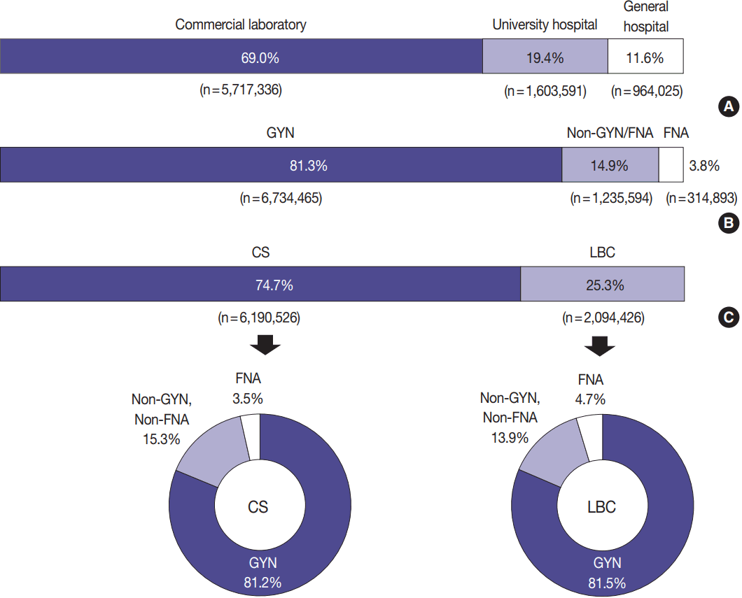
- The total number of cytopathologic examinations performed in 2015 was 8,284,952, of which 5,717,336 (69.0%) were performed in commercial laboratories, 1,603,591 (19.4%) in university hospitals, and 964,025 (11.6%) in general hospitals (Fig. 1A).
- Out of a total of 8,284,952 samples examined, 6,734,465 (81.3%) were classified as GYN, 314,893 (3.8%) as FNA, and 1,235,594 (14.9%) as non-GYN/non-FNA (Fig. 1B).
- In all, 6,190,526 (74.7%) cytology samples were prepared for conventional smear (CS) and 2,094,426 (25.3%) were prepared for LBC. Among the CS samples, 81.2% were GYN, 3.5% were FNA, and 15.3% were non-GYN/non-FNA. Among the LBC samples, 81.5% were GYN, 4.7% were FNA, and 13.9% were non-GYN/non-FNA (Fig. 1C).
- Statistics of GYN cytology
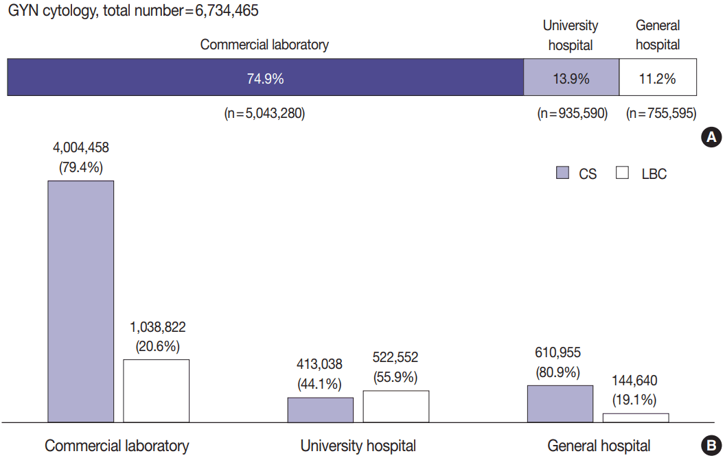
- The number of GYN cytology samples was 6,734,465. GYN cytology samples were examined mainly at commercial laboratories (74.9%, n = 5,043,280) and followed by university hospitals (13.9%, n = 935,590) and general hospitals (11.2%, n = 755,595) (Fig. 2A).
- GYN cytology samples in commercial laboratories and general hospitals were more frequently examined by CS than LBC, whereas university hospitals preferred LBC over CS (Fig. 2B).
- Statistics of FNA cytology
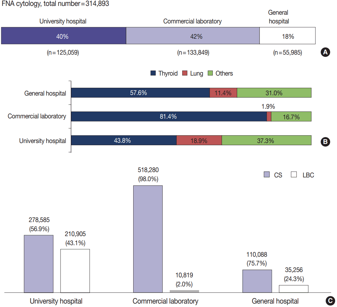
- A total of 314,893 FNA cytology samples were examined. Among them, 133,849 (42%) were examined in commercial laboratories, 125,059 (40%) in university hospitals, and 55,985 (18%) in general hospitals (Fig. 3A).
- The most common specimens were thyroid followed by lung (Fig. 3B). FNA Samples in university hospitals showed the highest ratio of LBC to CS followed by general hospitals and commercial laboratories in descending order (Fig. 3C).
- Statistics of non-GYN, non-FNA cytology
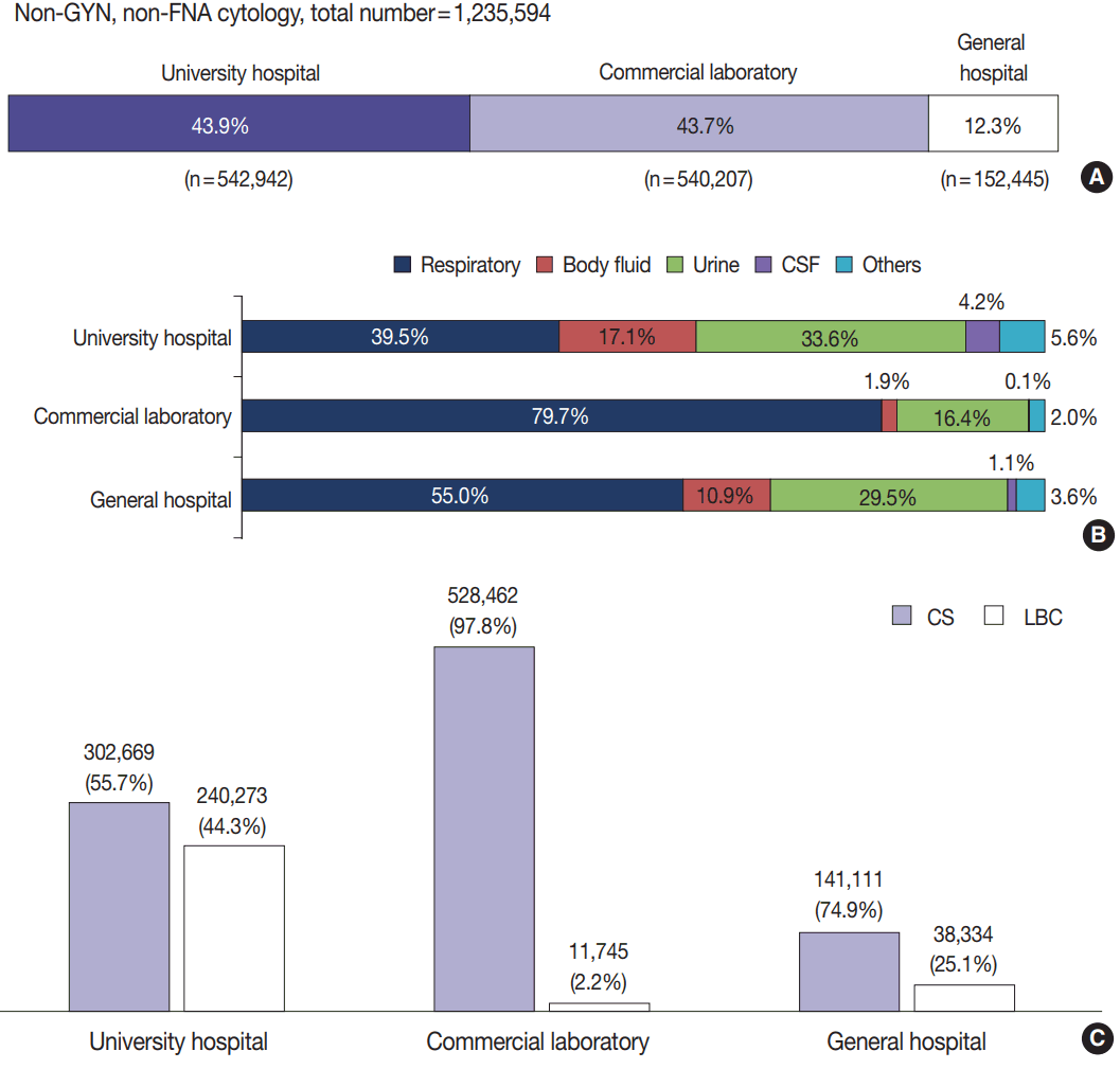
- A total of 1,235,594 non-GYN/non-FNA cytology samples were examined, consisting of 542,942 (43.9%) from university hospitals, 540,207 (43.7%) from commercial laboratories, and 152,445 (12.3%) from general hospitals (Fig. 4A). The proportion of each specimen type (respiratory, body fluid, urine, cerebrospinal fluid, etc.) is shown in Fig. 4B. The most common type of non-GYN/non-FNA specimen was respiratory cytology, regardless of the type of institution. The ratio of LBC to CS was higher in university and general hospitals than in commercial laboratories (Fig. 4C).
- Methods of LBC preparation
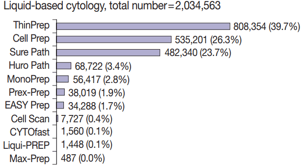
- A total of 11 methods for LBC preparation were used: ThinPrep (Hologic, Marlborough, MA, USA), Cell Prep (Biodyne, Seongnam, Korea), SurePath (BD Diagnostics, Sparks, MD, USA), Huro Path (CelltraZone, Seoul, Korea), MonoPrep (MonoGen, Arlington Heights, IL, USA), Prex-Prep, EASY Prep (YD Diagnostics, Yongin, Korea), Cell Scan (Cell & Tech Bio, Seoul, Korea), CYTO-fast (Hospitex Diagnostics, Firenze, Italy), Liqui-PREP (LGM International, Melbourne, FL, USA), and Max-Prep (Fig. 5). The number of medical institutions using each type of LBC preparation method is listed here in descending order: ThinPrep (68), SurePath (44), Cell Prep (17), EASY Prep (9), Huro Path (5), Cell Scan (4), and Prex-Prep (2). Only one institution used a preparation other than the aforementioned methods. The most commonly used LBC method was ThinPrep (39.7%) followed by Cell Prep (26.3%) and SurePath (23.7%); the others methods accounted for less than 5% of LBC cases. In GYN cytology specimens, ThinPrep (38.8%) was most commonly used followed by Cell Prep (30.0%) and SurePath (19.4%). In non-GYN/non-FNA cytology specimens, SurePath (49.9%) was most commonly used followed by ThinPrep (44.7%) and Cell Prep (2.6%). In FNA cytology specimens, ThinPrep (45.0%) was most commonly used followed by SurePath (33.6%), EASY Prep (12.9%), and Huro Path (4.5%).
- Prevalence of cytologic diagnoses of the uterine cervix
- Of the GYN cytology samples, 95% were diagnosed as negative, 3% as atypical squamous cells of undetermined significance (ASCUS), and 1% as low-grade squamous intraepithelial lesion (LSIL). In each type of institution, 1% of samples were considered to be unsatisfactory. The frequencies of unsatisfactory, negative, ASCUS, atypical squamous cells-cannot exclude high-grade squamous intraepithelial lesion, atypical glandular cells (AGC), AGC-favor neoplastic, LSIL, high-grade squamous intraepithelial lesion, adenocarcinoma in situ, squamous cell carcinoma, adenocarcinoma, etc. are shown in Table 2.
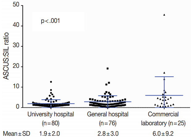
- The ratio of atypical squamous cells of undetermined significance to squamous intraepithelial lesion (ASCUS:SIL), representing the screening sensitivity, was significantly higher in commercial laboratories (6.0 ± 9.2) than in university hospitals (1.9 ± 2.0) and general hospitals (2.8 ± 3.0) (p < .001) (Fig. 6).
- Diagnostic accuracy of GYN cytology
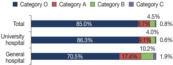
- Statistical data on diagnostic accuracy were obtained from 77 university hospitals and 54 general hospitals (Fig. 7). In university hospitals, a total of 44,044 GYN cytology cases were compared with matching histologic specimens with the following results: 9.1% in category A, 4.0% in category B, 0.6% in category C, and 86.3% in category O. In general hospitals, a total of 3,898 GYN cytology cases were compared with matching histologic specimens with the following results: 17.4% in category A, 10.2% in category B, 1.9% in category C, and 69.5% in category O. Accuracy data from commercial laboratories was not available.
RESULTS
- The survey response rate for this study was quite high. In all, 99.0% of medical institutions providing cytopathology service responded to this survey as mandated by the QI program of the Korean Society for Cytopathology.
- GYN cytology comprised 81.3% of all cases, which is due to the national cervical cancer screening program. Of the GYN cytology samples, 74.9% were examined at commercial laboratories, 13.9% at university hospitals, and 11.2% at general hospitals. In Korea, Pap smears are generally performed in primary care clinics and health promotion centers, and most of those specimens are then sent to commercial laboratories.
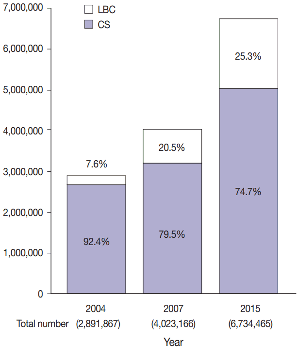
- The conventional Pap smear is a simple and effective method for screening cervical cancer. LBC was initially introduced in GYN cytology, and its use has increased continuously over the past two decades. According to a previous nationwide study in Korea, the use of LBC comprised 7.6% and 20.5% of all cytology cases in 2004 and 2007, respectively (Fig. 8) [1]. In 2015, LBC was used in 25.3% of all GYN cytology cases. In university hospitals, 55.9% of all GYN cytology were examined by LBC, whereas commercial laboratories used LBC less frequently (Fig. 2B).
- In the field of non-GYN cytology, LBC is only used for body fluid and thyroid aspiration samples by the National Health Insurance System in Korea. The proportion of LBC in non-GYN cytology cases was higher in university hospitals than in other types of institutions (Figs. 3C, 4C). The thyroid gland was the highest source of FNA samples (Fig. 3B). The high number of thyroid FNA cases in Korea closely correlates with the high rate of thyroid cancer screening, as Korea has the highest incidence of thyroid cancer in the world [4]. Although 11 methods of LBC were introduced in Korea, more than 95% of cases used either ThinPrep, Cell Prep, or SurePath. This is the first report to investigate the current status of the LBC methods used in Korea.
- The overall rates of unsatisfactory, negative, and ASCUS for GYN cytology cases were 0.6%, 94.6%, and 3.5% respectively. These results did not significantly differ among the three types of medical institutions. The ASCUS rate may have varied according to the cytopathologist performing the exam. The ASCUS:SIL ratio was less affected than the ASCUS rate by patient population, prevalence of disease, and the effect of screening [5-10]. Thus, the ASCUS:SIL ratio was used as a quality control reference [10]. In one study, cytotechnologists with an ASCUS:SIL ratio < 1.5 showed a significantly lower sensitivity than those with a ratio > 3.0.5 In our study, the mean ASCUS:SIL ratio was 1.9 in university hospitals, 2.8 in general hospitals, and 6.0 in commercial laboratories (Fig. 6). Thus, the mean screening sensitivity of commercial laboratories may be higher when compared with referral hospitals.
- Quality control of diagnostic accuracy was assessed by correlating cervical cytology with histologic results as mandated in university hospitals and general hospitals by the QI program of the Korean Society for Cytopathology. However, this correlation was not performed in most commercial laboratories. The concordance rate of cytologic and histologic diagnoses may vary depending upon the time difference between the two examinations [11-13]. Diagnostic correlations between cytology and histology are lower when the two examinations are performed simultaneously than when cytology precedes histologic examination [11,12]. During this study, we recognized that there was no standardization of cytologic-histologic correlation methods, including the type of histologic specimens, the intervals for correlation, and the statistical metrics used. In our study, cytologic-histologic correlation was performed in real time and/or retrospectively. The tissue samples for histologic correlation were obtained from either cervical biopsy, conization, or hysterectomy.
- We found that the volume of cytology cases, sample type, preparation method, access to paired tissue specimens, practice variables, and screening sensitivity all differed from institution to institution. In general, both the total volume of cytology cases and the proportion of cases using LBC have significantly increased. The screening sensitivity of GYN cytology was higher and more variable in commercial laboratories when compared with university and general hospitals. The results of this study can serve as basic data for the establishment of nationwide cytopathology policies and QI guidelines in Korean medical institutions.
DISCUSSION
Acknowledgments
Values are presented as number (%).
ASCUS, atypical squamous cells of undetermined significance; ASC-H, atypical squamous cells cannot exclude high-grade squamous intraepithelial lesion; LSIL, low-grade squamous intraepithelial lesion; HSIL, high-grade squamous intraepithelial lesion; AGC, atypical glandular cells.
- 1. Lee HK, Kim SN, Khang SK, Kang CS, Yoon HK. Quality control program and its results of Korean Society for Cytopathologists. Korean J Cytopathol 2008; 19: 65-71. Article
- 2. Kim Y, Jun JK, Choi KS, Lee HY, Park EC. Overview of the National Cancer screening programme and the cancer screening status in Korea. Asian Pac J Cancer Prev 2011; 12: 725-30. ArticlePubMed
- 3. Min KJ, Lee YJ, Suh M, et al. The Korean guideline for cervical cancer screening. J Gynecol Oncol 2015; 26: 232-9. ArticlePubMedPMC
- 4. Ahn HS, Kim HJ, Welch HG. Korea's thyroid-cancer “epidemic”: screening and overdiagnosis. N Engl J Med 2014; 371: 1765-7. ArticlePubMed
- 5. Renshaw AA, Deschênes M, Auger M. ASC/SIL ratio for cytotechnologists: a surrogate marker of screening sensitivity. Am J Clin Pathol 2009; 131: 776-81. ArticlePubMedPDF
- 6. Nascimento AF, Cibas ES. The ASC/SIL ratio for cytopathologists as a quality control measure: a follow-up study. Am J Clin Pathol 2007; 128: 653-6. ArticlePubMedPDF
- 7. Juskevicius R, Zou KH, Cibas ES. An analysis of factors that influence the ASCUS/SIL ratio of pathologists. Am J Clin Pathol 2001; 116: 331-5. ArticlePubMedPDF
- 8. Davey DD, Naryshkin S, Nielsen ML, Kline TS. Atypical squamous cells of undetermined significance: interlaboratory comparison and quality assurance monitors. Diagn Cytopathol 1994; 11: 390-6. ArticlePubMed
- 9. Davey DD, Nielsen ML, Naryshkin S, Robb JA, Cohen T, Kline TS. Atypical squamous cells of undetermined significance: current laboratory practices of participants in the College of American Pathologists Interlaboratory: comparison program in cervicovaginal cytology. Arch Pathol Lab Med 1996; 120: 440-4. ArticlePubMed
- 10. Clary KM, Davey DD, Naryshkin S, et al. The role of monitoring interpretive rates, concordance between cytotechnologist and pathologist interpretations before sign-out, and turnaround time in gynecologic cytology quality assurance: findings from the College of American Pathologists Gynecologic Cytopathology Quality Consensus Conference working group 1. Arch Pathol Lab Med 2013; 137: 164-74. ArticlePubMed
- 11. Jones BA, Novis DA. Cervical biopsy-cytology correlation. A College of American Pathologists Q-Probes study of 22 439 correlations in 348 laboratories. Arch Pathol Lab Med 1996; 120: 523-31. ArticlePubMed
- 12. Joste NE, Crum CP, Cibas ES. Cytologic/histologic correlation for quality control in cervicovaginal cytology: experience with 1,582 paired cases. Am J Clin Pathol 1995; 103: 32-4. ArticlePubMedPDF
- 13. Crothers BA, Jones BA, Cahill LA, et al. Quality improvement opportunities in gynecologic cytologic-histologic correlations: findings from the College of American Pathologists Gynecologic Cytopathology Quality Consensus Conference working group 4. Arch Pathol Lab Med 2013; 137: 199-213. ArticlePubMed
REFERENCES
Figure & Data
References
Citations

- Current state of cytopathology residency training: a Korean national survey of pathologists
Uiju Cho, Tae Jung Kim, Wan Seop Kim, Kyo Young Lee, Hye Kyoung Yoon, Hyun Joo Choi
Journal of Pathology and Translational Medicine.2023; 57(2): 95. CrossRef - Diagnostic proficiency test using digital cytopathology and comparative assessment of whole slide images of cytologic samples for quality assurance program in Korea
Yosep Chong, Soon Auck Hong, Hoon Kyu Oh, Soo Jin Jung, Bo-Sung Kim, Ji Yun Jeong, Ho-Chang Lee, Gyungyub Gong
Journal of Pathology and Translational Medicine.2023; 57(5): 251. CrossRef - Recent Application of Artificial Intelligence in Non-Gynecological Cancer Cytopathology: A Systematic Review
Nishant Thakur, Mohammad Rizwan Alam, Jamshid Abdul-Ghafar, Yosep Chong
Cancers.2022; 14(14): 3529. CrossRef - Re-Increasing Trends in Thyroid Cancer Incidence after a Short Period of Decrease in Korea: Reigniting the Debate on Ultrasound Screening
Chan Kwon Jung, Ja Seong Bae, Young Joo Park
Endocrinology and Metabolism.2022; 37(5): 816. CrossRef - Diagnostic distribution and pitfalls of glandular abnormalities in cervical cytology: a 25-year single-center study
Jung-A Sung, Ilias P. Nikas, Haeryoung Kim, Han Suk Ryu, Cheol Lee
Journal of Pathology and Translational Medicine.2022; 56(6): 354. CrossRef - Current status of cytopathology practice in Korea: impact of the coronavirus pandemic on cytopathology practice
Soon Auck Hong, Haeyoen Jung, Sung Sun Kim, Min-Sun Jin, Jung-Soo Pyo, Ji Yun Jeong, Younghee Choi, Gyungyub Gong, Yosep Chong
Journal of Pathology and Translational Medicine.2022; 56(6): 361. CrossRef - Systematic screening for cervical cancer in Dakar region: prevalence and correlation with biological and socio-demographic parameters
Dominique Diouf, Gora Diop, Cheikh Ahmadou Tidian Diarra, Aminata Issa Ngom, Khadija Niane, Moussa Ndiaye, Sidy Ka, Oumar Faye, Ahmadou Dem
Infectious Agents and Cancer.2020;[Epub] CrossRef - Continuous quality improvement program and its results of Korean Society for Cytopathology
Yoo-Duk Choi, Hoon-Kyu Oh, Su-Jin Kim, Kyung-Hee Kim, Yun-Kyung Lee, Bo-Sung Kim, Eun-Jeong Jang, Yoon-Jung Choi, Eun-Kyung Han, Dong-Hoon Kim, Younghee Choi, Chan-Kwon Jung, Sung-Nam Kim, Kyueng-Whan Min, Seok-Jin Yoon, Hun-Kyung Lee, Kyung Un Choi, Hye
Journal of Pathology and Translational Medicine.2020; 54(3): 246. CrossRef - Current status of cytopathology practices in Korea: annual report on the Continuous Quality Improvement program of the Korean Society for Cytopathology for 2018
Yosep Chong, Haeyoen Jung, Jung-Soo Pyo, Soon Won Hong, Hoon Kyu Oh
Journal of Pathology and Translational Medicine.2020; 54(4): 318. CrossRef - Current Status of and Perspectives on Cervical Cancer Screening in Korea
Sung-Chul Lim, Chong Woo Yoo
Journal of Pathology and Translational Medicine.2019; 53(4): 210. CrossRef - Cytomorphological Features of Hyperchromatic Crowded Groups in Liquid-Based Cervicovaginal Cytology: A Single Institutional Experience
Youngeun Lee, Cheol Lee, In Ae Park, Hyoung Jin An, Haeryoung Kim
Journal of Pathology and Translational Medicine.2019; 53(6): 393. CrossRef - Comparison Study of the Adequacy and Pain Scale of Ultrasound-Guided Fine-Needle Aspiration of Solid Thyroid Nodules with a 21- or 23-Gauge Needle for Liquid-Based Cytology: a Single-Center Study
Soo Jin Jung, Dong Wook Kim, Hye Jin Baek
Endocrine Pathology.2018; 29(1): 30. CrossRef - Thyroid Fine-Needle Aspiration Cytology Practice in Korea
Yoon Jin Cha, Ju Yeon Pyo, SoonWon Hong, Jae Yeon Seok, Kyung-Ju Kim, Jee-Young Han, Jeong Mo Bae, Hyeong Ju Kwon, Yeejeong Kim, Kyueng-Whan Min, Soonae Oak, Sunhee Chang
Journal of Pathology and Translational Medicine.2017; 51(6): 521. CrossRef
 PubReader
 ePub Link
-
 Cite this Article
- Cite this Article
-
- Close
- Download Citation
- Close
- Figure


Fig. 1.
Fig. 2.
Fig. 3.
Fig. 4.
Fig. 5.
Fig. 6.
Fig. 7.
Fig. 8.
| Histologic diagnosis | Cytologic diagnosis | Category |
|---|---|---|
| Squamous cell | ||
| Negative | Negative | O |
| ASCUS | A | |
| ASC-H | B | |
| LSIL | B | |
| HSIL | C | |
| Squamous cell carcinoma | C | |
| LSIL | Negative | B |
| ASCUS | A | |
| ASC-H | A | |
| LSIL | O | |
| HSIL | B | |
| Squamous cell carcinoma | B | |
| HSIL | Negative | C |
| ASCUS | B | |
| ASC-H | A | |
| LSIL | B | |
| HSIL | O | |
| Squamous cell carcinoma | A | |
| Squamous cell carcinoma | Negative | C |
| ASCUS | C | |
| ASC-H | B | |
| LSIL | B | |
| HSIL | A | |
| Squamous cell carcinoma | O | |
| Glandular cell | ||
| Negative | Negative | O |
| Atypical glandular cells | A | |
| Atypical glandular cells favor neoplastic | B | |
| Endocervical adenocarcinoma in situ | C | |
| Adenocarcinoma | C | |
| Adenocarcinoma in situ | Negative | B |
| Atypical glandular cells | A | |
| Atypical glandular cells favor neoplastic | A | |
| Endocervical adenocarcinoma in situ | O | |
| Adenocarcinoma | A | |
| Adenocarcinoma | Negative | C |
| Atypical glandular cells | B | |
| Atypical glandular cells favor neoplastic | B | |
| Endocervical adenocarcinoma in situ | A | |
| Adenocarcinoma | O | |
| Other | ||
| Other malignant neoplasm | Negative | C |
| ASCUS | B | |
| ASC-H | B | |
| LSIL | B | |
| HSIL | A | |
| Squamous cell carcinoma | A | |
| Atypical glandular cells | B | |
| Atypical glandular cells favor neoplastic | A | |
| Endocervical adenocarcinoma in situ | A | |
| Adenocarcinoma | A |
| Cytology | University hospital | Commercial laboratory | General hospital | Total |
|---|---|---|---|---|
| Unsatisfactory | 5,010 (0.5) | 30,271 (0.7) | 5,421 (0.7) | 40,702 (0.6) |
| Negative | 872,873 (93.6) | 4,382,832 (94.7) | 741,720 (95.5) | 5,997,425 (94.6) |
| ASCUS | 29,421 (3.2) | 171,549 (3.7) | 20,224 (2.6) | 221,194 (3.5) |
| ASC-H | 3,792 (0.4) | 6,640 (0.1) | 1,504 (0.2) | 11,936 (0.2) |
| LSIL | 13,262 (1.4) | 28,667 (0.6) | 5,253 (0.7) | 47,182 (0.7) |
| HSIL | 5,243 (0.6) | 6,010 (0.1) | 1,833 (0.2) | 13,086 (0.2) |
| Squamous cell carcinoma | 890 (0.1) | 500 (< 0.1) | 246 (< 0.1) | 1,636 (< 0.1) |
| AGC | 1,055 (0.1) | 2,350 (0.1) | 507 (0.1) | 3,912 (0.1) |
| AGC, favor neoplastic | 195 (< 0.1) | 292 (< 0.1) | 74 (< 0.1) | 561 (< 0.1) |
| Adenocarcinoma in situ | 51 (< 0.1) | 4 (< 0.1) | 19 (< 0.1) | 74 (< 0.1) |
| Adenocarcinoma | 348 (< 0.1) | 65 (< 0.1) | 64 (< 0.1) | 477 (< 0.1) |
| Others | 56 (< 0.1) | 628 (< 0.1) | 13 (< 0.1) | 697 (< 0.1) |
| Total | 932,196 (100) | 4,629,808 (100) | 776,878 (100) | 6,338,882 (100) |
ASCUS, atypical squamous cells of undetermined significance; ASC-H, atypical squamous cells ca nnot exclude high-grade squamous intraepithelial lesion; LSIL, low-grade squamous intraepithelial lesion; HSIL, high-grade squamous intraepithelial lesion.
Values are presented as number (%). ASCUS, atypical squamous cells of undetermined significance; ASC-H, atypical squamous cells cannot exclude high-grade squamous intraepithelial lesion; LSIL, low-grade squamous intraepithelial lesion; HSIL, high-grade squamous intraepithelial lesion; AGC, atypical glandular cells.

 E-submission


