E-submission
Articles
- Page Path
- HOME > J Pathol Transl Med > Volume 56(6); 2022 > Article
-
Review
Biomarker testing of cytology specimens in personalized medicine for lung cancer patients -
Hyojin Kim1,2
 , Jin-Haeng Chung,1,2
 -
Journal of Pathology and Translational Medicine 2022;56(6):326-333.
DOI: https://doi.org/10.4132/jptm.2022.10.17
Published online: November 9, 2022
1Department of Pathology and Translational Medicine, Seoul National University Bundang Hospital, Seongnam, Korea
2Department of Pathology, Seoul National University College of Medicine, Seoul, Korea
- Corresponding Author: Jin-Haeng Chung, MD, Department of Pathology and Translational Medicine, Seoul National University Bundang Hospital, Gumi-ro 173-Beon-gil 82, Bundang-gu, Seongnam 13620, Korea Tel: +82-31-787-3379, Fax: +82-31-787-4012, E-mail: 'chungjh@snu.ac.kr'
© 2022 The Korean Society of Pathologists/The Korean Society for Cytopathology
This is an Open Access article distributed under the terms of the Creative Commons Attribution Non-Commercial License (http://creativecommons.org/licenses/by-nc/4.0) which permits unrestricted non-commercial use, distribution, and reproduction in any medium, provided the original work is properly cited.
Abstract
- Every patient with advanced non–small cell lung cancer (NSCLC) should be tested for targetable driver mutations and gene arrangements that may open avenues for targeted therapy. As most patients with NSCLC in the advanced stage of the disease are not candidates for surgery, these tests have to be performed on small biopsies or cytology samples. A growing number of other genetic changes with targetable mutations may be treatable in the near future. To identify patients who might benefit from novel targeted therapy, relevant markers should be tested in an appropriate context. In addition, immunotherapy of lung cancer is guided by the status of programmed death-ligand 1 expression in tumor cells. The variety and versatility of cytological specimen preparations offer significant advantages for molecular testing; however, they frequently remain underused. Therefore, evaluating the utility and adequacy of cytologic specimens is important, not only from a lung cancer diagnosis, but also for the large number of ancillary studies that are necessary to provide appropriate clinical management. A large proportion of lung cancers is diagnosed by aspiration or exfoliative cytology specimens; thus, optimizing strategies to triage and best use the tissue for diagnosis and biomarker studies forms a critical component of lung cancer management. In this review, we discuss the opportunities and challenges of using cytologic specimens for biomarker testing of lung cancer and the role of cytopathology in the molecular era.
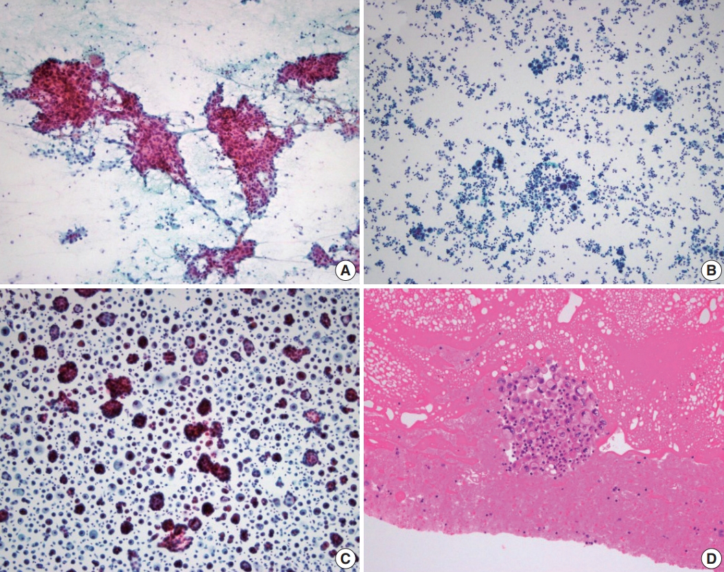
- The most common cytologic sampling methods in NSCLC cancer patients are fine needle aspiration of computed tomography–guided or electromagnetic navigation bronchoscopy–guided lung lesions and endobronchial ultrasound–guided lymph nodes and collection of exfoliative samples such as body fluid/effusions, bronchial brushing/washings, bronchoalveolar lavages, and sputum. Occasionally, minimally invasive aspiration samples from distant, deep-seated, or superficial metastatic lesions are also included. Cytological preparations that can be used for molecular studies include cell blocks (CBs), needle rinses, direct smears, cytospins, and liquid-based preparations (LBPs). To provide the best material for biomarker testing, the correct choice among different cytological preparations of the same sample should be considered. Representative microscopic images and advantages/ disadvantages of different cytological preparations are shown in Table 1 and Fig. 1, respectively.
- CBs are most commonly used for molecular diagnostic testing because they closely recapitulate FFPE specimens and generally do not require further validation; in addition, it is relatively easy to acquire multiple serial sections to perform immunocytochemical and molecular diagnostic assays [4]. However, on-site adequacy evaluation cannot be performed on CB, which leads to unpredictable results of cellularity and sometimes renders the CB paucicellular. Additionally, tumor cells are often widely spaced, resulting in low tumor cellularity per section area. In addition, the standard 4–5-μm CB sections do not represent the entire nuclei from the cell and are likely to have lower nucleic acid yields for molecular testing per cell than the whole cells obtained from other non–formalin-fixed cytologic preparations. To increase nucleic acid yield, not only providing more sections, but also macrodissecting the regions of highest tumor cellularity may be an option [5].
- Direct smears and cytospins that are either air-dried or ethanol-fixed are not formalin-fixed preparations, which have the obvious advantage of obtaining an excellent quality material with a higher nucleic acid yield than CBs [6,7]. Besides being suitable for DNA-based next-generation sequencing (NGS) analysis, direct smears may also be appropriate for RNA-based NGS testing [8]. In addition, they offer the advantage of on-site adequacy assessment and better triaging of the sample for diagnosis and ancillary studies. In cases in which all or most of the diagnostic material is on a single smear/cytospin preparation that will be used for biomarker testing, CAP guidelines allow for the sacrifice of diagnostic material when medically necessary; the diagnostic slide can be digitally scanned for the archives to mitigate the medicolegal constraints [9].
- Finally, LBPs represent a valuable alternative to conventional preparations to avoid inadequate management of the achieved material. The advantages of liquid-based cytology (LBC) specimens include optimal cell preservation, easy specimen transportation because of the stability of cells at room temperature, and minimal background debris and blood on slides [10-12]. Nucleic acid can be extracted from both rinse solutions, and cells can be scraped off the sides [13-15]. Of note, the properties of the different preservative solutions used in LBC may affect downstream molecular analysis. Some studies have indicated that cells preserved in CytoLyt (Cytyc Corp., Boxborough, MA, USA) solution provide higher DNA yields than those preserved in CytoRich Red fluid [16]. One study comparing cellularity and DNA yield between ThinPrep (Cytyc Corp.) slides (CytoLyt LBC) and direct smears reported greater cellularity and significantly higher average DNA yields in the latter [13], whereas a more recent study reported issues with long-term DNA stability and accelerated DNA degradation in LBC samples when compared with conventional smears [17].
WHICH CYTOLOGICAL SPECIMENS CAN BE USED?
- Polymerase chain reaction–based tests
- Molecular testing for genetic mutations, such as in EGFR, in cytologic specimens has been described using a variety of polymerase chain reaction (PCR)–based techniques, including direct sequencing, real-time PCR, pyrosequencing, and peptide nucleic acid–locked nucleic acid [14,18-21]. Different techniques have different limits of detection and reference ranges, and the choice of platform used for the detection of mutations remains a decision of the individual molecular laboratories performing the assay (Table 2). Although the CAP/IASLC/AMP guidelines recommend a technique used to detect mutations in specimens with >50% tumor fraction [2], more sensitive platforms capable of detecting mutations in specimens with <10% tumor are strongly encouraged. The adequacy of cytological samples for mutational analysis is another important factor that is assessed according to tumor cellularity and viability. The CAP/IASLC/AMP guidelines recommend testing from samples with as little as 20% tumor cellularity because current mutation testing uses PCR-based methods that are more sensitive than unmodified Sanger sequencing [2]. In our study for the detection of EGFR mutation using the cytologic samples, the following parameters were correlated with the most reliable EGFR mutation results using the pyrosequencing method (100% concordance with the corresponding histologic specimens) in cytologic samples: a DNA concentration >25 ng/μL, content of >30 tumor cells, or a tumor percentage >30% [22].
- Fluorescence in situ hybridization
- To detect gene rearrangements such as in ALK and ROS1, fluorescence in situ hybridization (FISH), which was verified as a break-apart probe in a clinical trial, was first certified as a companion diagnostic test [23]. Previously, FISH testing was recommended only for CBs, but the 2018 CAP/IASLC/AMP guidelines recommend the use of conventional cytologic preparations for FISH [2]. Several groups have reported the potential and usefulness of the probe in non-formalin cytological preparations, including Diff-Quik and Papanicolaou-stained smears, as well as LBC ThinPrep slides; some report better performance than that seen with CB sections [24-26]. The advantage of using smears or LBCs is that whole-cell nuclei are analyzed to eliminate signal loss due to truncating artifacts, as seen in FFPE sections, but the disadvantage is that thresholds for positive and negative cutoffs are established using FFPE histological materials [27]. Therefore, independent standardization and validation of each sample type are required.
- Immunocytochemistry
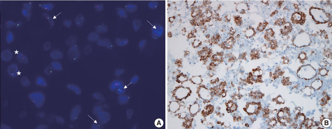
- After the ALK D5F3 CDx Assay (Ventana Medical Systems, Tucson, AZ, USA) was approved by the Food and Drug Administration (FDA), immunohistochemistry (IHC) has been established as a confirmatory diagnostic test rather than screening, supplementing the shortcomings of FISH in detecting ALK rearrangement. The 2018 CAP/IASLC/AMP guidelines recommend ALK IHC as a valid alternative to the FISH (Fig. 2) [2]. The FDA has approved the assay only for “routinely processed, paraffin-embedded specimens fixed in neutral-buffered formalin.” However, several studies have demonstrated the feasibility of ALK immunocytochemistry (ICC) for direct smears and LBPs [28,29]. The updated guidelines recommend using ROS1 IHC with D4D6 (Cell Signaling Technology, Danvers, MA, USA) only as a screening test that requires confirmation by a molecular or cytogenetic method [2]. A limited number of studies using ROS1 FISH in cytological specimens are available [30,31]. Studies on the use of ROS1 ICC in cytology preparations are currently limited in the literature [32,33].
- However, application of these assays to cytologic specimens requires meticulous validation because these assays are validated primarily on FFPE histological tissue samples. The lack of standardized processing protocols in cytology lead to a variety of preanalytic variables that can affect the antigenicity of antibodies used for predictive biomarker testing. CBs are most widely used for ICC; however, there is no standardized protocol for the type of collection media, pre-fixation, and processing techniques, and there is wide variation among pathology laboratories. Several recent studies have highlighted issues with immunostaining of specific markers that demonstrate reduced antigenicity and false-negative results, mostly related to ethanol or methanol-based fixatives used prior to CB preparation [34,35]. Non-CB cytological preparations present an even greater challenge for ICC validation. Immunostaining of ethanol-fixed smears or cytospins is used more frequently, with prior Papanicolau staining that can identify areas or cells of interest, or air-dried unfixed extra slides that can be used for ICC, usually after post-fixation step involving formalin or acetone [36,37]. In a recent meta-analysis of ALK ICC, the smear showed a slightly lower sensitivity than that of CB. These results are interpreted to indicate that the expression intensity of the antibody is low in alcohol-fixed smear slides because the expression of the antibody is optimized in FFPE [37].
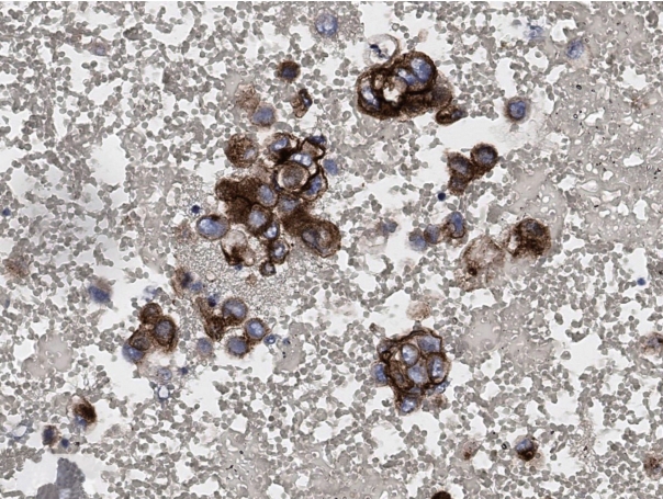
- Unfortunately, guidelines for PD-L1 testing have not yet been provided even in updated guideline [2,38]. Although cytology specimens were not included in the initial clinical validation studies for PD-L1, several groups have evaluated the feasibility of PD-L1 in cytology specimens and have demonstrated results that are comparable to those of paired histologic samples [39,40]. Fig. 3 shows representative microscopic findings of strong positive expression of PD-L1 stained in CB of a patient diagnosed with metastatic adenocarcinoma in pericardial fluid. Lozano et al. reported the variation in patterns of PD-L1 expression on cytological specimens; because entire cells were present on direct smear, tumor cells often demonstrated a folded cell membrane, demonstrating a thick and strong membranous positivity [41]. Taken together, ICC in cytologic specimens remains a number of challenges to be solved throughout the standardization of protocols that can control preanalytical variables, rigorous validation of staining results, and systematic training for interpretation.
WHAT TYPES OF BIOMARKER TESTING CAN BE PERFORMED ON CYTOLOGY SPECIMENS?
- Next-generation sequencing (NGS) is a fascinating tool that can analyze multiple genetic alterations simultaneously, even when applied to cytological samples with low DNA/RNA yields. The advantages of using cytology specimens for NGS include quicker fixation or, if the platform is validated, minimal/no fixation, improving the quality of the input nucleic acids. Several studies using cytological material, including CBs as well as non-FFPE substrates, have shown them to be equally effective in the genomic profiling of NSCLC by NGS analysis [42-46]. In fact, some studies have indicated better quality metrics when comparing NGS analysis in non-FFPE cytologic substrates versus FFPE materials [6,47]. However, studies of the application of NGS to cytology specimens generally have a retrospective design, and only samples characterized by at least 20% of tumor cells, which may not fully reflect current practice, were selected. Therefore, it is crucial to establish the minimum number of cells needed to allow an NGS approach from cytology sample in routine practice. In any case, sample requirement depends on target capture, gene panel, and platform types. Illumina NGS usually requires more cells and/or higher DNA input than Ion Torrent NGS; thus, the latter seems to be more efficient with the cytopathologist specimens [5]. Recently, it was shown that lowering the input DNA concentration below the manufacturer’s recommended threshold of 10 ng (>0.8 ng/μL) is feasible leading to a marked increase in the NGS success rate from 58.6% to 89.8% [5,48]. More important than DNA input is the percentage of neoplastic cells. The preferential amplification of a small number of DNA in a small amount of cancer cells may only be representative of non-neoplastic components, which may lead to false-negative results. Macrodissection or microdissection are especially important for enrichment of viable tumor cells [5,49].
Next-generation sequencing
- In this era of personalized medicine, biomarker testing of cytology preparations is a relatively new and rapidly developing field with great potential, especially in patients with advanced NSCLC. However, cytological specimens continue to be excluded from most biomarker-driven clinical trials, primarily because of the failure to exploit the variety of different specimen preparations and the lack of validation for different assays. The lack of standardization of specimen processing among laboratories is major limitation. Therefore, the implementation of strategies to optimize and standardize procedures for specimen acquisition, processing, and tissue extraction is critical to maximize the use of cytological samples for ancillary studies and to provide relevant information for inclusion in clinical trial design. At minimum, confirmation of validation for cytology preparations and close check of quantity and quality of submitted material is also expected.
CHALLENGES AND FUTURE DIRECTIONS
- In conclusion, biomarker testing can be used for a variety of cytologic specimen types and preparations. This is of utmost importance for NSCLC patients, where the cytology specimen may be the only sample available for diagnosis and ancillary studies. Therefore, a thorough understanding of the potential and the limitations of these substrates is required to properly classify and use them for molecular studies that can guide patient management.
CONCLUSION
-
Ethics Statement
Not applicable.
-
Availability of Data and Material
The datasets generated or analyzed during the study are available from the corresponding author on reasonable request.
-
Code Availability
Not applicable.
-
Author contributions
Conceptualization: HK, JHC. Writing—original draft: HK. Writing—review & editing: HK, JHC. Approval of final manuscript: all authors.
-
Conflicts of Interest
JHC, a contributing editor of the Journal of Pathology and Translational Medicine, was not involved in the editorial evaluation or decision to publish.
-
Funding Statement
No funding to declare.
Notes


- 1. Lindeman NI, Cagle PT, Beasley MB, et al. Molecular testing guideline for selection of lung cancer patients for EGFR and ALK tyrosine kinase inhibitors: guideline from the College of American Pathologists, International Association for the Study of Lung Cancer, and Association for Molecular Pathology. J Thorac Oncol 2013; 8: 823–59. ArticlePubMedPMC
- 2. Lindeman NI, Cagle PT, Aisner DL, et al. Updated molecular testing guideline for the selection of lung cancer patients for treatment with targeted tyrosine kinase inhibitors: guideline from the College of American Pathologists, the International Association for the Study of Lung Cancer, and the Association for Molecular Pathology. J Thorac Oncol 2018; 13: 323–58. PubMed
- 3. Camidge DR, Doebele RC, Kerr KM. Comparing and contrasting predictive biomarkers for immunotherapy and targeted therapy of NSCLC. Nat Rev Clin Oncol 2019; 16: 341–55. ArticlePubMedPDF
- 4. Roh MH. The Utilization of cytologic fine-needle aspirates of lung cancer for molecular diagnostic testing. J Pathol Transl Med 2015; 49: 300–9. ArticlePubMedPMCPDF
- 5. Roy-Chowdhuri S, Stewart J. Preanalytic variables in cytology: lessons learned from next-generation sequencing: the MD Anderson experience. Arch Pathol Lab Med 2016; 140: 1191–9. ArticlePubMedPDF
- 6. Hwang DH, Garcia EP, Ducar MD, Cibas ES, Sholl LM. Next-generation sequencing of cytologic preparations: an analysis of quality metrics. Cancer Cytopathol 2017; 125: 786–94. ArticlePubMedPDF
- 7. Harada S, Agosto-Arroyo E, Levesque JA, et al. Poor cell block adequacy rate for molecular testing improved with the addition of DiffQuik-stained smears: need for better cell block processing. Cancer Cytopathol 2015; 123: 480–7. ArticlePubMed
- 8. Velizheva NP, Rechsteiner MP, Wong CE, et al. Cytology smears as excellent starting material for next-generation sequencing-based molecular testing of patients with adenocarcinoma of the lung. Cancer Cytopathol 2017; 125: 30–40. ArticlePubMedPDF
- 9. Huang M, Wei S. Overview of molecular testing of cytology specimens. Acta Cytol 2020; 64: 136–46. ArticlePubMedPDF
- 10. Petriella D, Galetta D, Rubini V, et al. Molecular profiling of thinprep FNA samples in assisting clinical management of non-smallcell lung cancer. Mol Biotechnol 2013; 54: 913–9. ArticlePubMedPDF
- 11. Abedi-Ardekani B, Vielh P. Is liquid-based cytology the magic bullet for performing molecular techniques? Acta Cytol 2014; 58: 574–81. ArticlePubMedPDF
- 12. Zeppa P. Liquid-based cytology: a 25-year bridge between the pap smear and molecular cytopathology. Acta Cytol 2014; 58: 519–21. ArticlePubMedPDF
- 13. Bellevicine C, Malapelle U, Vigliar E, de Luca C, Troncone G. Epidermal growth factor receptor test performed on liquid-based cytology lung samples: experience of an academic referral center. Acta Cytol 2014; 58: 589–94. ArticlePubMedPDF
- 14. Malapelle U, de Rosa N, Bellevicine C, et al. EGFR mutations detection on liquid-based cytology: is microscopy still necessary? J Clin Pathol 2012; 65: 561–4. ArticlePubMed
- 15. Reynolds JP, Tubbs RR, Minca EC, et al. EGFR mutational genotyping of liquid based cytology samples obtained via fine needle aspiration (FNA) at endobronchial ultrasound of non-small cell lung cancer (NSCLC). Lung Cancer 2014; 86: 158–63. ArticlePubMed
- 16. Dejmek A, Zendehrokh N, Tomaszewska M, Edsjo A. Preparation of DNA from cytological material: effects of fixation, staining, and mounting medium on DNA yield and quality. Cancer Cytopathol 2013; 121: 344–53. PubMed
- 17. Kim WY, Oh SY, Kim H, Hwang TS. DNA degradation in liquidbased cytology and its comparison with conventional smear. Diagn Cytopathol 2016; 44: 450–8. ArticlePubMed
- 18. Lozano MD, Zulueta JJ, Echeveste JI, et al. Assessment of epidermal growth factor receptor and K-ras mutation status in cytological stained smears of non-small cell lung cancer patients: correlation with clinical outcomes. Oncologist 2011; 16: 877–85. ArticlePubMedPMCPDF
- 19. Billah S, Stewart J, Staerkel G, Chen S, Gong Y, Guo M. EGFR and KRAS mutations in lung carcinoma: molecular testing by using cytology specimens. Cancer Cytopathol 2011; 119: 111–7. PubMed
- 20. Malapelle U, Bellevicine C, De Luca C, et al. EGFR mutations detected on cytology samples by a centralized laboratory reliably predict response to gefitinib in non-small cell lung carcinoma patients. Cancer Cytopathol 2013; 121: 552–60. ArticlePubMed
- 21. Rekhtman N, Brandt SM, Sigel CS, et al. Suitability of thoracic cytology for new therapeutic paradigms in non-small cell lung carcinoma: high accuracy of tumor subtyping and feasibility of EGFR and KRAS molecular testing. J Thorac Oncol 2011; 6: 451–8. ArticlePubMed
- 22. Sun PL, Jin Y, Kim H, Lee CT, Jheon S, Chung JH. High concordance of EGFR mutation status between histologic and corresponding cytologic specimens of lung adenocarcinomas. Cancer Cytopathol 2013; 121: 311–9. ArticlePubMed
- 23. Lindeman NI, Cagle PT, Beasley MB, et al. Molecular testing guideline for selection of lung cancer patients for EGFR and ALK tyrosine kinase inhibitors: guideline from the College of American Pathologists, International Association for the Study of Lung Cancer, and Association for Molecular Pathology. Arch Pathol Lab Med 2013; 137: 828–60. ArticlePubMedPMC
- 24. Minca EC, Lanigan CP, Reynolds JP, et al. ALK status testing in nonsmall-cell lung carcinoma by FISH on ThinPrep slides with cytology material. J Thorac Oncol 2014; 9: 464–8. ArticlePubMed
- 25. Betz BL, Dixon CA, Weigelin HC, Knoepp SM, Roh MH. The use of stained cytologic direct smears for ALK gene rearrangement analysis of lung adenocarcinoma. Cancer Cytopathol 2013; 121: 489–99. ArticlePubMed
- 26. Proietti A, Ali G, Pelliccioni S, et al. Anaplastic lymphoma kinase gene rearrangements in cytological samples of non-small cell lung cancer: comparison with histological assessment. Cancer Cytopathol 2014; 122: 445–53. ArticlePubMed
- 27. Roy-Chowdhuri S, Aisner DL, Allen TC, et al. Biomarker testing in lung carcinoma cytology specimens: a perspective from members of the Pulmonary Pathology Society. Arch Pathol Lab Med 2016; 140: 1267–72. ArticlePubMedPDF
- 28. Savic S, Bode B, Diebold J, et al. Detection of ALK-positive nonsmall-cell lung cancers on cytological specimens: high accuracy of immunocytochemistry with the 5A4 clone. J Thorac Oncol 2013; 8: 1004–11. ArticlePubMed
- 29. Rosenblum F, Hutchinson LM, Garver J, Woda B, Cosar E, Kurian EM. Cytology specimens offer an effective alternative to formalinfixed tissue as demonstrated by novel automated detection for ALK break-apart FISH testing and immunohistochemistry in lung adenocarcinoma. Cancer Cytopathol 2014; 122: 810–21. ArticlePubMed
- 30. Bozzetti C, Nizzoli R, Tiseo M, et al. ALK and ROS1 rearrangements tested by fluorescence in situ hybridization in cytological smears from advanced non-small cell lung cancer patients. Diagn Cytopathol 2015; 43: 941–6. ArticlePubMed
- 31. Fernandez-Bussy S, Labarca G, Pires Y, Caviedes I, Burotto M. Molecular testing of EGFR, EGFR resistance mutation, ALK and ROS1 achieved by EBUS-TBNA in Chile. Arch Bronconeumol 2017; 53: 172–4. ArticlePubMed
- 32. Vlajnic T, Savic S, Barascud A, et al. Detection of ROS1-positive non-small cell lung cancer on cytological specimens using immunocytochemistry. Cancer Cytopathol 2018; 126: 421–9. ArticlePubMedPDF
- 33. Conde E, Hernandez S, Martinez R, et al. Assessment of a new ROS1 immunohistochemistry clone (SP384) for the identification of ROS1 rearrangements in patients with non-small cell lung carcinoma: the ROSING study. J Thorac Oncol 2019; 14: 2120–32. ArticlePubMed
- 34. Sauter JL, Grogg KL, Vrana JA, Law ME, Halvorson JL, Henry MR. Young investigator challenge: validation and optimization of immunohistochemistry protocols for use on cellient cell block specimens. Cancer Cytopathol 2016; 124: 89–100. ArticlePubMed
- 35. Gruchy JR, Barnes PJ, Dakin Hache KA. CytoLyt(R) fixation and decalcification pretreatments alter antigenicity in normal tissues compared with standard formalin fixation. Appl Immunohistochem Mol Morphol 2015; 23: 297–302. PubMed
- 36. Shidham VB, Chang CC, Rao RN, Komorowski R, Chivukula M. Immunostaining of cytology smears: a comparative study to identify the most suitable method of smear preparation and fixation with reference to commonly used immunomarkers. Diagn Cytopathol 2003; 29: 217–21. ArticlePubMed
- 37. Roy-Chowdhuri S. Immunocytochemistry of cytology specimens for predictive biomarkers in lung cancer. Transl Lung Cancer Res 2020; 9: 898–905. ArticlePubMedPMC
- 38. Chang S, Shim HS, Kim TJ, et al. Molecular biomarker testing for non-small cell lung cancer: consensus statement of the Korean Cardiopulmonary Pathology Study Group. J Pathol Transl Med 2021; 55: 181–91. ArticlePubMedPMCPDF
- 39. Skov BG, Skov T. Paired comparison of PD-L1 expression on cytologic and histologic specimens from malignancies in the lung assessed with PD-L1 IHC 28-8pharmDx and PD-L1 IHC 22C3pharmDx. Appl Immunohistochem Mol Morphol 2017; 25: 453–9. ArticlePubMed
- 40. Noll B, Wang WL, Gong Y, et al. Programmed death ligand 1 testing in non-small cell lung carcinoma cytology cell block and aspirate smear preparations. Cancer Cytopathol 2018; 126: 342–52. ArticlePubMedPDF
- 41. Lozano MD, Abengozar-Muela M, Echeveste JI, et al. Programmed death-ligand 1 expression on direct Pap-stained cytology smears from non-small cell lung cancer: comparison with cell blocks and surgical resection specimens. Cancer Cytopathol 2019; 127: 470–80. ArticlePubMedPDF
- 42. Karnes HE, Duncavage EJ, Bernadt CT. Targeted next-generation sequencing using fine-needle aspirates from adenocarcinomas of the lung. Cancer Cytopathol 2014; 122: 104–13. ArticlePubMed
- 43. Baum JE, Zhang P, Hoda RS, et al. Accuracy of next-generation sequencing for the identification of clinically relevant variants in cytology smears in lung adenocarcinoma. Cancer Cytopathol 2017; 125: 398–406. ArticlePubMedPDF
- 44. Buttitta F, Felicioni L, Del Grammastro M, et al. Effective assessment of egfr mutation status in bronchoalveolar lavage and pleural fluids by next-generation sequencing. Clin Cancer Res 2013; 19: 691–8. ArticlePubMedPDF
- 45. Reynolds JP, Zhou Y, Jakubowski MA, et al. Next-generation sequencing of liquid-based cytology non-small cell lung cancer samples. Cancer Cytopathol 2017; 125: 178–87. ArticlePubMedPDF
- 46. Treece AL, Montgomery ND, Patel NM, et al. FNA smears as a potential source of DNA for targeted next-generation sequencing of lung adenocarcinomas. Cancer Cytopathol 2016; 124: 406–14. ArticlePubMedPMC
- 47. Roy-Chowdhuri S, Chen H, Singh RR, et al. Concurrent fine needle aspirations and core needle biopsies: a comparative study of substrates for next-generation sequencing in solid organ malignancies. Mod Pathol 2017; 30: 499–508. ArticlePubMedPDF
- 48. Roy-Chowdhuri S, Goswami RS, Chen H, et al. Factors affecting the success of next-generation sequencing in cytology specimens. Cancer Cytopathol 2015; 123: 659–68. ArticlePubMed
- 49. Bellevicine C, Malapelle U, Vigliar E, Pisapia P, Vita G, Troncone G. How to prepare cytological samples for molecular testing. J Clin Pathol 2017; 70: 819–26. ArticlePubMed
References
Figure & Data
References
Citations
- Molecular testing of cytology specimens: Issues in specimen adequacy and clinical utility
Ghulam Ghous, Komal Ijaz, Magda Esebua, Lester J. Layfield
Diagnostic Cytopathology.2024; 52(2): 123. CrossRef - The updated College of American Pathologists principles of analytic validation of immunohistochemical assays: A step forward for cytopathology
Sinchita Roy‐Chowdhuri
Cancer Cytopathology.2024;[Epub] CrossRef
 PubReader
 ePub Link
-
 Cite this Article
- Cite this Article
-
- Close
- Download Citation
- Close
- Figure
-