Angiomyomatous Hamartoma of Popliteal Lymph Node: An Unusual Entity
Article information
Angiomyomatous hamartoma (AMH) of the lymph node is characterized by partial replacement of normal nodal parenchyma by disorganized blood vessels and smooth muscle cells with or without adipose tissue within a fibrous stroma. Inguinal and femoral lymph nodes are commonly involved [1-3], while popliteal lymph node involvement is uncommon [4,5]. We report a rare case of AMH of the popliteal lymph node in a young patient with a clinical diagnosis of Baker’s cyst.
CASE REPORT
An 18-year-old male presented with pain and swelling in the left popliteal fossa lasting 3 years. The swelling slowly increased in size. There was no history of any trauma, fever, tuberculosis, weight loss, or chronic illness. A clinical diagnosis of Baker’s cyst was made without any imaging studies. The lesion was operated on under local anesthesia. The swelling persisted, however, and physical examination revealed a 1.5 cm-sized, mildly tender, non-reducible, firm mass in the left popliteal fossa. An magnetic resonance imaging (MRI) scan revealed a 1.5×1.0-cm soft tissue mass close to the popliteal blood vessels without encasing them (Fig. 1A, B). No cyst was seen. The radiological differential diagnoses offered were benign neoplasm and pseudotumor. The left popliteal fossa was explored again and a firm, soft tissue mass loosely adherent to the popliteal blood vessels was identified and excised. The postoperative period was uneventful. Hematoxylin and eosin–stained sections revealed a lymph node with partial replacement of the parenchyma from the hilum to the cortex by fibrous tissue containing several irregular blood vessels of varying sizes, interspersed with spindle cells and smooth muscle cells (Fig. 2A, B). Mature adipose tissue infiltration was seen in a small area near the hilum of the lymph node (Fig. 2C). The capsule was thickened. The subcapsular and medullary sinuses were obliterated. Cortical lymphoid tissue showed variable atrophy. Immunohistochemistry with a primary antibody against smooth muscle actin (SMA; 1:400, Thermo Scientific, Waltham, MA, USA) demonstrated smooth muscle cells in the blood vessel walls and in the stromal tissue (Fig. 2D, E). The rich vascularity of the lesion was highlighted by CD34 antibodies (1:100, Diagnostic BioSystems, Pleasanton, CA, USA) (Fig. 2F). A diagnosis of AMH of the lymph node was made.

Axial T1- (A), sagittal T2-weighted (B) magnetic resonance imaging scans showing a well-circumscribed lesion with heterogeneous signal intensity in the soft tissues in close proximity to the popliteal blood vessels (arrows).
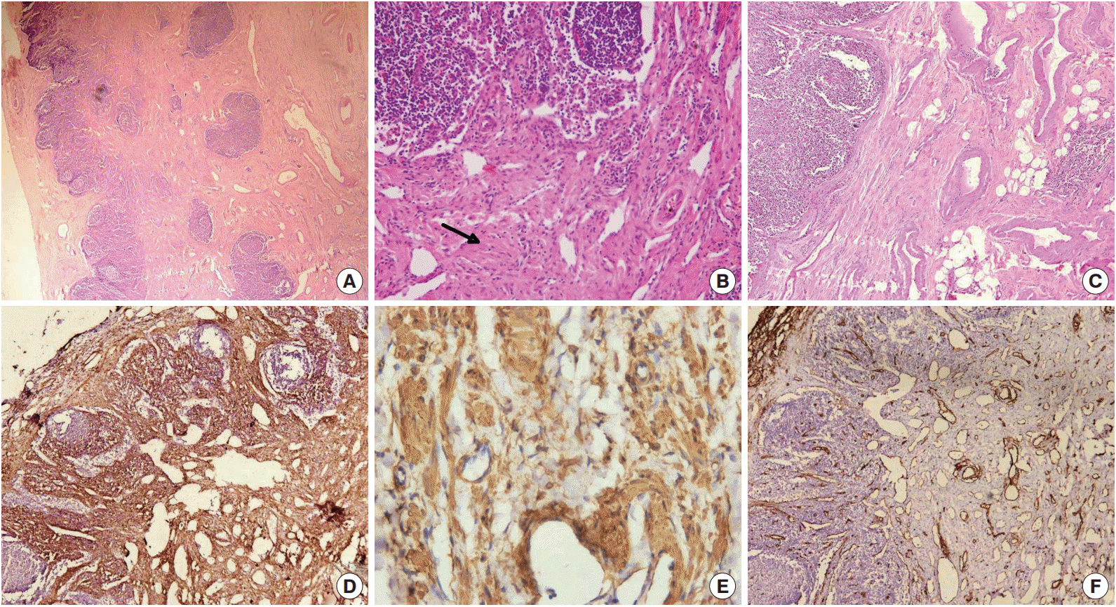
Photomicrograph showing partial replacement of lymph nodal parenchyma by several disorganized vascular channels in a fibrocollagenous stroma and smooth muscle cells (arrow) (A, B); thick-walled blood vessels and adipocytes in nodal hilum (C). (D, E) Immunohistochemical stain with smooth muscle actin antibodies demonstrating smooth muscle in the blood vessel wall and the stroma. (F) CD34 immunostain highlighting the rich vascularity of the lesion.
DISCUSSION
Angiomyomatous hamartoma of the lymph node was first described by Chan et al. [1] in 1992 as a distinctive vascular hamartomatous lesion that primarily occurs in inguinal and femoral lymph nodes [2,3]. Occasional cases have been reported in popliteal and cervical lymph nodes [4,5]. Patients may present with painless or painful swelling [4]. Painful lesions in the popliteal fossa require careful evaluation because a number of non-neoplastic and neoplastic lesions can mimic this entity [6]. Baker’s cyst results from herniation of the synovial membrane through the posterior capsule of the knee joint or by an escape of synovial fluid through an anatomic bursa next to the semimembranosus or gastrocnemius muscle. Baker’s cysts are usually diagnosed by physical and radiological examination between the semimembranosus and medial head of the gastrocnemius [7]. Sometimes it is difficult to differentiate a Baker’s cyst from other causes of posterior knee pain, and the differential clinical diagnosis of a Baker’s cyst can include arterial aneurysm or tortuous blood vessels in the popliteal fossa, deep vessel thrombosis, adipose tissue or tumor [8]. The best imaging for evaluation of a popliteal cyst is MRI as it helps in localizing the cyst and any other internal derangements [9]. In the index case the initial misdiagnosis of Baker’s cyst was due to the presence of painful swelling in the popliteal fossa and the diagnosis was based on physical examination alone. No radiological investigation was done prior to the initial surgery. To the best of our knowledge, to date only twenty-nine cases of AMH of the lymph node have been reported in the literature [3]. This is the third documented case in a popliteal lymph node. Mauro et al. [4] reported a case of AMH of the popliteal lymph node which presented with pain in the posterior knee in a 41- year-old male. The second case was reported by Prusac et al. [5] in a 14-year-old boy who presented with a right popliteal mass and right leg edema. In both cases, MRI scans revealed mass lesions with heterogeneous signal intensity. The index case also showed similar radiologic features. The radiologic diagnosis of AMH of the lymph node is usually a hemangioma or a tumor [4,5]. In our case, the MRI scan findings suggested a benign tumor or pseudotumor. Microscopically, AMH is characterized by disorganized blood vessels and smooth muscle cells in a collagenous stroma with or without adipose tissue. Our case showed a small area of adipose tissue near the hilum of the lymph node. Histopathological differential diagnosis of AMH includes lymphangiomyomatosis, leiomyomatosis, and angiomyolipoma of the lymph node [10]. Nodal lymphangiomyomatosis occurs exclusively in women and is histologically characterized by the presence of smooth muscle cells forming fascicles and sheets around anastomosing ectatic vascular spaces, resulting in a pericytomatous pattern. Histologically smooth muscle cells are plumper, with lighter/clear cytoplasm, and sclerosis is absent. Nodal leiomyomatosis resembles leiomyoma and is made up of a proliferation of compact bundles of smooth muscle cells with an insignificant vascular component. Angiomyolipoma of the lymph node shows an epithelioid appearance, hypercellularity, pleomorphism, prominent perivascular arrangement and positivity for melanoma associated antigen human melanoma black 45. Our index case did not show ec-static vessels, pleomorphism or hemangiopericytoma like patterns. Adipose cells are known to occur as a significant component in a number of vasoproliferative lesions, such as nodal hemangioma, intramuscular hemangioma (infiltrative angiolipoma) and angiolipomatous hamartoma associated with Castleman’s disease. It has been suggested that all cases of AMH of the lymph node with a significant adipose tissue component should be termed angiomyolipomatous hamartoma [10]. The pathogenesis of this lesion is unclear. A possible explanation is that AMH represents a vascular and smooth muscle proliferative response to chronic impairment of nodal lymphatic flow or to previous nodal inflammation [3].
Lesions in the popliteal fossa should be evaluated carefully, especially when associated with pain. The treatment depends on the type of lesion. Radiological investigation is mandatory for proper characterization. Histopathological examination will confirm this unusual benign entity that is managed surgically.
Notes
Conflicts of Interest
No potential conflict of interest relevant to this article was reported.