Focal Hematopoietic Hyperplasia of Rib: A Rare Pseudotumor and Review of Literature
Article information
Primary tumors of the rib are uncommon, comprising 5%– 10% of all bony tumors. Benign tumors constitute 63% of this group [1]. Common benign entities include osteochondroma, enchondroma, and fibrous dysplasia, while common malignant tumors of the ribs include plasmacytoma, chondrosarcoma, osteosarcoma, and Ewing’s sarcoma [1]. Focal hematopoietic hyperplasia (FHH) is an unusual benign lesion characterized by an expansive osteolytic mass involving the rib in all reported cases [2-4]. Most cases were detected incidentally on routine chest radiography performed for unrelated reasons. The lesion is typically composed of hypercellular marrow containing all three lineages of hematopoietic cells with interspersed fatty marrow; no hematopoietic dyspoiesis or malignancy is seen. Though clinical signs and radiological features of the lesion often simulate a true neoplasm, in reality it is a pseudotumor. Follow-up investigation has failed to show any recurrence or neoplastic transformation after resection of the lesion [2]. To the best of our knowledge, to date only four cases have been reported in the English literature [2-4]. We report a case of FHH in the right second rib in a 26-year-old female who underwent surgery after radiological diagnosis of osteochondroma.
CASE REPORT
A 26-year-old female patient presented with upper backache and pain in the right side of the neck for 2 months in duration. There was no history of trauma, tuberculosis, hypertension or diabetes. Physical examination, and hematological and biochemical investigation did not reveal any abnormalities. Routine chest radiography showed a well-defined, expansile sclerotic mass in the right second rib (Fig. 1A). Subsequent computed tomography revealed a 5.0×4.5-cm mass in the body of the rib with internal linear calcification. There was cortical bulging and thinning of the inner surface without cortical destruction (Fig. 1B, C). The patient underwent surgery with a radiological diagnosis of osteochondroma. The mass was completely excised, fixed in 10% buffered formalin and processed for paraffin embedding after decalcification in 5% nitric acid. Microscopic examination revealed an inner shell with essentially normal trabeculae and a focal haphazard arrangement (Fig. 2A). Hypercellular marrow containing normal hematopoietic cells of all three lineages intermixed with fatty marrow in the inter-trabecular spaces was observed (Fig. 2B, C). No dyspoiesis or hematological malignancy was seen, nor was there any fibrosis. Postoperative follow-up of the patient was uneventful.
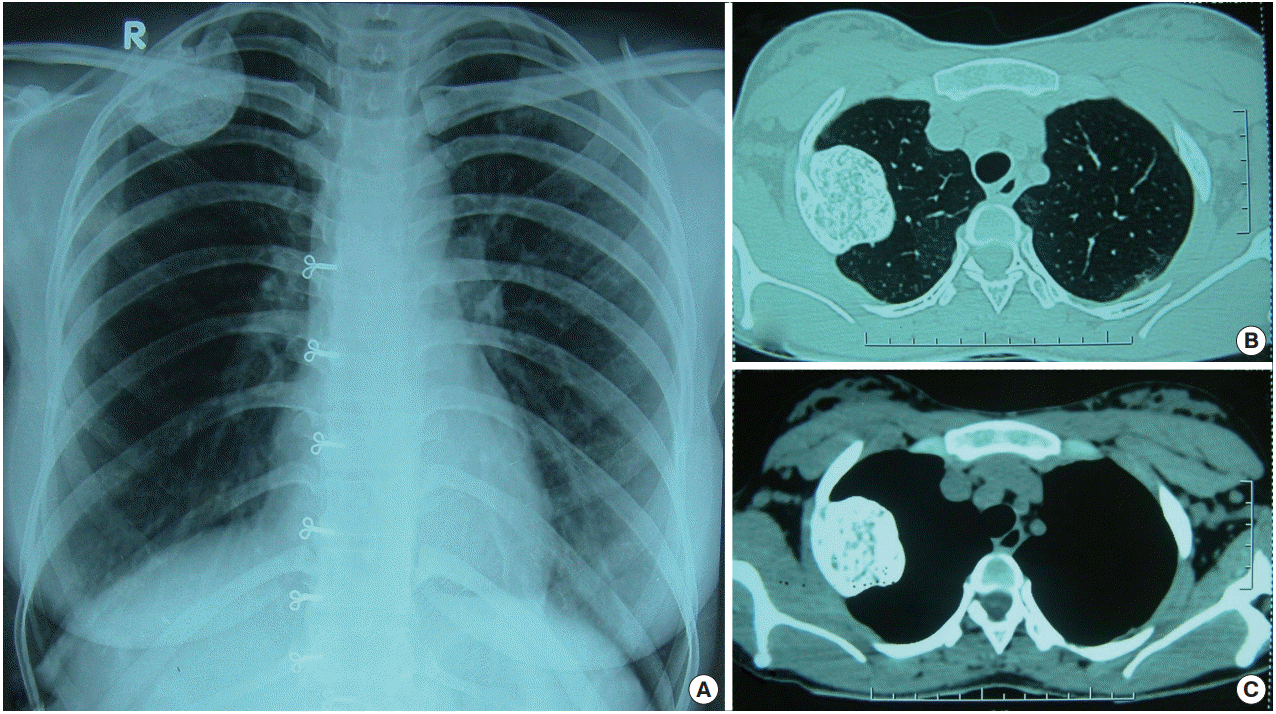
Radiologic features of focal hematopoietic hyperplasia. (A) Chest radiograph showing a well-defined sclerotic bony lesion arising from the right second rib and projecting into the right upper zone. (B, C) Axial computed tomography images of the lung and mediastinal window demonstrating a well-defined sclerotic osseous lesion arising from the lateral aspect of the right second rib. The lesion has an intrathoracic, extrapleural component, which indents the upper lobe of right lung.
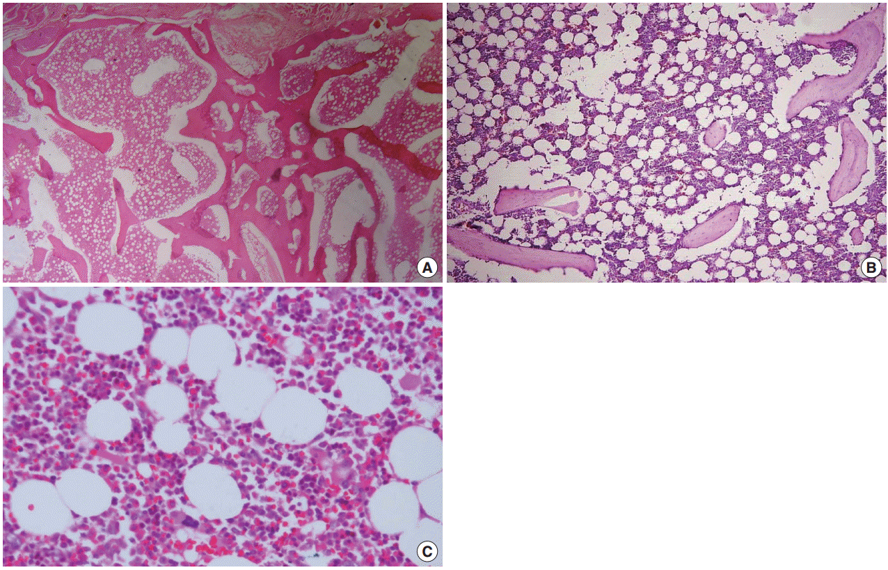
Microscopic features of focal hematopoietic hyperplasia. (A) Photomicrograph showing an inner shell with essentially normal-thickness bony trabeculae with a focal haphazard arrangement. (B) Hypercellular marrow containing normal hemopoietic cells with merging fatty marrow in the intertrabecular spaces. (C) Higher magnification showing normal hematopoietic cells of all lineages.
DISCUSSION
The term “focal hematopoietic hyperplasia of rib” or “hematopoietic pseudotumor” was first coined by Edelstein and Kyriakos in 1984 [2]. They reported two cases of incidentally-detected lesions in a 66-year-old female and a 71-year-old male during routine radiologic investigation for other reasons. The typical radiologic manifestation of the pseudotumor is an expansive, solitary, osteolytic mass of the rib with internal ill-defined linear hyperdense areas and calcification. There is expansion and thinning of the bony cortex without destruction [2-4]. Histologically, the lesion is characterized by focal hyperplasia of normal hematopoietic elements including myeloid, erythroid and megakaryocytic cells. Hyperplastic marrow merging with fatty marrow is a characteristic finding, while no hematopoietic dyspoiesis or malignancy has been reported [2,3]. Diagnosis is often made after excision of the lesion. The clinical and radiological manifestations of this lesion are like that of a true neoplasm, and, consequently, the differential diagnoses on radiology commonly considered are aneurysmal bone cyst, osteochondroma, chondrosarcoma, fibrous dysplasia, plasmacytoma, and metastatic tumor [2]. Only four cases of FHH have been reported in the literature [2-4]. A diagnosis was made based on postoperative specimens in three cases; only one case was diagnosed based on radiology, with confirmation by fine needle aspiration cytology [4]. Similar to the other reported cases, the present case was also detected incidentally on routine chest radiography performed for the investigation of upper backache. A radiological diagnosis of osteochondroma was initially considered, and a definite diagnosis was made after microscopic examination.
Certain hematologic disorders such as chronic anemia, thalassemia, leukemia and myelofibrosis can demonstrate increased hematopoiesis with marrow hyperplasia, fatty marrow and thin bony trabeculae [5]. In chronic hemolysis like thalassemia, medullary expansion of multiple bones including the craniofacial bones, vertebrae, pelvic bones and ribs is common [5]. In this condition, the ribs usually show prominent cortical expansion in the posterior aspect and increased normoblastic erythropoiesis, and the patients typically present with anemia, hepatosplenomegaly and skeletal deformity [6]. In contrast, anemia and hepatosplenomegaly are not associated with FHH and no such clinical features were present in our case. In myelofibrosis, the trilineage proliferation of hematopoietic cells, including normoblasts, granulocyte precursors and megakaryocytes, is accompanied by a varying degree of marrow fibrosis [7]. Our case revealed trilineage proliferation of marrow cells without any fibrosis. The majority of the patients with myelofibrosis present clinically with anemia and hepatosplenomegaly; expanding bone mass is not a typical clinical manifestation of myelofibrosis.
Myelolipoma can also demonstrate similar morphology to that seen in our case [8]. The most common location of myelolipoma is the adrenal gland [8]; however, a number of extra-adrenal myelolipomas have been reported in soft tissue, mostly in presacral locations, as well as in the retroperitoneum, pelvis, stomach, musculofascial tissue and in the nose [9]. Only three cases of intraosseous myelolipoma have been described in the literature [10]. Sundaram et al. [10] reported two cases occurring in the roof of the acetabulum and the proximal femur in 35-year-old and 51-year-old patients, respectively. According to the available literature, a classical intraosseous myelolipoma radiologically appears as an osteolytic and or osteosclerotic intramedullary lesion without any cortical expansion. In contrast, the typical radiologic features of FHH are an expansive, osteolytic lesion with internal calcification, as seen in our case. Microscopically FHH exhibits hypercellularity of marrow elements for the age as compared to myelolipoma wherein the cellularity is normal.
Two possible mechanisms that have been proposed in the pathogenesis of FHH include 1) the reactive process after trauma and inflammation, and 2) developmental anomaly [2]. Our patient had no history of trauma or any preceding infection in this location. She had no other abnormality on physical examination to suggest a developmental anomaly.
Irrespective of these putative mechanisms, FHH of the rib is a rare non-neoplastic lesion with a characteristic radiologic appearance. The microscopic features are similar to hyperplastic marrow and myelolipoma, and a high index of clinical and radiological suspicion is required for the diagnosis of this pseudotumor. Radiologists should be aware of this incidentally-detected lesion of the rib. Histopathologists need to recognize this pseudotumor keeping in mind the differential diagnosis of intraosseous myelolipoma. Awareness of this entity will help to prevent unnecessary biopsy and surgical procedures.
Notes
Conflicts of Interest
No potential conflict of interest relevant to this article was reported.