Clinical and Prognostic Significances of Cytokeratin 19 and KIT Expression in Surgically Resectable Pancreatic Neuroendocrine Tumors
Article information
Abstract
Background:
Pancreatic neuroendocrine tumors (PanNETs) are malignant endocrine neoplasms that present diverse clinical behaviors. Therefore, identification of biomarkers of PanNETs is important for stratification of the prognosis of PanNET patients. Recently, cytokeratin 19 (CK19) and KIT expression were reported to have prognostic significance in PanNET patients.
Methods:
To identify their prognostic significance, CK19 and KIT protein expression were assessed in 182 surgically resected PanNETs and compared with clinicopathologic factors.
Results:
Of 182 PanNETs cases, CK19 and KIT expression was noted in 97 (53.3%) and 16 (8.8%) cases, respectively. PanNET patients with CK19 expression had larger tumors (p=.006), higher World Health Organization (WHO) grade (p=.002) and pT classification (p<.001), increased distant metastasis (p=.004), and lymphovascular (p=.012) and perineural (p=.019) invasion. Similarly, those with KIT expression had larger tumors (p=.030), higher WHO grade (p=.001), advanced pT classification (p<.001), distant metastasis (p=.001), and lymphovascular invasion (p=.014). The 5-year survival rate for PanNET patients with KIT expression was significantly lower (62%) than that of patients without KIT expression (77%, p=.011), as determined by univariate but not by multivariate analyses.
Conclusions:
CK19 and KIT expression correlate with higher metastatic potential and advanced disease stage, and KIT expression is associated with worse survival in PanNET patients.
Pancreatic neuroendocrine tumors (PanNETs) are malignant tumors of endocrine origin from the pancreas and comprise 1.3% to 2.8% of all pancreatic neoplasms [1]. The incidence and prevalence of PanNETs have increased over the past decades according to data from the Surveillance, Epidemiology, and End Results (SEER) study [2]. Compared with ductal adenocarcinoma of the pancreas, PanNETs are generally considered to have indolent behaviors with diverse clinical features ranging from benign to highly malignant [3,4]. Therefore, predicting the clinical behavior of PanNETs is very difficult.
A recent whole-exome sequencing study demonstrated unique driver mutations in PanNETs, such as MEN1, ATRX, and DAXX [5]. In addition, although the frequencies are low, several genes in the mammalian target of rapamycin pathway, including PTEN, TSC2, and PIK3CA, were also reported to be involved in the tumorigenesis of PanNETs [5]. The expression status of some of these proteins was shown to be related to the survival of PanNET patients. For example, the losses of ATRX and DAXX were associated with worse survival. On the other hand, loss of PTEN expression was associated with better survival in PanNET patients [6,7].
The new World Health Organization (WHO) grading scheme and the TNM staging system from the American Joint Committee on Cancer (AJCC) and the International Union for Cancer Control (UICC) provide reliable guidelines for the prognosis and treatment of PanNET patients [4,8]. However, except for pathologic grade and TNM stage, few prognostic biomarkers for PanNETs have been reported. Hence, the identification of prognostic biomarkers for PanNETs will provide more precise information regarding PanNET patient survival after surgical resection [2,9-13]. Several previous studies have reported the prognostic significance of cytokeratin 19 (CK19), KIT, cyclooxygenase 2, and CD99 in PanNET patients [2,9-13]. However, there have not been any validation studies for these markers, except for CK19 expression [2,9,11,12]. The aims of this study were to determine the clinical and prognostic significance of CK19 and KIT expression in surgically resected PanNET patients using tissue microarray immunohistochemical staining.
MATERIALS AND METHODS
After approval (2014-0580) from the Institutional Review Board, 182 patients with primary PanNETs who underwent surgical resection at our institution from 1995 to 2013 were selected from the files of the Department of Pathology. Medical records were reviewed to evaluate clinical data, such as age, sex, symptoms, and follow-up data. Pathologic information, including tumor size, extension, metastases to regional lymph nodes, distant metastases, and lymphovascular and perineural invasions were carefully reviewed. Hematoxylin and eosin–stained slides were independently reviewed by three pathologists (S.-M.H., J.Y.K., and E.-M.S.). All PanNET cases were confirmed by immunohistochemical staining using neuroendocrine markers, synaptophysin, chromogranin, and/or CD56. Immunohistochemical staining for synaptophysin and chromogranin was performed in 144 and 138 cases, respectively. All 144 cases (100%) were positive for synaptophysin, and 113 of 138 (81.9%) cases were positive for chromogranin. All PanNET cases were re-classified into grades 1, 2, or 3 based on mitotic counts (per 10 high-power fields) and the Ki-67 labeling index according to the scheme of the 2010 WHO classification [8]. Tumor extension was assessed based on the T classification of the 2010 AJCC/UICC cancer staging system.
Tissue microarrarys (TMAs) were constructed using three 2-mm-diameter tumor cores from donor blocks using a manual tissue microarryer (Uni TMA Co., Ltd., Seoul, Korea). The sections of TMAs were stained using an automatic immunohistochemistry staining device (Benchmark XT, Ventana Medical System, Tucson, AZ, USA). Briefly, 5-µm-thick formaldehydefixed paraffin-embedded tissue sections were transferred onto adhesive slides and dried at 62°C for 30 minutes. Standard heat epitope retrieval was performed for 30 minutes in ethylene diamine tetraacetic acid, pH 8.0, in the autostainer. The samples were then incubated with antibodies against KIT (1:400, Dako-Cytomation, Glostrup, Denmark) and CK19 (1:100, Cell Marque, Rocklin, CA, USA). The sections were subsequently incubated with biotinylated anti-mouse immunoglobulins, peroxidase-labeled streptavidin (LSAB kit, DakoCytomation), and 3,30-diaminobenzidine. Negative control samples were processed without the primary antibody. Slides were counterstained with Harris hematoxylin. Nuclear labeling of intra-tumoral mast cells was used as an internal positive control for KIT immunohistochemical staining. Normal pancreatic acinar cells, ductal epithelial cells, and islet cells were negative for KIT staining, while mast cells in the pancreatic parenchyma were positive (Fig. 1A). Membranous immunolabeling in normal pancreatic ductal epithelial cells and centroacinar cells was used as an internal positive control for CK19 staining. Normal pancreatic islet cells and acinar cells were negative for CK19 staining (Fig. 1B). Sections demonstrating >5% positivity for KIT- and CK19-labeled tumor cells were considered to be positive, as previously described [2,13].
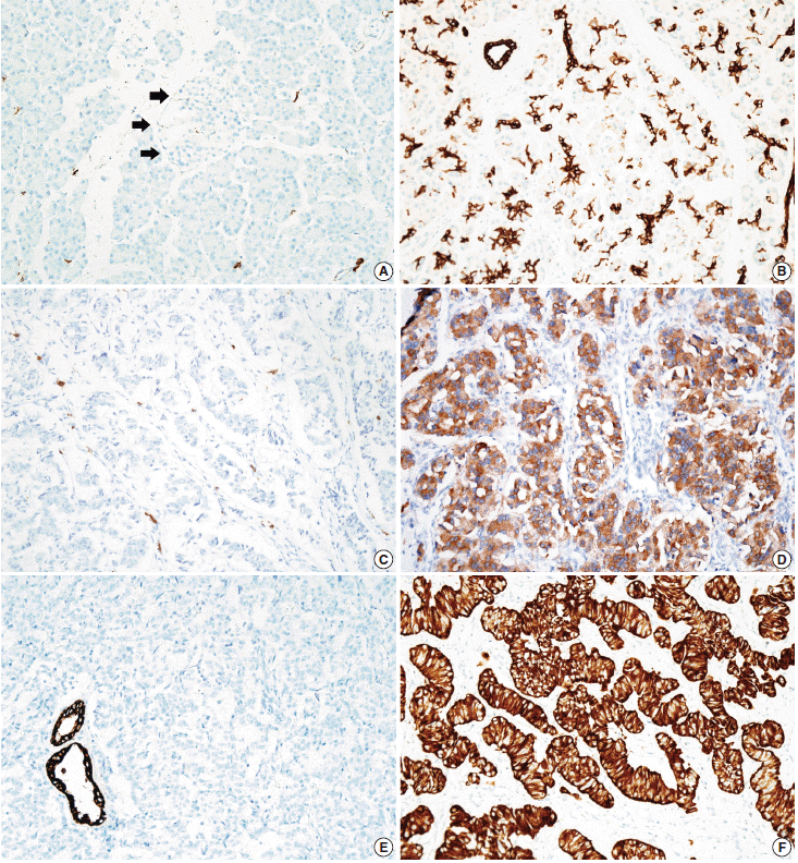
Representative images of KIT and cytokeratin 19 (CK19) staining. (A) Normal acinar cell, ductal epithelial cells, and islet cells (arrows) of the pancreas are negative for KIT staining, while mast cells in the pancreatic parenchyma are positive for KIT staining. (B) Membranous CK19 immunolabeling is noted in the normal centroacinar cell and the ductal epithelial cells, while islet cells are not stained. Negative (C) and positive (D) KIT expression in a pancreatic neuroendocrine tumor. (E) Negative CK19 expression is observed in a pancreatic neuroendocrine tumor, while entrapped normal pancreatic ductal cells are positive for CK19. (F) Positive CK19 expression in a pancreatic neuroendocrine tumor.
SPSS ver. 18.0 (SPSS, Chicago, IL, USA) was used for the statistical analysis. The overall survival time was calculated from the date of diagnosis of PanNET to that of death from any cause. The overall survival rate was calculated with the Kaplan-Meier method, and the associations between overall survival rate and clinicopathologic factors were compared using the log-rank test. Correlations between KIT and CK19 expression and other prognostic factors were analyzed using chi-square and Fisher exact tests. Possible prognostic factors associated with survival probability were calculated by the Cox’s proportional hazard regression model. A p<.05 was considered statistically significant.
RESULTS
Of a total of 182 PanNET patients, 44.5% (81 cases) were men, 55.5% (101 cases) were women, and the mean age was 51.4±13.10 years. The mean tumor size was 3.06±2.31 cm. Tumors were classified as functional tumors if they were associated with the distinct clinical manifestations of hormonal alterations, including hypoglycemia, sweating, or syncope; 45 cases (24.7%) were consequently classified as functional PanNETs. There were 71 head (39.0%), 7 uncinate (3.8%), 29 body (15.9%), 67 tail (36.8%), 2 head to body (1.1%), and 6 body to tail (3.3%) tumors. Using the TNM staging system, PanNETs were classified as pT1 in 74 (40.7%), pT2 in 47 (25.8%), and pT3 in 61 (33.5%) cases. Because only surgically resected PanNETs were included in the study, pT4 tumors were not included. Lymphovascular and perineural invasion was noted in 48 (26.4%) and 27 (14.8%) cases, respectively. Evaluation of the lymph nodes was possible in 99 cases (54.4%), and lymph node metastasis was present in 29 cases (15.9%). Distant metastasis was observed in 36 cases (19.8%), and liver was the most common site of these distant metastases (32 cases, 88.9%). Adequate follow-up of survival information was available in 181 patients, with a median follow-up period of 30 months (range, 1 to 142 months), and 153 patients (84.1%) were alive and 28 patients (15.3%) had died; 9 of these patients died of an unrelated cause.
KIT expression was observed in 16 of 182 PanNETs (8.8%) (Fig. 1D) and CK19 expression was noted in 97 cases (53.3%) (Fig. 1F). All 16 PanNET cases that showed KIT expression were also positive for CK19. The associations between KIT or CK19 expression and clinicopathological factors are summarized in Table 1. KIT expression was more frequently observed in larger PanNETs (p=.030), those with extrapancreatic extension (p<.001), advanced pT classification (p<.001), distant metastasis (p=.001), higher WHO grade (p=.001), and lymphovascular invasion (p=.014). There was no statistically significant correlation between KIT expression and sex, age, functional status, lymph node metastasis, expression of synaptophysin and chromogranin, or perineural invasion.
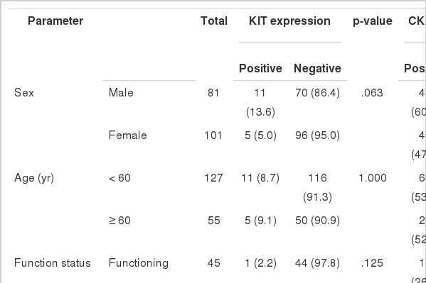
Correlations between KIT or CK19 expression and clinicopathological factors
CK19 expression was more commonly observed in functioning PanNETs (p<.001), larger tumors (p=.006), extrapancreatic extension (p<.001), advanced pT classification (p<.001), lymph node metastasis (p=.009), distant metastasis (p=.004), higher WHO grade (p=.002), and lymphovascular (p=.012) and perineural (p=.019) invasion. There was no statistically significant correlation between CK19 expression and sex, age, or expression of synaptophysin and chromogranin. The overall survival rate of PanNET patients with KIT expression was significantly lower (5-year survival rate, 62%) than that of patients without KIT expression (77%, p=.011) (Fig. 2A). However, the overall survival rate of PanNET patients with CK19 expression (5-year survival rate, 74%) was not significantly different from that of patients lacking CK19 expression (79%, p=.242) (Fig. 2B).
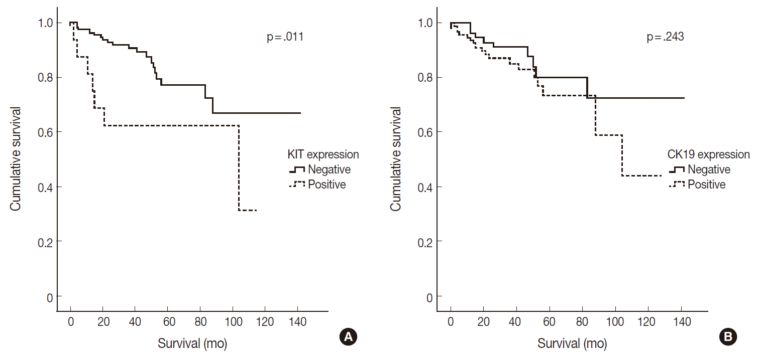
Kaplan-Meier survival analysis of pancreatic neuroendocrine tumor patients according to KIT (A) and cytokeratin 19 (CK19) (B) expression status. (A) The overall 5-year survival rate for pancreatic neuroendocrine tumor (PanNET) patients with KIT expression is significantly lower (62%) than that of patients without KIT expression (77%, p=.011). (B) The overall 5-year survival rate for PanNET patients with CK19 expression (74%) is not significantly different from that of those without CK19 expression (79%, p=.243).
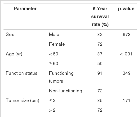
The relationships between the survival of PanNET patients and other clinicopathologic factors are summarized in Table 2. Of these clinicopathologic factors, increased age (p<.001), extrapancreatic extension (p=.003), increased pT classification (p=.009), lymph node metastasis (p=.026), distant metastasis (p=.003), and higher WHO grade (p<.001) were correlated with worse survival. In contrast, overall survival was not correlated with sex, functional status, or lymphovascular and perineural invasion.
Univariate analysis of overall survival by clinicopathologic features with PanNETs
Multivariate analysis was performed to assess which factors remained independent predictors of survival after adjusting for factors that were found to be significant by univariate analysis. Only WHO grade remained as an independent prognostic factor (p=.001) (Table 3). Conversely, KIT expression, pT classification, and lymph node and distant metastases were not found to be independent prognostic factors in the present model.
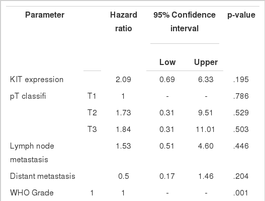
Multivariate analysis of overall survival by clinicopathologic features with pancreatic neuroendocrine tumors
DISCUSSION
KIT is a cytoplasmic membrane-bound receptor tyrosine kinase and a stem cell marker. Activation of KIT signaling has been found to mediate cell survival, migration, and proliferation [14]. Signaling from KIT is crucial for normal hematopoiesis, pigmentation, fertility, and gut movement. Deregulated KIT kinase activity has been observed in a number of pathological conditions, including small cell lung cancer, breast cancer, colon cancer, melanoma, gastrointestinal stromal tumors, and allergy [14]. KIT is involved in β-cell development and survival and in regulation of glucose metabolism of the endocrine pancreas via receptor phosphorylation [15-19]. Normally, KIT expression in the endocrine pancreas is present during the embryonic and early fetal periods; however, it is not expressed in the adult pancreas [15,19]. Thus, KIT expression in the endocrine pancreas is considered to be a stem cell feature. Several pancreatic neoplasms with KIT expression have been reported, including ductal adenocarcinomas, intraductal papillary mucinous neoplasms, mucinous cystic neoplasms, serous cyst adenomas, and neuroendocrine tumors, although the proportion of pancreatic neoplasms expressing KIT was small [20,21].
Several previous studies have reported KIT expression in PanNETs, with 22% to 46% of PanNETs expressing KIT [2,13,22]. In the present study, we observed KIT expression in 8.8% (16/182) of PanNET cases. This is a significantly lower percentage than reported in previous studies and might be related to ethnicity, as all previous studies were performed with Caucasians while all the cases included in the present study were Koreans. KIT expression is generally associated with worse prognosis in cancers of other organs, including colorectal, stomach, bile duct, and endometrial cancers, and malignant melanomas of the vulva [23-27]. In contrast, KIT expression in patients with neuroblastomas and hepatocellular carcinomas is an indicator of good survival [28,29]. There have been controversies with respect to the prognostic significance of KIT expression in PanNETs [2,13]. Zhang et al. [13] reported that KIT expression was an independent worse prognostic factor for PanNETs, while Han et al. [2] observed no significant survival differences based on KIT expression. PanNETs with KIT expression resulted in significantly worse survival than those without KIT expression in the present study; a finding that concurs with those of the previous study of Zhang et al. [13]. A plausible explanation for the worse prognosis in PanNET cases with KIT expression is that these PanNETs may possess stem cell features. The mechanisms of KIT expression in PanNETs are unknown. One previous study demonstrated no KIT mutations in exons 9 and 11 in any of the 21 examined PanNETs expressing KIT [13]. Plausible explanations for KIT overexpression without mutations in exons 9 and 11 include KIT mutations that occur in other exons.
CK19 is one of a 20-member cytokeratin family that encompasses the intermediate filaments of epithelial cells. In the early embryonic stage, endocrine cells in the pancreas strongly express CK19. This expression is gradually lost such that most islet cells no longer express CK19 by 41 gestational weeks, whereas all ductal and acinar cells maintain CK19 labeling [30,31].
Previous studies have demonstrated CK19 expression in 49% to 70% of PanNETs [2,9,12,13]. In the present study, CK19 expression was found in 53.3% (97/182 cases) of PanNETs, which is concordant with the results of previous studies. In previous studies, CK19-expressing PanNETs were associated with aggressive pathologic features, including increased tumor size, mitoses, lymphovascular invasion, and necrosis [9-12]. In accordance with the results of these previous studies, we also observed that PanNETs expressing CK19 were associated with larger tumor size, higher WHO grade, higher TNM stage, and lymphovascular and perineural invasions. Several previous studies have demonstrated that PanNET patients with CK19 expression had decreased survival rates [9-12]. However, in contrast to the results of previous studies, we did not observe any prognostic significance of CK19 expression in PanNET patients, although CK19 expression was related with aggressive clinical behavior in PanNET patients.
In summary, we observed that 1) subsets of PanNETs express KIT or CK19, 2) KIT and CK19 expression was associated with aggressive behavior of PanNETs, and 3) KIT expression was correlated with decreased survival of PanNET patients but was not an independent prognostic factor.
Notes
Conflicts of Interest
No potential conflict of interest relevant to this article was reported.