Soft Tissue Roasi-Dorfman Disease with Features of IgG4-Related Disease in a Patient with a History of Acute Myeloid Leukemia
Article information
Rosai-Dorfman disease (RDD), also known as sinus histiocytosis with massive lymphadenopathy, was first described by Rosai and Dorfman in 1969 [1]. A subtype of this disease shows overlapping features with IgG4-related disease [2]. Soft tissue involvement of RDD is very rare, and there has been no report of RDD associated with acute myeloid leukemia to date. Here, we describe a case of soft tissue RDD (STRDD) with features of IgG4-related disease in a patient with a history of acute myeloid leukemia.
CASE REPORT
Authorization for the use of the case information and materials was obtained from the Institutional Review Board of the Yonsei University College of Medicine (4-2015-0612).
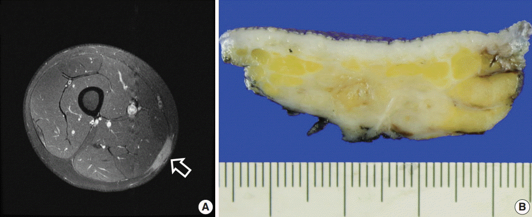
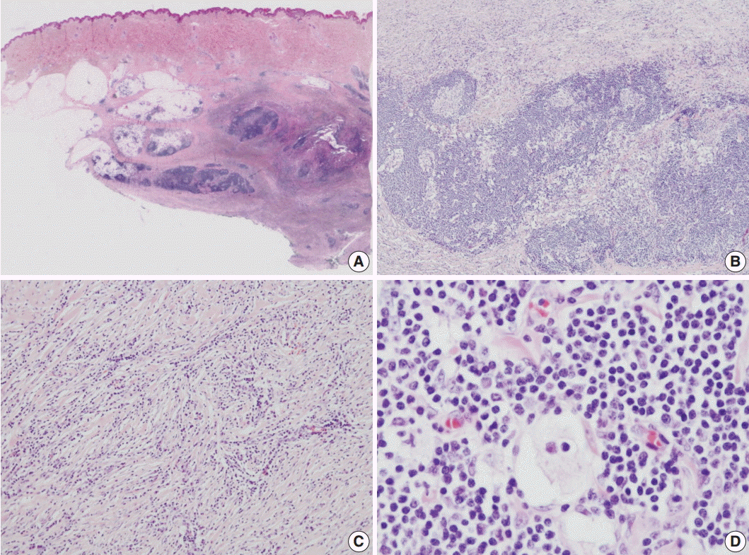
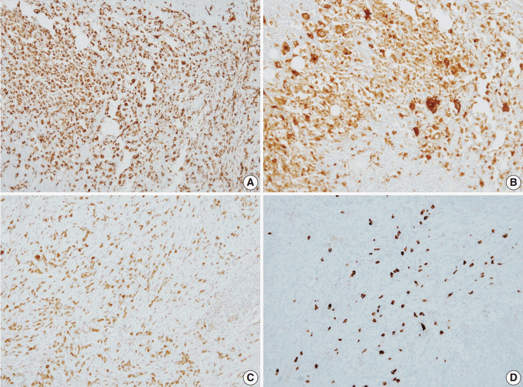
A 12-year-old male patient presented with a recently developed, hard, non-tender mass in the right thigh. He had been diagnosed with acute leukemia 4 years earlier and has been in complete remission after receiving standard chemotherapy and bone marrow transplantation. Magnetic resonance imaging showed a 3.8×2.8 cm enhanced soft tissue mass in the subcutaneous layer on the posteromedial side of the right mid-thigh that was suggestive of leukemic infiltration (Fig. 1A). Grossly, the excised lesion showed an irregular, tan-white appearance with sclerotic areas (Fig. 1B). Microscopic examination revealed dense lymphoplasmacytic and histiocytic infiltration into sclerotic stroma involving the subcutaneous layer and deep dermis (Fig. 2A, B). Dilated sinuses were filled with large histiocytes containing intact lymphocytes (emperipolesis) (Fig. 2C, D), and these histiocytes were positive for CD68 and S100 protein (Fig. 3A, B). CD117- and CD34-positive atypical blastic cell infiltration was not identified in any of the submitted sections (data not shown). Up to 70 IgG4-positive plasma cells per high power field were noted, and the IgG4/IgG ratio was 25% in the most IgG4-positive area (Fig. 3C, D). No evidence of recurrence has been found in imaging workups during a 12-month follow-up.

Imaging and macroscopic findings of soft tissue Rosai-Dorfman disease with features of IgG4-related disease. (A) Magnetic resonance imaging shows a 3.8×2.8 cm enhanced soft tissue mass (arrow) in the subcutaneous layer on the posteromedial side of the right mid-thigh, suggestive of leukemic infiltration. (B) In the excised specimen, a 3.0-cm irregular tan-white mass-like lesion is observed in the subcutis.

Microscopic findings of soft tissue Rosai-Dorfman disease with features of IgG4-related disease. (A) In the scanning view, the infiltrative lesion involving the deep dermis and subcutaneous layer is noted. (B) In the low-power view, dense lymphoplasmacytic infiltration with scattered histiocytes is noted. (C) Diffuse lymphoplasmacytic infiltration is noted in the sclerotic stroma. (D) Dilated sinuses are filled with histiocytes containing intact lymphocytes (emperipolesis).

Immunohistochemical staining results of soft tissue Rosai-Dorfman disease with features of IgG4-related disease. (A) Immunohistochemical staining for CD68 demonstrates histiocytes. (B) Immunohistochemical staining for S100 protein demonstrates emperipolesis. (C) Plasma cell IgG immunoreactivity. (D) Frequently identified IgG4-positive cells.
DISCUSSION
RDD frequently involves extranodal sites, observed in 43% of cases. However, isolated extranodal involvement is unusual, and soft tissue involvement is even more rare [3]. Aside from a few case reports, only a few large-scale studies have assessed STRDD. In a previous study, most STRDD presented as a rapidly growing mass and was frequently found in trunk and proximal extremities [4]. Multicentricity was occasionally demonstrated in STRDD; however, associated lymphadenopathy was not common. Other studies have reported female predominance for STRDD over a wide range of ages with variable-sized masses [5,6]. A higher recurrence rate has been suggested for STRDD with multicentric lesions compared to solitary lesions [7]. The present case was a solitary lesion at diagnosis, and the patient is receiving regular follow-up.
Various degrees of sclerosis and IgG4-positive plasma cells have been noted in the RDD literature [8]. A recent study reported that about 30% of RDD cases showed stromal sclerosis and increased number of IgG4-positive plasma cells, sharing similar histologic features to IgG4-related disease [2]. These results assumed that RDD with features of IgG4-related disease could possibly be on the spectrum of IgG4-related disease or the certain phase of RDD [2,8]. However, a general consensus regarding cut-off values for IgG4-positive plasma cells and the IgG4/IgG ratio has not been established for patients with RDD [2,9]. The patient described here had dense lymphoplasmacytic infiltrations in a sclerotic stroma with increased number of IgG4-positive plasma cells.
The differential diagnoses showing similar histologic features to RDD include inflammatory myofibroblastic tumor (IMT), Langerhans cell histiocytosis (LCH), and leukemic infiltration [3]. Some IMTs show compact spindle cell proliferation with fibrosis and diffuse inflammatory cell infiltration, similar histologic features to RDD. IMTs show membranous expression of anaplastic lymphoma kinase (ALK) in immunohistochemistry. We could not find ALK reactivity in this case (data not shown) and excluded the possibility of IMT.
LCH shows histiocytic aggregation in the background of mixed inflammatory cells, a similar morphologic feature to RDD. However, we could not identify the histologic characteristics of LCH exhibiting Langerhans cells with elongated nuclei and occasional nuclear grooves positive for S100 protein and CD1a.
Considering our patient’s clinical history of acute myeloid leukemia, leukemic infiltration can also be considered as a differential diagnosis. Leukemic infiltration shows infiltration of atypical blastic cells in the fibrotic background, with frequent subcutaneous involvement [10]. We assessed the possibility of leukemic involvement via immunohistochemical staining for CD117 and CD34, but did not observe immunoreactive atypical blastic cells.
RDD is a rare disease entity and can have overlapping histologic features with IgG4-related disease. STRDD is even more rare and presents as a rapidly growing mass, making it difficult to be differentiated from other malignancies. In this case report, we described a case of STRDD with characteristic histologic features of IgG4-related disease in a patient with a history of acute myeloid leukemia.
Notes
Conflicts of Interest
No potential conflict of interest relevant to this article was reported.