Fibrosarcoma of bone masquerading as fibrous dysplasia: diagnostic pitfall and molecular correlates of aggressive behavior
Article information

Abstract
Low-grade fibro-osseous lesions can be challenging to classify. A 33-year-old man presented with synchronous lesions involving the ilium and T2 vertebra. Because of an impending pathologic fracture, he received several doses of denosumab. Initial biopsies showed bland fibro-osseous lesions lacking GNAS or MDM2 alterations and were favored to represent polyostotic fibrous dysplasia. Subsequent iliac curettage revealed a fascicular spindle cell proliferation with subtle atypia and focal ossification, leading to a revised diagnosis of low-grade fibrosarcoma. Re-biopsy of the T2 lesion demonstrated a similar spindle cell proliferation without ossification. Sequencing identified copy number gains involving KIT, PDGFRA, and TERT. At 16-month follow-up, two metastatic pulmonary nodules developed, one responsive to chemotherapy. The patient remains alive with disease at 27 months after presentation. This case highlights a diagnostic pitfall in which low-grade fibrosarcoma with denosumab-associated ossification may mimic fibrous dysplasia and underscores the prognostic value of molecular profiling beyond histologic grading.
INTRODUCTION
Fibro-osseous lesions of bone represent a heterogeneous group of neoplasms with variable biologic potential, ranging from benign fibrous dysplasia (FD) to malignant entities such as low-grade osteosarcoma and fibrosarcoma. Histologic classification is challenging because of overlapping bland morphologic features and the lack of diagnostic immunohistochemical markers. FD is a benign fibro-osseous lesion that typically involves the medullary cavity of the craniofacial bones or the femur and may present with symptoms such as pain or pathologic fracture [1]. FD is associated with activating GNAS mutations. Depending on the degree of mosaicism, patients may present with a broad clinical spectrum, ranging from monostotic and polyostotic forms to McCune-Albright syndrome [1]. Radiologically, FD often demonstrates a smooth, ground-glass matrix with cortical thinning. Histologically, FD is characterized by curvilinear trabeculae of woven bone without osteoblastic rimming, set within a background of bland fibroblastic proliferation. Although rare, FD can undergo malignant transformation, most commonly to osteosarcoma and less frequently to fibrosarcoma [2].
Primary fibrosarcoma of bone is a rare malignant neoplasm of fibroblastic origin, accounting for <5% of all primary malignant bone tumors. It shows a strong predilection for long bones, with the femur being the most common site, followed by the tibia and pelvis [3]. Patients typically present with a symptomatic bone lesion with pain and sometimes a pathologic fracture. Mean age at presentation is approximately 38 years (range, 8 to 84 years), with nearly equal sex distribution [3]. On imaging, fibrosarcoma of bone typically presents as an osteolytic lesion without intrinsic mineralization. Histologically, fibrosarcoma is characterized by a fascicular proliferation of spindle cells without evidence of specific lines of differentiation [4]. Prognostically, primary fibrosarcoma of bone shows a high metastatic rate (~68%) [3]. Therefore, although surgery is the mainstay of treatment, chemotherapy may be warranted, particularly for tumors with high-risk features, including age older than 40 years, tumor location in the axial skeleton, and high-grade tumor (grade 3 or 4) [3].
Distinguishing FD from low-grade fibrosarcoma largely relies on the identification of curvilinear bony trabeculae in FD, as the spindle cell component can appear similar in both entities. Denosumab, a RANKL inhibitor, is used in the treatment of osteoporosis and cancer-related bone complications [5]. It is well known that denosumab induces ossification in giant cell tumors of bone [6,7]. A similar phenomenon has also been documented in FD, aneurysmal bone cyst, osteoblastoma, and central giant cell granuloma [8-11]. In cases of fibrosarcoma previously treated with denosumab, secondary ossification may be mistaken for curvilinear bony trabeculae suggestive of FD. We present such a case to raise awareness of this diagnostic pitfall, which, to the best of our knowledge, has not previously been reported. In addition, this case highlights the diagnostic and prognostic value of molecular profiling beyond histologic evaluation.
CASE REPORT
A 33-year-old man presented to an outside hospital with acute-on-chronic groin pain following a powerlifting competition. His past medical history was significant for papillary thyroid carcinoma, status post total thyroidectomy. Magnetic resonance imaging (MRI) demonstrated a large lytic lesion in the ilium and an additional lesion in the T2 vertebra, placing the patient at risk for pathologic fracture. To improve skeletal stability, he received several doses of denosumab. Serial imaging demonstrated a treatment effect, with new ossification developing along the cortical margins of both lesions. The subsequent iliac biopsy showed a fibro-osseous lesion, whereas the T2 biopsy revealed an atypical fibrous lesion. No overtly malignant features were identified. Molecular studies performed at the outside hospital were negative for GNAS alterations by sequencing and MDM2 amplification by fluorescence in situ hybridization. Overall, the findings were favored to represent polyostotic FD.
Due to persistent pain, the patient was referred to our institution for further evaluation and management. Computed tomography (CT) revealed a 5.6-cm lytic lesion in the ilium involving the weight-bearing dome of the left acetabulum, with subtle cortical breakthrough. Positron emission tomography–CT demonstrated that the lesion was hypermetabolic (Fig. 1A, B). The T2 vertebral lesion showed expansile growth with soft tissue extension and corresponding enhancement (Fig. 1C, D). Additional smaller lesions were identified within the T1 vertebral body and the T4 transverse process.
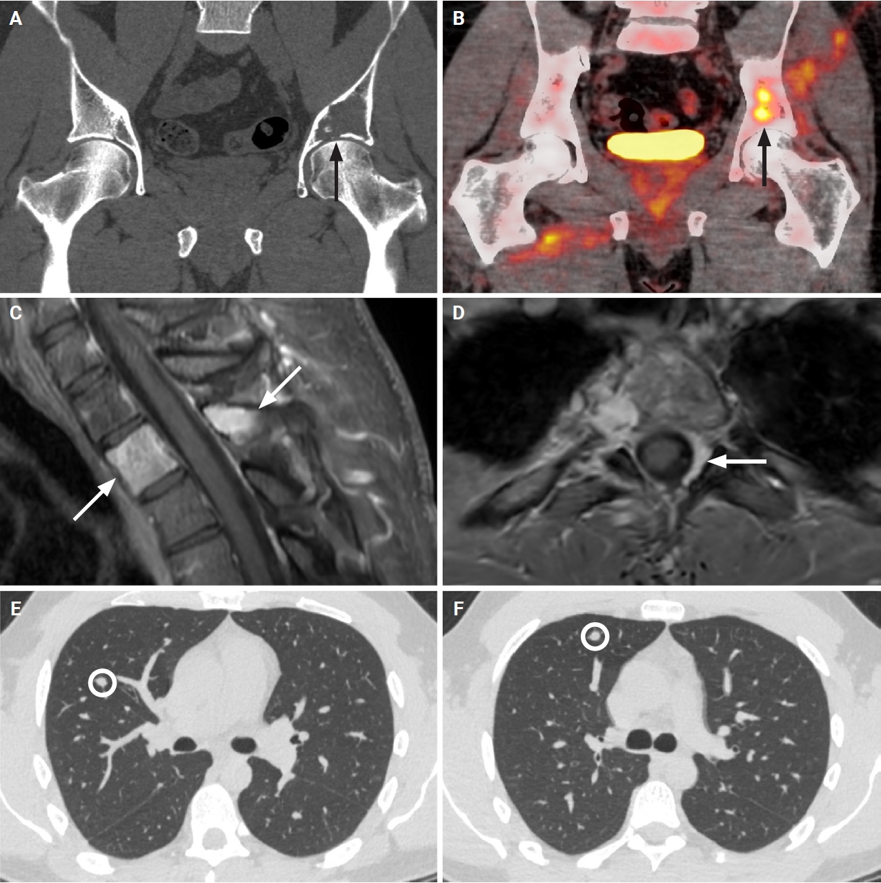
Coronal computed tomography (CT) of the pelvis at presentation shows a lytic lesion involving the weight-bearing dome of the acetabulum, with evidence of cortical destruction (A). The lesion is hypermetabolic on subsequent 18F-fluorodeoxyglucose positron emission tomography–CT with a maximum standardized uptake value of 6.6 (B). Sagittal and axial T1-weighted fat-suppressed post-contrast magnetic resonance images of the thoracic spine show abnormal enhancement of the T2 vertebral body and spinous process, with abnormal epidural enhancement especially on the left reflecting extra-osseous extension (C, D). Additional smaller lesions are present in the T1 vertebral body and the left T4 transverse process (not shown). CT of the chest without contrast demonstrates two right upper lobe lung nodules, the larger of which subsequently was biopsied (E, F) (A–D, arrows indicating lesions; E, F, circles highlighting lesions).
The patient underwent curettage of the left iliac lesion. Microscopically, the tumor demonstrated predominantly fascicular proliferations of spindle cells with focal mature-appearing bone spicules lacking osteoblastic rimming, reminiscent of FD (Fig. 2A–C). In the ossified areas, the stromal spindle cells appeared bland, without significant nuclear atypia or mitotic activity (Fig. 2C). However, in the nonossified areas, the spindle cells exhibited increased cellularity (Fig. 2D), along with variable hyperchromasia and occasional mitotic activity (Fig. 2E, F). Immunohistochemical studies showed that the spindle cells were negative for smooth muscle actin, desmin, nuclear β-catenin, SOX10, STAT6, ERG, pan-keratin (AE1/AE3, CAM5.2, and MNF116), p40, PAX8, thyroid transcription factor 1, and BRAF V600E. A subset of tumor cells was positive for p63. SATB2 (special AT-rich sequence-binding protein 2) staining was focal and weak, predominantly confined to the osseous component. In the nonossified areas, SATB2 was mostly negative (Supplementary Fig. S1).
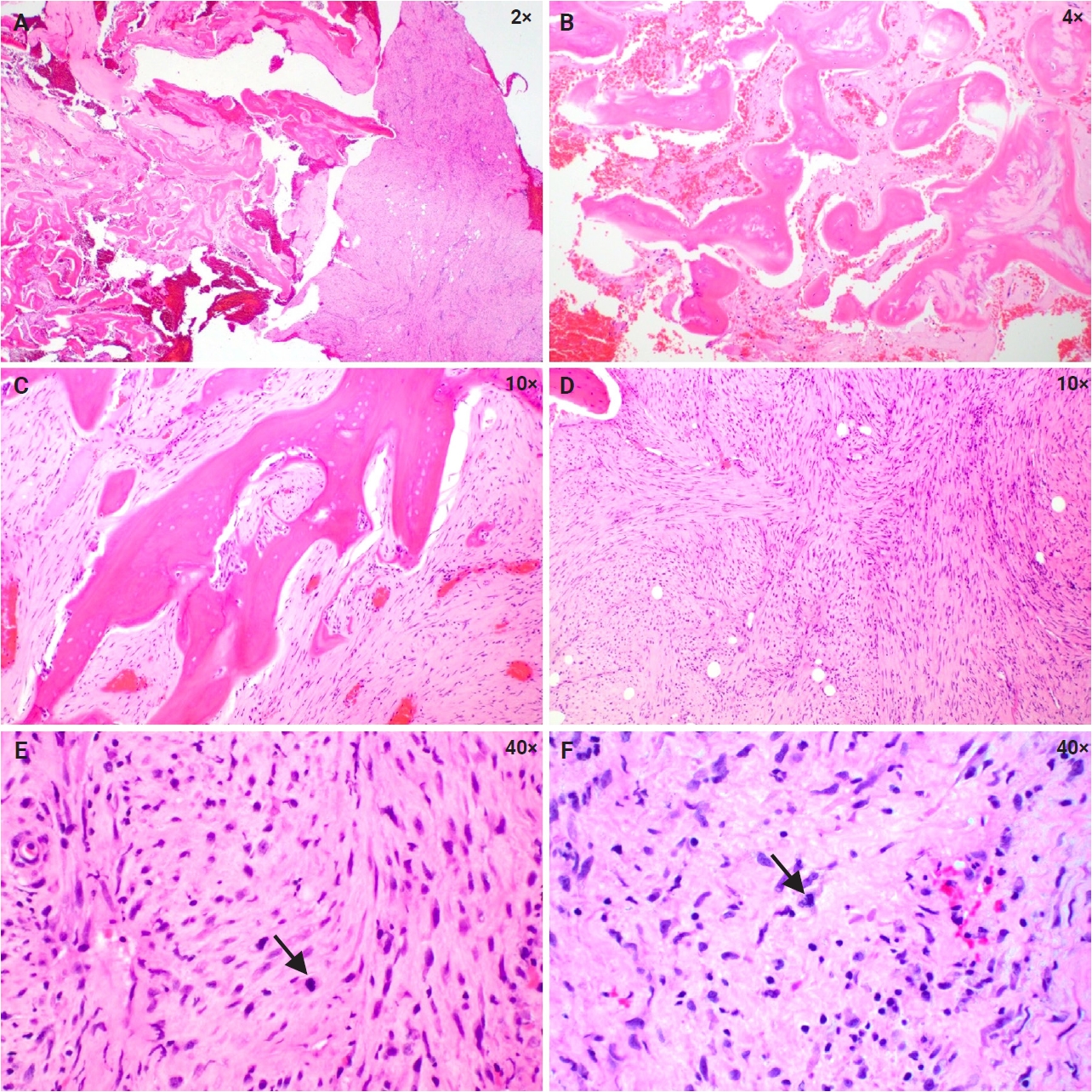
Histology of the iliac tumor. The focal fibrous dysplasia–like area shows bony spicules surrounded by bland spindle cells without osteoblastic rimming (A–C). The majority of the specimens demonstrate a cellular spindle cell proliferation with mild cytologic atypia and rare mitoses, arranged in a fascicular or herringbone pattern (D–F) (E–F, arrows indicating mitoses).
Despite the histologic features reminiscent of FD, the diagnosis was revised to low-grade fibrosarcoma based on the aggressive radiologic findings, subtle cytologic atypia, lack of immunophenotypic evidence of specific lineage differentiation, and absence of molecular evidence supporting FD or low-grade osteosarcoma. Radiofrequency ablation and re-biopsy of the T2 vertebral lesion demonstrated a spindle cell proliferation similar to that of the iliac tumor, except for the absence of ossification and the presence of small background native bone fragments, consistent with fibrosarcoma (Fig. 3A, C, E). The synchronous presentation of the iliac and vertebral lesions makes it difficult to distinguish osseous metastases from multifocal disease.
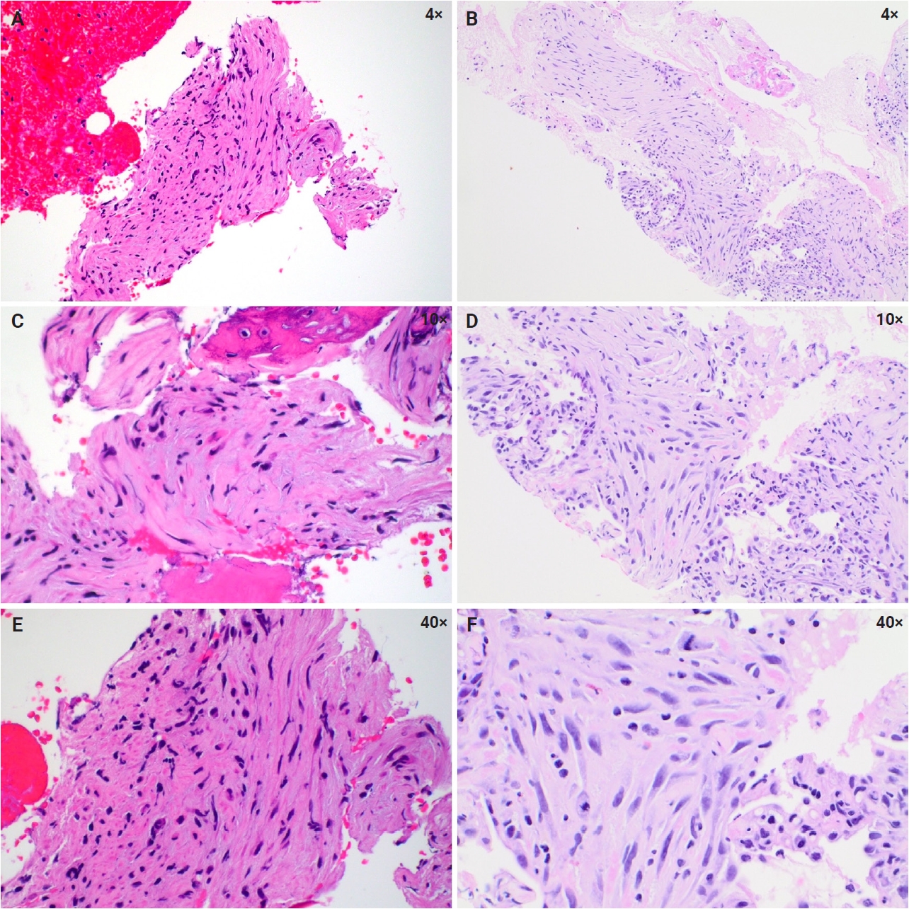
Histology of the spinal and lung tumors. The spinal lesion shows a proliferation of spindle cells with hyperchromatic nuclei (A, C, E). The lung lesion demonstrates a similar spindle cell proliferation with more pronounced cytologic atypia and mitoses (B, D, F).
The iliac specimen was submitted for the Tempus xT CDx assay, a targeted hybrid capture–based next-generation sequencing panel analyzing 648 cancer-related genes, including GNAS and MDM2 (Tempus Labs, Inc., Chicago, IL, USA) (Supplementary Table S1). Copy number gains involving KIT, PDGFRA, and TERT were identified. Somatic variants of uncertain significance included TAF1 (p.W276C), RHOA (p.P75S), TUSC3 (p.R117H), and APC (p.L1522F). Tempus RNA sequencing demonstrated no gene rearrangements or reportable aberrant splicing events. Given the patient’s history of two malignancies at a young age, the possibility of an underlying genetic predisposition was considered. The patient had no significant family history of cancer, except for kidney cancer in his maternal grandfather and prostate cancer in his paternal grandfather. A 77-gene CancerNext-Expanded panel (Ambry Genetics, Aliso Viejo, CA, USA) (Supplementary Table S2) performed on peripheral blood identified a germline MITF variant of uncertain clinical significance.
Sixteen months after the initial presentation, chest CT demonstrated two new solid nodules in the right upper lobe of the lung, measuring 6 mm and 7 mm, respectively (Fig. 1E, F). Biopsy of one of the lung nodules revealed a spindle cell proliferation characterized by more pronounced atypia, increased mitotic activity, and no evidence of ossification (Fig. 3B, D, F), consistent with metastatic fibrosarcoma. Chemotherapy with gemcitabine and docetaxel was initiated. One lung nodule adjacent to the fiducial marker showed marked shrinkage, whereas the other failed to respond. Follow-up pelvic MRI demonstrated edema and postsurgical changes. Twenty-seven months after presentation, the patient remains alive with the disease. Definitive treatment of the lung metastases is being planned, either by surgical resection or ablation.
DISCUSSION
The relative proportions of fibrous and osseous components in FD vary. The iliac curettage specimen in our patient was superficially reminiscent of FD due to the presence of bony spicules, despite an exuberant fibrous component. Notably, the patient had previously been treated with denosumab, which is known to induce secondary bone formation as early as two months after initiation of therapy [6,7]. In giant cell tumor of bone, denosumab-induced ossification often demonstrates osteoblastic rimming. Of note, the morphologic spectrum of denosumab-related changes is broad and varies with treatment duration and tumor type [7,10]. Early lesions in denosumab-treated giant cell tumor of bone lack osteoblastic rimming and can closely resemble FD [7]. Similarly, in denosumab-treated aneurysmal bone cyst and osteoblastoma, osteoblastic rimming is absent or inconspicuous, again mimicking FD [10]. Although a pre-treatment biopsy was unavailable for comparison in our case, serial imaging suggested that the new ossification observed on follow-up studies was consistent with a treatment effect. However, this potential pitfall of treatment-related histologic change was not initially considered, and the bony spicules were therefore interpreted as an intrinsic osseous component of a spindle cell proliferation, leading to a misdiagnosis of FD.
Radiologically, the diagnosis of FD should be questioned in this case based on the aggressive imaging findings, i.e., cortical breakthrough. Microscopically, although subtle, the presence of hyperchromasia and mitotic activity argues against a benign process. Genetically, no GNAS mutation was detected to support a diagnosis of FD. In FD, false-negative results can occur due to somatic mosaicism or technical issues [1,12]. Even in cases of malignant transformation, GNAS mutations are typically retained [13]. However, the tumor tested negative for GNAS mutation on two separate occasions and was positive for alternative genetic alterations, making false-negativity less likely. Ultimately, the malignant nature of the tumor was confirmed by the development of pulmonary metastases. Interestingly, the lung biopsy exhibited more pronounced cytologic atypia and increased mitotic activity, which may reflect disease progression or sampling bias.
Another diagnostic pitfall associated with denosumab-induced ossification is misinterpretation as osteoid, potentially leading to a misdiagnosis of osteosarcoma. Low-grade central osteosarcoma (LGCOS) is excluded based on the following constellation of findings: (1) Tumor localization: LGCOS most commonly arises in the appendicular skeleton, with only rare involvement of axial bones. Reported cases in sites such as the iliac bone demonstrate diagnostic molecular features, including positivity for MDM2 and cyclin-dependent kinase 4 (CDK4) [14], both of which are absent in the current case. In contrast, in a large Mayo Clinic series of 92 patients, the pelvis was the third most common site of primary fibrosarcoma of bone, accounting for 15% of cases [3]. (2) Radiographic correlation: LGCOS characteristically shows coarsened trabeculation on imaging, corresponding histologically to long, thick, parallel bony trabeculae. In contrast, the lesions in this case were initially lytic, with a differential diagnosis that included a myeloproliferative process and metastatic papillary thyroid carcinoma rather than osteosarcoma. Following several doses of denosumab, new ossification developed along the cortical margins of both lesions, interpreted as a treatment-related change. (3) Clinical course: LGCOS is locally aggressive but has low metastatic potential [15-17]. When metastasis occurs, it is usually associated with recurrence and dedifferentiation [15-17]. In this case, pulmonary metastases developed 16 months after initial presentation, without prior recurrence or histologic evidence of dedifferentiation. By comparison, primary fibrosarcoma of bone shows a high metastatic rate (approximately 68%) at a median of 9 months (range, 1 to 51 months), which aligns with the observed clinical course [3]. (4) Immunophenotype: Although not entirely specific, SATB2 is a highly sensitive osteoblastic marker, and osteosarcoma typically shows strong, diffuse nuclear staining [18,19]. In this case, focal, weak SATB2 staining, predominantly confined to the osseous component, argues against a diagnosis of osteosarcoma [19]. Importantly, SATB2 expression has been reported in a range of non-osteogenic bone tumors, including approximately 45% of fibrosarcomas [18,20]. (5) Molecular profile: LGCOS lacking MDM2 amplification is genetically heterogeneous and poorly defined. Reported alterations include CDK4 amplification, loss of RB1, amplification of 6p12–p21, and gains of 8q21–q24, 10p15, 12q13–q15, and 16q23–q24 [21,22], none of which are identified in this case. To the best of our knowledge, copy number gains involving KIT, PDGFRA, and TERT have not been described in LGCOS.
Although extremely rare, primary myoepithelioma of bone has been reported [23]. Myoepithelial tumor can show pure spindle cell morphology without epithelioid or plasmacytoid cells, regarded as spindle cell myoepithelioma [24]. In our case, patchy p63 positivity raised the consideration of myoepithelioma. However, the lack of expression of epithelial markers (pan-keratin) and more specific myoepithelial markers (smooth muscle actin, SOX10, and p40) argues against this possibility. Another sarcoma that is also a diagnosis of exclusion is undifferentiated pleomorphic sarcoma, which can similarly show a negative immunoprofile for essentially all lineage-specific markers, as in our case. However, it is noted that adult fibrosarcoma should not show more than a moderate degree of nuclear pleomorphism, whereas undifferentiated pleomorphic sarcoma usually shows marked nuclear pleomorphism, a key cytologic feature absent in our case [25,26].
Fibrosarcoma of bone, like its soft tissue counterpart, is a diagnosis of exclusion [3]. It is defined as a primary malignant fibroblastic neoplasm exhibiting a fascicular or herringbone architecture and relatively monotonous cytology, without morphologic, immunophenotypic, or genetic evidence of another specific malignancy. The frequency of fibrosarcoma of bone was historically estimated to account for 2%–4% of all primary bone tumors. However, advances in ancillary studies, particularly molecular testing, have led to the reclassification of many tumors into specific sarcoma subtypes, resulting in a lower true incidence of fibrosarcoma. Bahrami and Folpe reported that 84% of previously diagnosed adult-type fibrosarcomas in somatic soft tissue could be reclassified, and they concluded that true fibrosarcoma is exceedingly rare and should be diagnosed with great caution [4].
The molecular landscape of bone fibrosarcoma, although less extensively characterized than that of some other sarcomas, has been explored using cytogenetic approaches. Array comparative genomic hybridization analysis of 19 specimens revealed frequent deletions at 6q, 8p, 9p, 10, 13q, and 20p, as well as gains at 1q, 4q, 5p, 8q, 12p, 15q, 16q, 17q, 20q, 22q, and Xp [27]. Co-amplification of KIT and PDGFRA, located in proximity on chromosome 4q12, was detected in the majority of patients [27]. Another independent study suggested involvement of the autocrine PDGF receptor signaling loop in bone fibrosarcoma [28]. The TERT copy number gain detected in our patient is in line with gains involving chromosome 5p, which harbors the TERT locus [27]. Taken together, the molecular profile of copy number gains in KIT, PDGFRA, and TERT supports the histologic diagnosis of fibrosarcoma.
So far, the only recognized high-risk feature in our case is tumor location in the axial skeleton [3]. Notably, despite its low-grade histologic appearance and treatment, the tumor followed an aggressive clinical course more consistent with a high-grade sarcoma, as evidenced by multiple osseous lesions and the development of lung metastasis within 16 months of the initial presentation. Gene expression studies have demonstrated that TERT amplification results in markedly increased TERT expression, even exceeding levels associated with the more common TERT promoter mutations [29]. Increased TERT expression and telomerase activity have been linked to aggressive tumor behavior across multiple cancer types, including fibrosarcoma, and may represent a key mechanism in sarcoma progression [29-31]. This association is further supported by evidence that TERT regulates fibrosarcoma growth both in vitro and in vivo [32]. In addition, the alternative lengthening of telomeres phenotype, which is associated with poor prognosis in liposarcoma, has also been reported in fibrosarcoma [33]. Taken together, these findings raise the possibility that TERT copy number gain may have contributed to the aggressive clinical behavior of a histologically deceptive fibrosarcoma in this case, similar to what has been reported in well-differentiated thyroid carcinomas [34]. Although morphologic assessment remains the gold standard for diagnosis, the World Health Organization classification systems increasingly incorporate molecular features as defining diagnostic criteria across multiple tumor types, including peripheral nerve sheath tumors and hematologic malignancies [35,36]. This case illustrates how this evolving paradigm may also influence risk stratification of fibro-osseous lesions beyond histologic grading and potentially inform therapeutic decision-making.
Fibrosarcoma of bone presents a significant diagnostic challenge because of its rarity and the often subtle cytologic atypia seen in low-grade cases. Awareness of the potential diagnostic pitfall created by denosumab-induced ossification is essential to prevent misinterpretation. Accurate diagnosis requires a multidisciplinary approach that integrates clinical history, including prior treatment, radiologic findings, histopathologic features, and molecular data. Molecular profiling may provide important prognostic insight when the histologic appearance is deceptively low grade.
Supplementary Information
The Data Supplement is available with this article at https://doi.org/10.4132/jptm.2026.05.29.
Notes
Ethics Statement
This study was conducted in accordance with the 1964 Helsinki Declaration and its later amendments. A single-case report conducted at the authors’ institution does not constitute “human subjects research” and therefore does not require formal IRB review. Formal written informed consent was not required as the data are fully de-identified.
Availability of Data and Material
The data that support the findings of this study are available from the corresponding author upon reasonable request.
Code Availability
Not applicable.
Author Contributions
Conceptualization: LL. Data curation: WJS. Formal analysis: WJS, BKH, LL. Investigation: WJS, BKH, LL. Methodology: WJS, LL. Project administration: LL. Resources: BKH, LL. Supervision: LL. Validation: BKH, LL. Visualization: WJS, BKH. Writing—original draft: WJS. Writing—review & editing: LL. Approval of final manuscript: all authors.
Conflicts of Interest
The authors declare that they have no potential conflicts of interest.
Funding Statement
No funding to declare.
Acknowledgments
The authors thank Dr. Andrew L. Folpe (Mayo Clinic, Rochester, MN, USA) for expert review of the iliac biopsy.