Ultrasound-guided cryoablation of recurrent thyroid cancer: radiologic-pathologic correlation
Article information

Abstract
Cryoablation is a minimally invasive thermal ablation modality that destroys tumor cells through intracellular ice crystal formation and osmotic injury. Although it offers theoretical advantages, its application in recurrent papillary thyroid cancer (PTC) remains limited, and the radiologic-pathologic findings of treatment response have not been well characterized. A 55-year-old male patient with biopsy-proven recurrent PTC in the left supraclavicular fossa and a history of total thyroidectomy, neck dissection, and advanced colorectal cancer underwent a single-session ultrasound-guided cryoablation. The 20-minute procedure was completed without complications. Immediately post-ablation, the lesion volume increased by over 200% with marked hypoechogenicity and obliteration of abnormal feeding vessels on microvascular imaging. Pathology demonstrated disrupted tumor cell membranes, necrotic changes, and myxoid peritumoral stromal alteration, consistent with direct and indirect injuries caused by cryoablation. At the first-month follow-up, repeat biopsy showed no viable tumor, with subacute inflammatory infiltrates, histiocytic aggregates, and progressive fibrosis. On ultrasound, a volume reduction rate of 71.4% was achieved at one month follow-up and 97.1% at three months follow-up. This case report provides the first radiologic-pathologic findings of both immediate and delayed effects of cryoablation in recurrent PTC, supporting its role as a promising minimally invasive palliative therapy.
INTRODUCTION
Cryoablation employs the Joule-Thomson effect to rapidly decrease tissue temperature and form ice, resulting in direct cancer cell destruction via localized hypothermic effect and indirect destructive effects via alterations to the microenvironment and osmolarity. Specifically, during ice formation, intracellular ice crystals may cause direct damage to intracellular organelle membranes and plasma membranes, severely damaging the cellular structures. In the subsequent thawing process, the osmolarity difference between melting extracellular ice and intracellular fluid results in osmotic shifts, promoting fluid influx into cells and ultimately causing cellular rupture [1]. Cryoablation may offer advantages such as reduced procedural pain and possible immunomodulatory effects, suggesting it could be a useful minimally invasive option for head and neck lesions, including recurrent thyroid cancer [2,3]. Nevertheless, the application of cryoablation for head and neck lesions remains limited, mainly for anatomical reasons. Few studies have explored the efficacy of cryoablation in treating recurrent papillary thyroid cancer (PTC), with the recently published case series by Sag et al. and Simon et al. being the only reports to date [4,5]. Furthermore, the radiologic-pathologic findings of cryoablation treatment response have not been well characterized. This case report aims to present both immediate and delayed radiologic-pathologic findings associated with palliative cryoablation in recurrent PTC.
The cryoablation was performed under ultrasound guidance (Canon Aplio i800, Canon Medical Systems, Otawara, Japan). Thorough pre-procedure scanning of the target lesions and surrounding structures was performed to avoid injuries to anatomical structures, such as the trachea and large vessels. The following procedural parameters were evaluated: procedure time in minutes (defined as time duration from hydrodissection to final cryoablation probe retraction), pain measured by a three-point Likert scale, complication rate, volume reduction rate (VRR), and pathology outcomes at first-month follow-up. Specifically, pain severity was assessed by a 3-point Likert scale: mild (1), moderate (2), and severe pain (3), and VRR was calculated with the following equation: [(pre-ablation volume – post-ablation volume)/pre-ablation volume] × 100. Complications were assessed in accordance with the Society of Interventional Radiology Clinical Practice Guidelines [6].
Cryoablation was performed using a single-probe 17.5 cm length, 17-gauge cryoprobe (IceSeed, Boston Scientific, Marlborough, MA, USA) (Supplementary Fig. S1). Based on the manufacturer’s data, the estimated volume of tumoricidal zone at –40°C measures approximately 8 mm × 10 mm, oriented coaxially about the center of the cryoablation probe. Therefore, the volume and shape of each target nodule were estimated pre-procedurally. Following local anesthesia with 1% lidocaine, hydrodissection was carried out using lukewarm saline for “cold-sink” effect to prevent hypothermic injuries to the common carotid artery, subclavian vein, and nerve structures [7]. The cryoprobe was advanced so that at least 3 mm of the tip penetrated through the target nodule, accounting for the non-tumoricidal distal 2–3 mm of the probe [5]. A slight increase in freezing power to create “tethering” was performed to facilitate a lever-pull maneuver, increasing the distance between the target nodule and important anatomical structures (Fig. 1). The freezing protocol involved five cycles of freezing and thawing, beginning at 60% power with sequential increases of 10%. Specifically, freezing power escalated in 10% increments, with each cycle comprising approximately one minute of freezing followed by one minute of thawing until reaching 100% power (Fig. 1). Passive thawing was employed, as it is considered more effective in destroying cancer cells compared to active thawing [8]. Procedural endpoints, based on the mechanism of cryoablation (intracellular ice crystal formation, osmotic injury, and microvascular thrombosis), included destruction of abnormal microvasculature and hypoechogenicity in the target lesion.

Schematic illustration of cryoablation for recurrent thyroid cancer. (A) Pre-procedural planning is performed to optimize probe placement for complete coverage of the target nodule, which is in proximity to the subclavian vein (arrow). (B) Hydrodissection is performed to create a thermal barrier and to prevent thrombosis in the subclavian vein (arrow). (C) An ice ball forms instantaneously upon activation. (D) “Tethering” is performed to facilitate a lever-pull maneuver, further increasing the distance between the target nodule and the subclavian vein. (E) Passive thawing is applied before the freezing cycle is re-initiated.
CASE REPORT
A 55-year-old male patient with advanced colorectal cancer and core-needle biopsy (CNB)–confirmed recurrent PTC underwent single-session cryoablation in the left supraclavicular region. He had a history of total thyroidectomy and neck dissection for PTC. Having already undergone multiple surgeries and chemotherapy for advanced colorectal cancer and PTC, the patient was hesitant to undergo additional surgery. The patient fully understood and provided informed consent for cryoablation, selecting it over radiofrequency ablation (RFA) due to its theoretical advantage in reducing procedure-related pain and complications [9]. The recurrent PTC in the left supraclavicular fossa was initially measured at 3 mm × 6 mm × 7 mm, corresponding to an approximate volume of 0.07 mL. The pre-procedure microvascular imaging demonstrated abnormal feeding vessels. Procedure time was approximately 20 minutes, and no complications occurred. Pain was minimal both during and after cryoablation, scored 1 on a 3-point Likert scale. There was no damage to the left subclavian vessels and left lung. The abnormal vascularity feeding the recurrent PTC markedly regressed on microvascular imaging (Fig. 2). Approximately one month after the cryoablation, the recurrent PTC demonstrated a VRR of 71.4%, indicating a significant decrease in nodule volume. The therapeutic impact was further evident at the three-month follow-up, when the VRR improved to 97.1%. Follow-up positron emission tomography–computed tomography (PET-CT) revealed a marked reduction of high fluorodeoxyglucose (FDG) uptake in the left supraclavicular fossa, confirming the therapeutic efficacy of cryoablation.
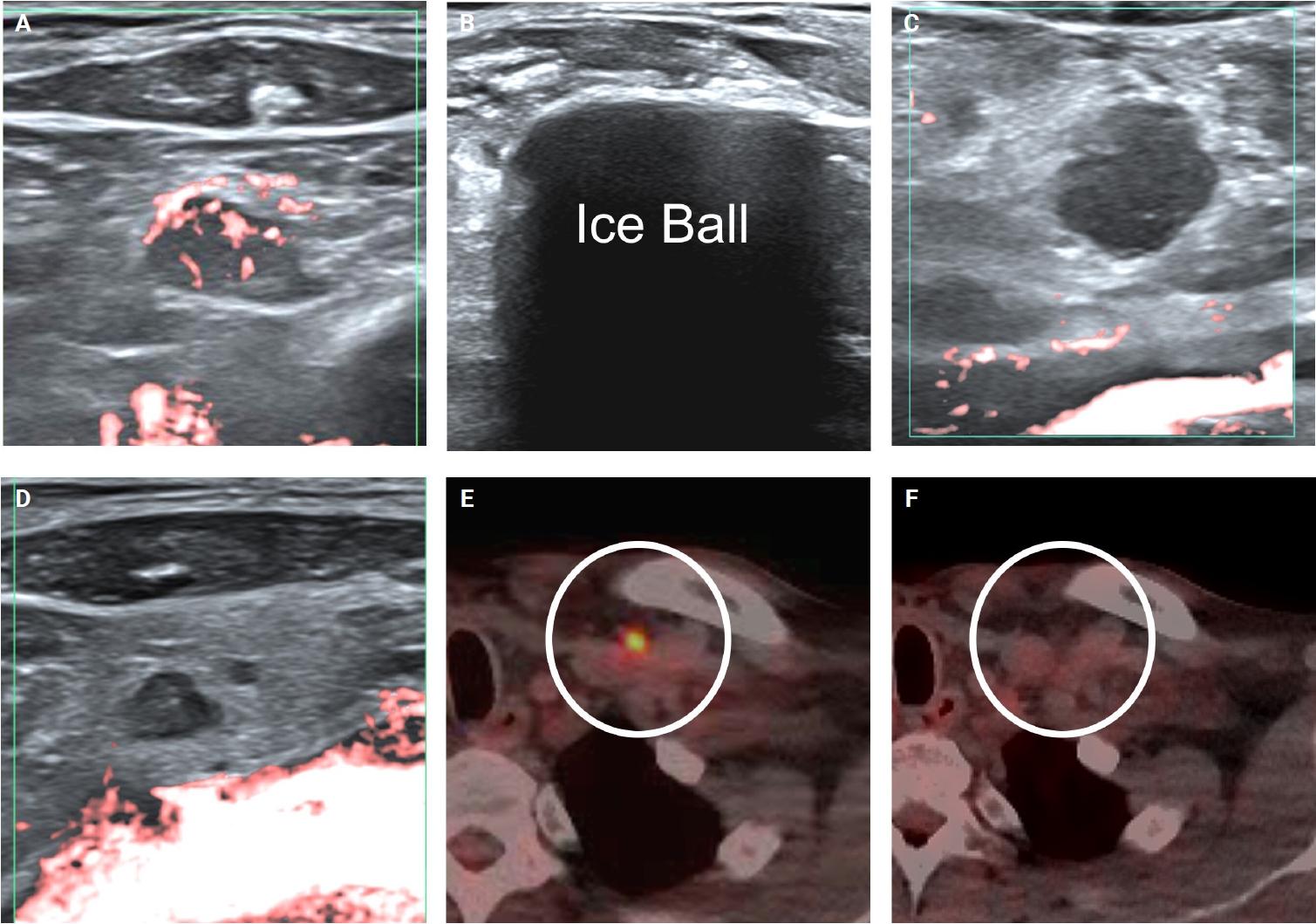
Ultrasound-guided cryoablation of recurrent papillary thyroid cancer at supraclavicular fossa. (A, B) A 55-year-old male patient underwent palliative-intent cryoablation for biopsy-proven recurrent thyroid cancer at the left supraclavicular fossa. The initial lesion measured 0.3 cm (anteroposterior) × 0.6 cm (transverse) × 0.7 cm (craniocaudal), corresponding to a volume of approximately 0.07 mL. Prominent abnormal vascularity (i.e., feeding arteries) was visualized on microvascular imaging. (C) Immediately post-cryoablation, abnormal vascularity was obliterated, and the lesion volume increased more than 200% (from 0.07 mL to 0.23 mL). (D) At approximately 1-month follow-up, a 71.4% volume reduction was achieved. The left subclavian vein remained patent, with no delayed complications. (E) Pre-procedural fluorodeoxyglucose (FDG) uptake (circle) was observed at the left supraclavicular fossa. (F) After cryoablation, the nodule with avid FDG uptake was markedly reduced (circle). At the third month follow-up, a 97.1% volume reduction was achieved.
Radiologic-pathologic correlation
Immediately after cryoablation, the volume of the ablated lesion increased from 0.07 mL to 0.23 mL (over 200% volume increase). This finding, combined with the marked hypoechogenicity observed on ultrasound, may be attributed to edema and fluid influx into the cancer cells. Both direct injury and indirect injury resulted from repeated ice formation and from osmotic shifts driven by the osmolarity difference between melting extracellular ice and intracellular space. Immediate post-procedure pathology confirmed cells with necrotic changes and disruption of the tumor cell membranes, with distinct borders of malignant clusters and scattered single tumor cells. In addition, myxoid stromal alteration was observed in the peritumoral microenvironment, suggestive of cryoablation-induced edema (Fig. 3).
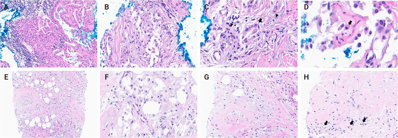
Pathology perspective of cryoablation of recurrent papillary thyroid cancer. (A) Pre-cryoablation hematoxylin and eosin (H&E) section demonstrated papillary growth with nuclear atypia, accompanied by lymphocytic infiltrates. (B–D) Sections were obtained immediately after cryoablation. (B) Tumor cell membranes appear disrupted, with distinct borders of malignant clusters and scattered single tumor cells. (C) A residual malignant follicle (arrow) shows degenerative changes of nuclei and disrupted follicular architecture. Myxoid stromal alteration is evident in the peritumoral microenvironment, suggestive of cryoablation-induced edema (arrowhead). (D) Cells with necrotic changes were identified (arrow). (E–H) Representative H&E sections showing histologic changes at 1-month post-cryoablation. (E) Subacute inflammatory infiltrates with progressive stromal fibrosis are present. (F) Focal histiocytic aggregates reflect a subacute inflammatory response. (G) Karyorrhectic nuclear debris is present in some areas, along with residual stromal edema. (H) More advanced fibrosis is evident, with the presence of hemosiderin-laden macrophages (arrows), indicative of prior hemorrhage.
At the 1st month follow-up, marked volume reduction of the recurrent PTC was achieved. Repeat CNB was performed, which demonstrated no evidence of a viable tumor and advanced fibrotic changes. Subacute inflammatory infiltrates, histiocytic aggregates, and progressive stromal fibrosis were observed. Therefore, the small hypoechoic residual nodule noted on ultrasound likely represents scar formation rather than residual or recurrent tumor. This ultrasound finding was also supported by the absence of abnormal feeding vessels on microvascular imaging and by the marked regression of FDG uptake on PET-CT.
DISCUSSION
This case report is the first to detail both the immediate and delayed effects of cryoablation on recurrent PTC, as observed through radiologic and pathologic findings. The initial increase in tumor volume following cryoablation on ultrasound, combined with evidence of necrotic cells and myxoid stromal changes in the microenvironment, supported the direct injury (intracellular ice crystal formation) and indirect injury (fluid influx due to osmotic gradients). However, this report has several limitations. First, it describes a single case of cryoablation-treated recurrent PTC with only short-term follow-up; although cryoablation was associated with reduced pain and demonstrated therapeutic efficacy over three months, longer-term outcomes remain to be determined. Second, CNB is inherently subject to sampling error and may not fully represent the target lesion. Third, immediate post-ablation CNB specimens often exhibit substantial freezing artifacts that can hinder accurate histopathologic analysis. Nevertheless, given the marked regression of FDG uptake on PET-CT, the decreased lesion size on follow-up ultrasound, and the CNB findings, the residual tiny hypoechoic focus most likely represents fibrotic change rather than viable tumor.
Cryoablation offers several notable advantages. Foremost, it is associated with reduced pain and bleeding during the procedure. This modality may be particularly beneficial for patients undergoing multiple ablation procedures without sedation, especially when targeting tumors with significant feeding vessels. In addition, cryoablation results in less hypothermic cellular damage to anatomical structures compared to RFA, as demonstrated by animal studies involving renal cells [1]. This advantage is especially valuable in the head and neck, where critical structures are densely situated. Consistent with this, Sag et al. [5], in a series of 15 patients with recurrent PTC, reported no vascular injury even when the cryoablation zone was in direct contact with vessels. In our case, hydrodissection, together with tethering and tilting maneuvers, was employed to further protect critical structures such as the subclavian vein. Despite these benefits, cryoablation has certain limitations. The most significant challenge is inadequate visualization of the target and probe due to substantial posterior acoustic shadowing caused by ice-ball formation. Moreover, there is a lack of long-term studies validating the efficacy of cryoablation, resulting in an absence of established guidelines regarding optimal power settings and freezing durations. Lastly, cryoablation is less cost-effective and requires frequent replenishment of argon gas, which may pose obstacles when compared to the readily available conventional heat-based ablation techniques.
Since published reports on cryoablation of head and neck lesions, including recurrent PTC, remain very limited, no direct comparison has been made between cryoablation and other thermal ablation techniques such as RFA. Comparative studies are available, however, for other organs such as non–small cell lung cancer. A meta-analysis demonstrated that cryoablation yields better prognostic outcomes and a lower recurrence rate than RFA for tumors smaller than 3 cm [10]. Furthermore, evidence suggests that cryoablation may elicit antitumor cytotoxic antibody production and activate cytotoxic T-cell immunity, thereby enhancing anti-tumor immune responses in lung cancer [10]. This effect may contribute to tumor destruction and reduce the likelihood of recurrence.
Cryoablation may serve as a useful minimally invasive palliative option for recurrent PTC. Future work should focus on establishing standardized protocols and refined techniques that ensure safe, predictable, and cost-effective treatment. Finally, long-term research, including a comparative study to RFA, will be essential to evaluate the therapeutic outcomes of cryoablation. In conclusion, cryoablation employs localized hypothermic effect to induce both direct and indirect damage to cancer cells, as evidenced by ultrasound and pathological findings.
Supplementary Information
The Data Supplement is available with this article at https://doi.org/10.4132/jptm.2026.06.04.
Notes
Ethics Statement
This case report was approved by Korea University Anam Hospital Institutional Review Board (IRB registration: K2025-1595-003). Informed consent was waived.
Availability of Data and Material
The datasets generated or analyzed during the study are provided in the manuscript.
Code Availability
Not applicable.
Author Contributions
Conceptualization: JHS, YK. Investigation: all authors. Supervision: JHS. Writing—original draft: JHS, YK. Writing—review & editing: all authors. Approval of final manuscript: all authors.
Conflicts of Interest
The authors declare that they have no potential conflicts of interest.
Funding Statement
No funding to declare.
Acknowledgments
Special thanks to Boston Scientific and Canon Medical Systems Korea.