Sentinel lymph node biopsy in melanoma: pathologic evaluation and diagnostic considerations
Article information

Abstract
Sentinel lymph node biopsy (SLNB) is a widely used staging procedure in melanoma that provides important prognostic information and guides clinical management. The sentinel lymph node (SLN), defined as the first lymph node in the lymphatic drainage pathway from a primary tumor, represents the most likely site of early regional metastasis. Accordingly, identification of metastatic melanoma within SLNs has significant implications for staging and risk stratification and is associated with worse clinical outcomes. The SLNB procedure involves preoperative and intraoperative lymphatic mapping techniques, including radiotracer localization with or without blue dye injection, which allow identification of SLNs. Histopathologic evaluation includes careful gross examination, serial sectioning, and immunohistochemical analysis using melanocytic markers such as SOX10, Melan-A, HMB45, and occasionally PRAME to detect metastatic disease. In addition, prognostic features such as tumor burden, extranodal extension, and microanatomic location of metastases within the lymph node further refine risk assessment. This review provides an overview of SLNB in melanoma, with emphasis on clinical indications, histopathologic evaluation, and key diagnostic and prognostic considerations.
INTRODUCTION
Sentinel lymph node biopsy (SLNB) is a key component of melanoma staging. The sentinel lymph node (SLN) is defined as the first lymph node in the lymphatic drainage pathway from a primary tumor and represents the most likely site of early regional metastasis [1]. Identification of metastatic melanoma within SLNs provides critical prognostic information and plays a central role in risk stratification and clinical decision-making [1,2].
The SLNB procedure relies on preoperative and intraoperative lymphatic mapping techniques to identify and excise SLNs for pathologic evaluation [3,4]. Radiotracer lymphoscintigraphy and intraoperative blue dye injections are used to localize sentinel nodes (Fig. 1A, B). Nodes demonstrating radiotracer uptake are referred to as hot nodes, whereas nodes stained with blue dye are referred to as blue nodes [5].
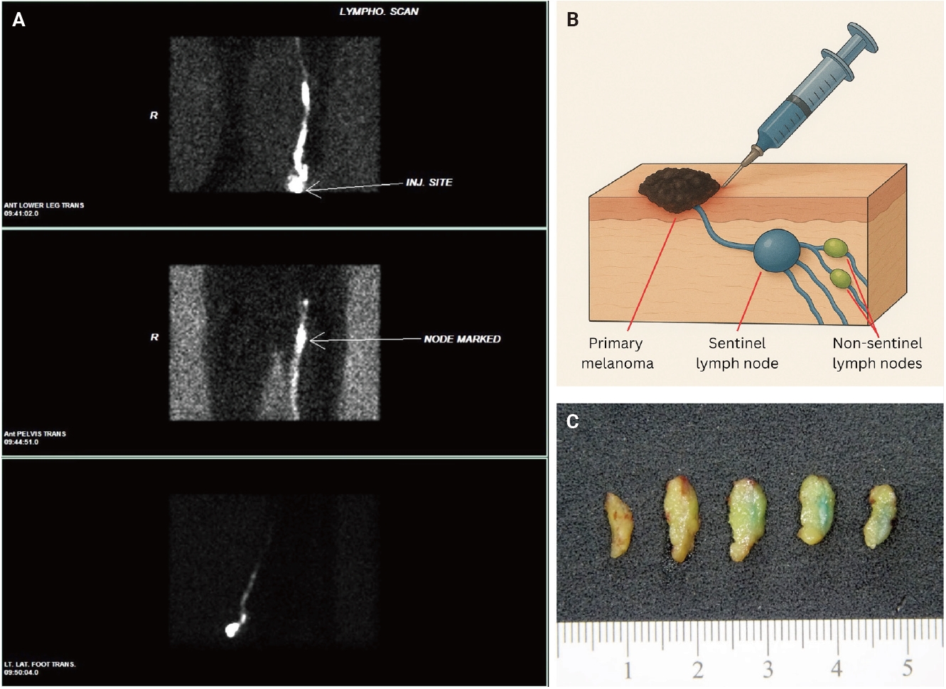
Sentinel lymph node biopsy workflow. (A) Preoperative lymphoscintigraphic identification of sentinel node. (B) Intraoperative identification of sentinel node. (C) Gross image of serially sectioned sentinel node with focal areas of blue dye permeation.
The presence of metastatic melanoma within SLNs is a key determinant of staging and prognosis. In the final report of the Multicenter Selective Lymphadenectomy Trial I (MSLT-I), sentinel node status was one of the strongest predictors of melanoma-specific survival in patients with intermediate-thickness melanoma. Patients with positive SLNs have significantly worse outcomes than those with negative nodes, and nodal status directly influences decisions regarding adjuvant therapy, surveillance, and clinical trial eligibility [2].
CLINICAL INDICATIONS FOR SLN BIOPSY
SLNB is recommended for patients with melanomas at increased risk of nodal metastasis. Current guidelines recommend SLNB for T1b melanomas, defined as 0.8 to 1.0 mm in Breslow thickness or less than 0.8 mm with ulceration, as well as tumors of stage T2 and higher [6,7].
In selected patients, SLNB may also be considered for T1a melanomas when clinicopathologic features suggest an increased risk of nodal metastasis, including lymphovascular invasion, increased mitotic activity, young age, anatomically complex sites such as the head and neck, or concern for incomplete microstaging due to a transected biopsy or positive deep margin. Current risk-based guidelines generally discourage SLNB when the estimated likelihood of SLN positivity is less than 5%, recommend discussion or consideration when the likelihood is 5% to 10%, and support offering SLNB when the likelihood exceeds 10% [7]. Emerging gene expression profiling (GEP) assays have been evaluated as adjunctive tools for refining recurrence risk and SLN positivity risk, particularly in thin and intermediate-thickness melanomas. However, current consensus statements emphasize that available evidence does not support replacing clinicopathologic assessment or standard SLNB indications with GEP testing in routine practice [8]. SLNB is not routinely recommended for thin melanomas without high-risk features and may be deferred in patients with significant comorbidities or limited life expectancy [6,7].
In addition to its role in staging, SLNB has been associated with improved regional disease control, with lower rates of nodal recurrence observed in patients undergoing the procedure [2].
RADIOLOGIC EVALUATION AND LYMPHATIC MAPPING
Preoperative lymphatic mapping is performed to identify SLNs prior to surgical excision. Lymphoscintigraphy is typically performed following peritumoral injection of a radiotracer, allowing visualization of lymphatic drainage pathways and identification of SLNs as focal areas of radiotracer uptake [9].
Additional imaging modalities may be used to evaluate regional lymph nodes for metastatic disease. Ultrasound can demonstrate suspicious features such as lymph node enlargement, cortical thickening, and loss of the fatty hilum [10]. Positron emission tomography/computed tomography (PET/CT) may demonstrate fluorodeoxyglucose (FDG)-avid nodal or distant metastatic disease in higher-stage melanoma; however, its sensitivity is limited for microscopic or small-volume regional nodal disease. FDG-PET detection of melanoma lymph node metastases is limited by tumor volume, with one report demonstrating a 90% sensitivity threshold at approximately ≥78 mm³ and markedly reduced sensitivity below this threshold [11]. Therefore, PET/CT cannot exclude clinically occult SLN metastasis and is not a substitute for histopathologic evaluation when SLNB is clinically indicated.
GROSS EXAMINATION
Accurate pathologic evaluation begins with careful gross examination. SLNs may demonstrate blue dye on the external or cut surface as a result of intraoperative mapping (Fig. 1C). Nodes involved by metastatic melanoma may appear enlarged or firm and may show melanin pigmentation or necrosis, although small-volume metastases are often not grossly apparent.
Common grossing approaches include longitudinal bisection through the hilum, where lymphatic channels are concentrated, or bread loafing into approximately 1- to 2-mm sections to maximize tissue surface area for evaluation [12-14]. When feasible, SLNs are submitted in their entirety because metastatic deposits may be small, focal, or present only on deeper levels [13,14].
Serial sectioning is critical to improve detection of small metastatic deposits. Examination of multiple histologic levels increases the likelihood of identifying occult metastatic melanoma, particularly when disease is present as isolated tumor cells or small subcapsular aggregates [14]. Frozen section evaluation is generally discouraged in melanoma SLNs because sensitivity is limited for small-volume metastatic disease, and tissue loss during frozen section processing may compromise permanent section evaluation [15].
HISTOPATHOLOGIC EVALUATION
Microscopic evaluation of SLN biopsies includes assessment of hematoxylin and eosin-stained sections in conjunction with immunohistochemical studies, with additional levels and stains used as needed to detect small or morphologically subtle metastatic deposits [13].
Metastatic melanoma cells typically resemble those in the primary tumor and may be characterized by enlarged, atypical nuclei, prominent nucleoli, and abundant cytoplasm. However, the histologic appearance of metastatic melanoma in SLNs can be variable with respect to both architecture and cytology. Metastatic deposits may appear as isolated tumor cells, small subcapsular or sinusoidal clusters, expansile nodules, or more diffuse parenchymal involvement. Cytologic features may mirror the primary melanoma, but they can be subtle or obscured by dense background inflammation, histiocytic reaction, pigment deposition, fibrosis, or treatment-related regression. Correlation with the morphology and immunophenotype of the primary melanoma is especially useful in diagnostically challenging cases.
Immunohistochemical analysis plays an essential role in identifying metastatic melanoma. Commonly used markers include SOX10, Melan-A, HMB45, and PRAME. A panel approach improves diagnostic sensitivity and specificity, particularly for small tumor deposits or morphologically subtle disease. SOX10 is a sensitive nuclear marker for melanocytic differentiation and is useful for highlighting small deposits of melanoma in SLNs [16,17]. Melan-A and HMB45 may help confirm melanocytic differentiation in conventional melanoma, although their expression may be reduced or absent in some melanoma subtypes [18].
Selection of immunohistochemical stains should be guided by the morphology and immunophenotype of the primary melanoma when available. For example, desmoplastic melanomas often demonstrate reduced expression of Melan-A and HMB45, making SOX10 a particularly useful screening marker [16]. PRAME may serve as a useful adjunct when the primary melanoma demonstrates PRAME expression; however, negative staining does not exclude metastatic disease because PRAME expression can be variable across melanomas [18-20]. Knowledge of the immunophenotype of the primary melanoma is necessary when evaluating small or diagnostically challenging melanocytic deposits within SLNs.
MICROANATOMIC LOCATION OF METASTASIS
The microanatomic distribution of metastatic deposits within the SLN carries prognostic significance. The Dewar classification describes patterns of involvement including subcapsular, parenchymal, and combined metastases (Fig. 2) [21].

Microanatomic patterns of metastatic melanoma in sentinel lymph nodes. (A) Dewar classification overview. (B) Subcapsular and parenchymal involvement: H&E (left), SOX10 (middle), and Melan-A (right). (C) Variant pattern of subcapsular and parenchymal involvement, showing nodular subcapsular growth with extension into the lymph node parenchyma. (D) Extensive nodal involvement by melanoma. (E) Extranodal extension: H&E (left), SOX10 (middle), and Melan-A (right).
Subcapsular metastases are generally associated with more favorable outcomes compared with deeper parenchymal involvement. In contrast, parenchymal, multifocal, or extensive nodal involvement has been associated with a greater likelihood of additional non-SLN metastases [21]. More extensive disease, particularly in the presence of extranodal extension (ENE), is associated with more aggressive tumor behavior [22].
TUMOR BURDEN AND PROGNOSTIC FACTORS
Tumor burden within the SLN is commonly assessed using the Rotterdam criteria (Table 1), which stratify metastases by the size of the largest metastatic deposit. Lower tumor burden is associated with improved outcomes and reduced risk of regional recurrence, whereas larger metastatic deposits are associated with increased risk of additional nodal involvement and worse prognosis [23]. ENE is an adverse prognostic feature in sentinel node-positive melanoma (Fig. 2E). In a cohort of 1,047 patients with positive SLNs, the 5-year overall survival rate was lower in patients with ENE compared with those without ENE, 62.5% versus 71.7%, respectively, and the 5-year disease-free survival rate was similarly lower, 54.0% versus 64.0% [22].

Rotterdam classification of sentinel node tumor burden
Standardized reporting is important because SLN findings directly affect staging and management. Key elements include the total number of SLNs examined, the number involved by metastatic melanoma, the size of the largest metastatic deposit, microanatomic location of tumor within the lymph node, and the presence or absence of ENE [24].
The American Joint Committee on Cancer 8th edition staging system distinguishes clinically occult nodal disease detected by SLNB from clinically detected nodal disease and incorporates the number of involved regional lymph nodes into pathologic stage assignment [25]. Satellite, microsatellite, and in-transit metastases also contribute to stage III subclassification when present. For the practicing pathologist, accurate documentation of these findings provides essential information for multidisciplinary treatment planning.
DIAGNOSTIC PITFALLS
Distinguishing metastatic melanoma from benign intranodal melanocytic proliferations represents a key diagnostic challenge in SLN evaluation. Benign nodal nevi are typically composed of small, cytologically bland melanocytes arranged in nests or cords and are most often located within the lymph node capsule or trabeculae (Fig. 3A). In contrast, metastatic melanoma more commonly involves the subcapsular sinus or parenchyma and demonstrates cytologic atypia, including nuclear enlargement, pleomorphism, and prominent nucleoli [19].

Differential diagnoses in melanoma sentinel lymph nodes. (A) Benign intranodal nevus: H&E (left), SOX10 (right). (B) Tumoral melanosis/melanophages: H&E (upper left, lower right), SOX10 (upper right), HMB45 (lower left). (C) Sinus histiocytes.
Immunohistochemistry can aid in this distinction but must be interpreted in the appropriate morphologic context. Both benign nodal nevi and metastatic melanoma are typically positive for melanocytic markers such as SOX10 and Melan-A, limiting their utility as discriminators despite their value in identifying small melanocytic deposits [17,19]. HMB45 is often weak or negative in nodal nevi and more frequently positive in metastatic melanoma; however, its expression is variable and should not be used as a standalone discriminator. Therefore, HMB45 negativity may support a benign nodal nevus only when the morphologic and anatomic findings are also concordant [19].
PRAME has emerged as a useful adjunct in the evaluation of nodal melanocytic proliferations. In diagnostically challenging nodal melanocytic deposits, PRAME immunohistochemistry can help distinguish metastatic melanoma from nodal nevi [19]. However, PRAME should not be interpreted in isolation because a subset of melanomas may demonstrate absent or only focal staining [20]. PRAME expression is best used as part of a broader assessment that integrates morphology, microanatomic location, comparison with the primary tumor when available, and the overall immunohistochemical profile.
Tumoral melanosis represents an important pitfall and is characterized by aggregates of melanin-laden macrophages without identifiable viable melanoma cells, often in the setting of regression or treatment effect (Fig. 3B). These cells are negative for melanocytic markers such as SOX10 and Melan-A while positive for histiocytic markers including CD68 and CD163 [26,27]. In the modern treatment era, similar findings may be encountered in lymph nodes or resection specimens after neoadjuvant immunotherapy or targeted therapy, where the tumor bed may show variable combinations of residual viable melanoma, tumoral melanosis, fibrosis, necrosis, and immune-related regression. Standardized pathologic assessment of post-neoadjuvant melanoma specimens emphasizes estimation of residual viable tumor, necrosis, and regression to support reproducible response assessment and clinicopathologic correlation [28]. Awareness of histiocytic staining patterns is important, as pigment-laden macrophages may mimic melanoma on routine sections and may show nonspecific staining artifacts. Careful correlation with nuclear morphology and a focused immunohistochemical panel helps avoid overinterpretation. Reactive histiocytic proliferations may also be encountered within lymph nodes and can mimic metastatic melanoma, particularly when present in sheets or associated with cytologic atypia or pigment deposition (Fig. 3C). In contrast to melanoma, these cells lack true melanocytic differentiation and are negative for melanocytic markers while showing positivity for histiocytic markers such as CD68 and CD163. Careful correlation with morphology and immunohistochemistry is essential to avoid overinterpretation.
CONTEMPORARY CONSIDERATIONS IN SLN EVALUATION
Management of patients with positive SLNs has evolved over time. Historically, completion lymph node dissection was routinely considered for patients with melanoma metastatic to SLNs. However, the Multicenter Selective Lymphadenectomy Trial II (MSLT-II) demonstrated that immediate completion lymph node dissection improved regional disease control but did not improve melanoma-specific survival compared with active nodal surveillance in patients with sentinel node-positive melanoma [29]. As a result, many patients with positive SLNs are now managed with nodal observation rather than immediate completion lymph node dissection.
Because neoadjuvant immunotherapy and targeted therapy are increasingly used in clinically detectable stage III melanoma, standardized recognition and reporting of treatment-related changes are becoming increasingly important in nodal and regional resection specimens [28].
Molecular adjuncts, including circulating tumor DNA analysis and GEP, are being studied for melanoma risk stratification and disease monitoring. While GEP assays may provide additional prognostic information, current consensus statements do not support their routine use as a replacement for SLNB selection based on clinicopathologic risk factors [8,30]. Circulating tumor DNA-based approaches also show promise for minimal residual disease assessment and recurrence monitoring, although their role in early-stage melanoma management continues to evolve [31].
CONCLUSION
SLNB plays a central role in the staging and management of melanoma and remains one of the most important prognostic tools in clinical practice. Accurate evaluation of SLNs requires a systematic approach that integrates careful gross examination, thorough histologic assessment, serial sectioning, and appropriate use of immunohistochemistry. Recognition of key prognostic features, including tumor burden, microanatomic distribution, and ENE, is essential for accurate staging and risk stratification.
Equally important is awareness of potential diagnostic pitfalls, including benign nodal nevi, tumoral melanosis, and histiocytic mimickers, which may lead to overdiagnosis if not carefully interpreted. Although the management of positive SLNs has changed in the modern systemic therapy era, precise pathologic assessment remains critical for staging, prognostication, treatment planning, and clinical trial enrollment.
Notes
Ethics Statement
Not applicable.
Availability of Data and Material
Data sharing not applicable to this article as no datasets were generated or analyzed during the study.
Code Availability
Not applicable.
Author Contributions
Conceptualization: DB, BL. Investigation: DB, BL. Methodology: DB, BL. Supervision: BL. Visualization: DB, BL. Writing—original draft: DB. Writing—review & editing: DB, BL. Approval of final manuscript: all authors.
Conflicts of Interest
The authors declare that they have no potential conflicts of interest.
Funding Statement
No funding to declare.