Diagnostic performance of fine needle aspiration cytology in the preoperative identification of mucoepidermoid carcinoma: a cross-sectional diagnostic analytical study
Article information

Abstract
Background
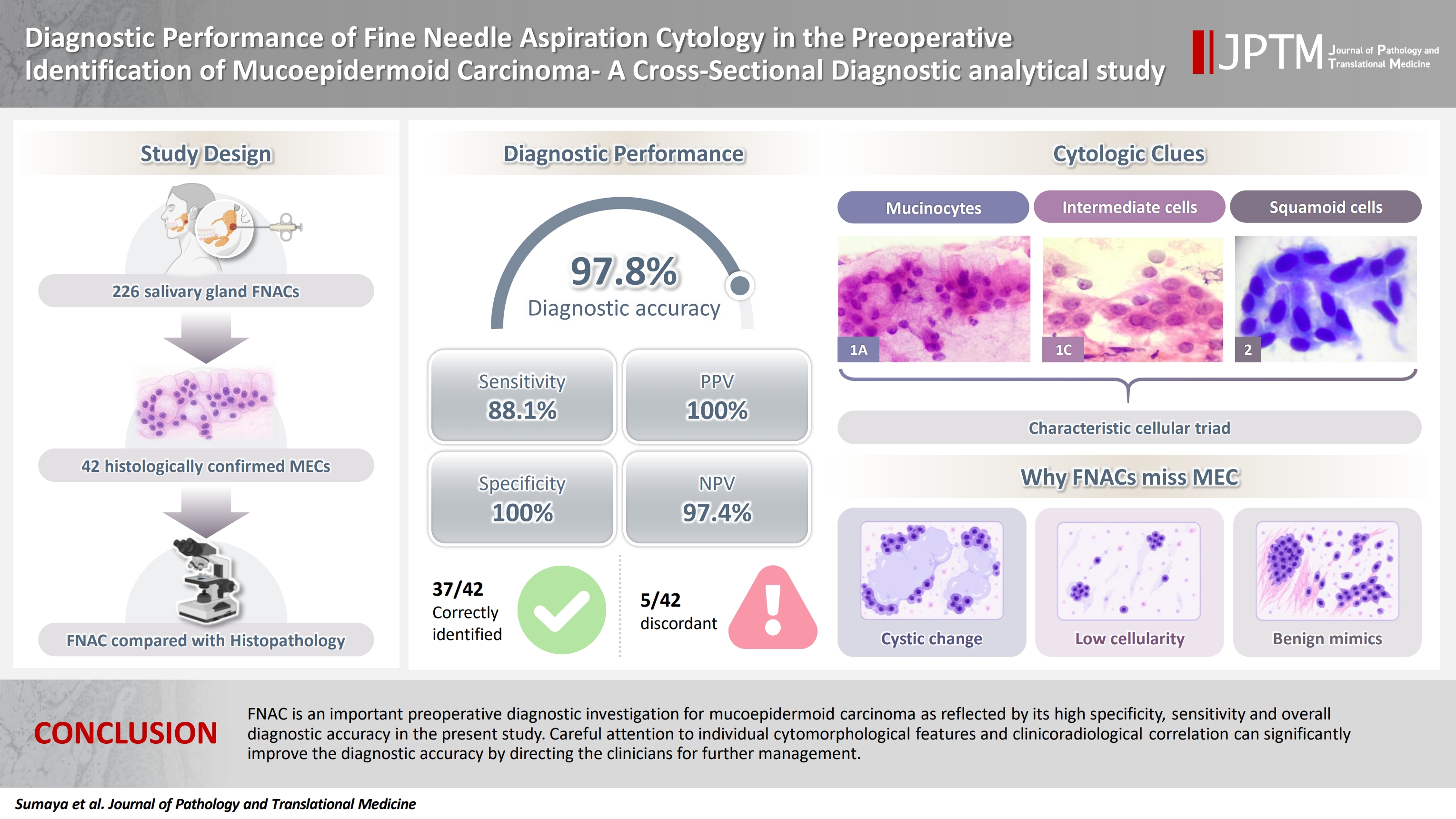
Mucoepidermoid carcinoma (MEC), the most common malignant tumor of the salivary gland, accounts for 5%–10% of all salivary gland tumors. MEC shows extensive cytomorphological diversity, making preoperative diagnosis challenging. Hence, the present study was conducted to assess the diagnostic accuracy of fine-needle aspiration cytology (FNAC) in diagnosing MEC preoperatively and to discuss the cytological pitfalls in diagnosing mucoepidermoid carcinoma.
Methods
The present study was a cross-sectional diagnostic analytical study conducted over a period of five years, which included confirmed cases of MEC. Each case was classified according to the Milan System for Reporting Salivary Gland Cytopathology (MSRSGC) and individual cytomorphological features were noted. All cases were compared by considering histopathology as the gold standard for diagnosis. Sensitivity, specificity, positive predictive value (PPV), and negative predictive value (NPV) of FNAC in diagnosing MEC were calculated to evaluate the diagnostic accuracy of FNAC.
Results
All 42 cases of MEC were reclassified according to MSRSGC as follows: category II, one case; category III, one case; category IVA, two cases; category IVb, one case; category V, four cases; and category VI, 33 cases. Discordant cases (5 cases) were mainly due to cystic change, low cellularity, and overlapping features with benign lesions. The sensitivity, specificity, PPV, NPV, and diagnostic accuracy were found to be 88.1%, 100%, 100%, 97.4%, and 97.8%, respectively.
Conclusions
FNAC is an important preoperative diagnostic investigation for MEC as reflected by its high specificity, sensitivity, and overall diagnostic accuracy in the present study. Careful attention to individual cytomorphological features and clinicoradiological correlation can significantly improve the diagnostic accuracy by directing the clinicians for further management.
INTRODUCTION
Salivary gland tumors constitute approximately 2%–6% of all head and neck malignancies, with mucoepidermoid carcinoma (MEC) being the most common malignant tumor of salivary gland origin [1]. MEC accounts for 5%–10% of all salivary gland tumors, with a predominant occurrence in the parotid gland, followed by the submandibular and minor salivary glands [2-6]. The clinical presentation of MEC varies widely, ranging from asymptomatic slow-growing masses in low-grade tumors to aggressive, rapidly enlarging lesions in high-grade cases. Given its diverse biological behavior, timely and accurate diagnosis of MEC is critical for determining appropriate treatment strategies and optimizing patient outcomes [7-10].
Fine-needle aspiration cytology (FNAC) is a widely used, minimally invasive, cost-effective, and highly specific diagnostic technique for evaluating salivary gland lesions. It is particularly useful in distinguishing benign from malignant tumors when combined with clinical and radiological findings [11]. However, despite its advantages, FNAC has notable limitations, including a risk of false-negative results, especially in low-grade MEC [2,7,11-13]. These false negatives primarily arise due to morphological variability within MEC, where tumor cells exhibit overlapping cytological and architectural patterns that can mimic benign cystic lesions [2,8]. Additionally, when MEC presents as a partially cystic tumor, FNAC may fail to sample the solid component, and the material yielded is hypocellular, consisting mainly of mucoid secretions, debris, and inflammatory cells, leading to misinterpretation and delayed diagnosis [7-9]. Studies by Edwards and Wasserman [14], Frable and Frable [15], and Mavec et al. [16] have highlighted that a significant proportion of false-negative FNAC in salivary gland tumors are associated with cystic lesions, emphasizing the need for improved diagnostic awareness.
The challenges of diagnosing MEC by cytology have been discussed in a few studies; hence, the present study was conducted to assess the diagnostic accuracy of MEC and discuss the pitfalls in diagnosing mucoepidermoid carcinoma.
MATERIALS AND METHODS
The present study was a cross-sectional diagnostic analytical study conducted over a period of 5 years, from January 2021 to December 2025, in the Department of Pathology at the Adichunchanagiri Institute of Medical Sciences, Adichunchanagiri University. Convenience sampling technique was employed, and all consecutive cases of MEC during the study duration were included. The study included patients of all age groups and either gender. Patients with a clinical or cytological suspicion of MEC, confirmed or suspected through FNAC or histopathological examination, were included. Patients with a history of recurrent MEC cases, cases in which slides were not available, and those who had undergone chemotherapy/radiotherapy were excluded from the study. During the study period, a total of 226 patients underwent salivary gland FNAC and following the above inclusion criteria, 42 cases were included in the present study.
Clinical data, including age, sex, laterality, site of the lesion such as parotid, submandibular, sublingual palate, oral cavity, and other minor salivary glands, were retrieved from the medical records. The institution follows a standard protocol for FNAC, which was performed after obtaining written informed consent from all patients. The procedure was conducted with the patient in either the supine or sitting position, ensuring patient comfort during the procedure. FNAC was performed using a 23-gauge needle attached to a 10-mL syringe by trained cytopathologists. In cases of cystic nodules, the cyst contents were aspirated, centrifuged, and slides were prepared from the sediment for microscopic examination. For each patient, a minimum of two passes were made to ensure an adequate sample. The collected material was spread onto slides, and the following staining techniques were used. Slides fixed in 90% ethanol were stained with Papanicolaou and hematoxylin and eosin (H&E) stains. Air-dried slides were stained using the Giemsa stain.
All FNAC slides from 42 cases were retrieved. The FNAC smears were retrospectively reviewed by two independent cytopathologists with more than 10 years of experience in cytopathology.
Each case was classified according to the Milan System for Reporting Salivary Gland Cytopathology (MSRSGC). The cytomorphological features studied were cellularity, type of cells (mucus cells, clear cells, oncocytic cells, and squamoid cells), mitosis, mucin, and necrosis. All cases at FNAC were stratified as low-grade and high-grade categories based on cytological findings. Diagnostic accuracy was calculated across tumor grades, as reported in previous studies.
Following clinical evaluation and FNAC, patients underwent surgical procedures, and the excised specimens were submitted for histopathological examination. The specimens were fixed in 10% formalin, processed, and embedded in paraffin blocks. The sections were cut, mounted on slides, and stained with H&E for microscopic examination. Histopathological diagnoses were made under a light microscope, and cases were graded as low, intermediate, or high-grade MEC based on the Armed Forces Institute of Pathology (AFIP) grading system, which includes criteria such as intracystic component <20% (2), neural invasion (2), necrosis (3), 4 or more mitoses (3), and anaplasia (4). Tumors were graded as low (0–4), intermediate (5–6), and high (≥7) [3]. The FNAC findings of all 42 cases were compared with histopathological results, considering histopathology as the gold standard for diagnosis. Cases were categorized into the following diagnostic groups. Cases were assigned to be true positive when MEC was correctly diagnosed on FNAC, true negative in the absence of MEC, false positive (FP) when MEC was incorrectly diagnosed on FNAC and false negative (FN) when MEC was not diagnosed on FNAC.
Data were analyzed using SPSS ver. 19 software (SPSS Inc., Chicago, IL, USA). Descriptive statistics, including mean, percentage, proportion, and standard deviation, were calculated to summarize demographic and clinical data. Sensitivity, specificity, positive predictive value (PPV), and negative predictive value (NPV) of FNAC in diagnosing MEC were calculated to evaluate the diagnostic accuracy of FNAC. Diagnostic accuracy was also calculated across tumor grades at cytology.
For all diagnostic accuracy indicators, a 95% confidence interval was calculated. In order to derive meaningful measures of diagnostic performance irrespective of the disease prevalence, positive and negative likelihood ratios were calculated. Since the present study did not have any FP cases, a continuity correction was incorporated to obtain the positive likelihood ratio (LR+) and confidence interval.
McNemar's test was applied to determine the concordance between the paired categorical data comprising various cytomorphological features on cytology and histopathology. Percentage agreement was calculated as the proportion of concordant cases among total cases.
RESULTS
A total of 226 cases that presented to the cytology department between January 2021 and December 2025 with palpable salivary gland swelling and who underwent FNAC constituted the study population. Among these 226 cases, 42 cases suspected or confirmed as MEC on histopathology were included in the study. In the present study, females outnumbered males (28/42, 66.7% vs. 14/42, 33.3%) with a female-to-male ratio of 2:1. The mean age of the patients in the present study was 42.2 ± 2.00 years, with the youngest patient being 12 years and the oldest being 62 years old. The parotid (28 cases, 66.7%) was the most commonly involved salivary gland in the present study, followed by the submandibular gland (12/42, 28.6%) and minor salivary gland (2/42, 4.8%). Right-sided salivary glands were affected more frequently (22/42, 52.4%) as compared to the left side (20/42, 47.6%), as summarized in Supplementary Table S1.
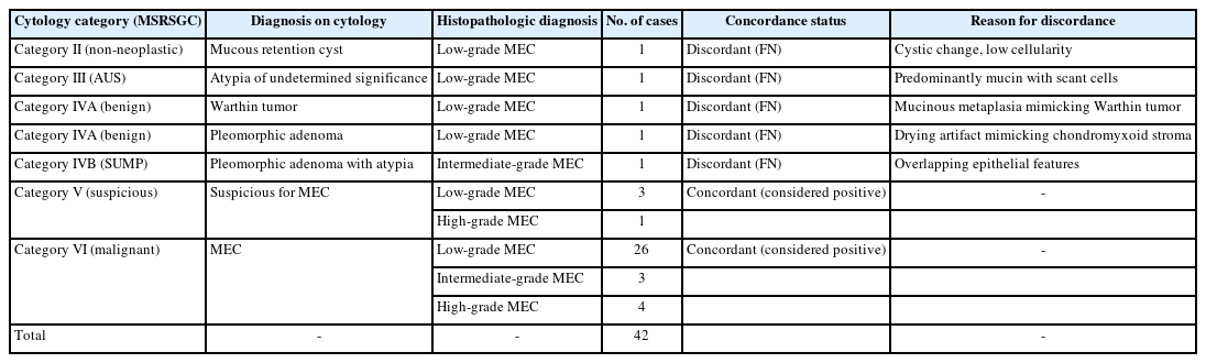
In the present study, all 42 cases were cytologically classified according to MSRSGC as follows: category II (non-neoplastic), 1 case; category III (atypia of undetermined significance [AUS]), 1 case; category IVA (benign), 2 cases; category IVB (salivary gland neoplasm of uncertain malignant potential [SUMP]), 1 case; category V (suspicious of malignancy), 4 cases; and category VI (malignant), 33 cases (Table 1).
Cytohistological correlation of cases
Taking cases in category V and category VI as malignant, the total number of cases diagnosed as malignant on cytology was 37/42 (88.1%). These cases were further categorized as low-grade MEC (33/42 cases, 78.6%) and high-grade MEC (9/42 cases, 21.4%) based on cytomorphological features such as mucinocytes, intermediate cells, mucinous background, oncocytes, squamoid cells, clear cells, lymphocytes, necrosis, nuclear pleomorphism, and atypical mitosis (Figs. 1A–E, 2, Supplementary Table S2). The high-grade cytology group included cases that were subsequently classified as intermediate-grade (4 cases) and high-grade (5 cases) on histopathology. In the present study, a high degree of concordance was noted between various cytological features on cytology and histopathology, such as the presence of mucinocytes, intermediate cells, necrosis, and nuclear pleomorphism. Similarly, squamoid cells, oncocytes, and clear cells showed a moderate degree of concordance, which may be due to variations in the sampling technique and cytology smear preparations. Background features such as mucinous dirty background and lymphocytic infiltrate were analyzed descriptively, as these are smear-dependent and less reliable indicators of histological correlation. Grade-based stratification on cytology showed a high sensitivity of 87.9% for low-grade MEC and 88.9% for high-grade MEC. The high-grade MEC group included cases diagnosed as intermediate and high grade at histopathology. A fairly comparable diagnostic performance was demonstrated between the two groups (Table 2). For statistical analysis, intermediate-grade and high-grade MEC on histopathology were combined into a single high-grade category (n = 9).
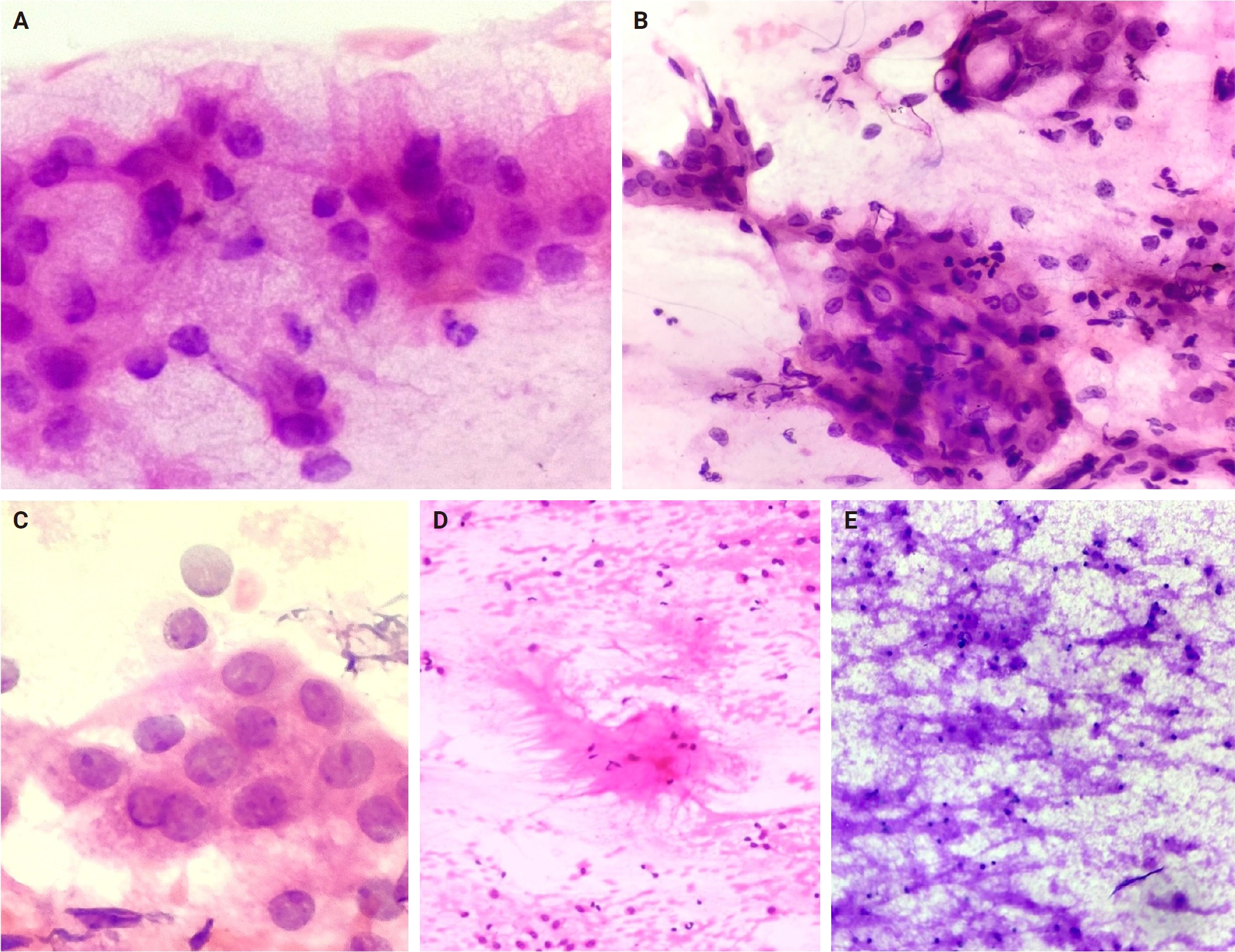
(A) Fine-needle aspiration (FNA) smear shows mucin-secreting vacuolated cells in a clean mucinous background. (B) FNA smear shows a cluster of clear cells with intracytoplasmic mucin vacuoles. (C) FNA smear shows intermediate cells in clusters. (D) FNA smear showing mucin with peripheral fibrillary appearance due to drying artifact, mimicking fibrillary stroma of pleomorphic adenoma. (E) FNA smear showing dirty mucinous background in low-grade mucoepidermoid carcinoma.
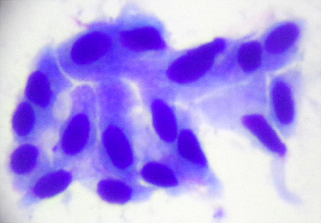
Fine-needle aspiration smear showing a cluster of squamoid cells in high-grade mucoepidermoid carcinoma.

Grade-wise sensitivity of FNAC for MEC
All the cases of MEC on histopathology were further categorized according to the AFIP grading system as shown in Supplementary Table S3. Out of the total 42 cases, 5/42 (11.9%) cases showed high-grade morphology, whereas 4/42 (9.5%) depicted intermediate grade morphology. The maximum number of cases (33/42, 78.6%) showed low-grade morphology as per the AFIP grading system for MEC. Five cases (11.9%) showed discordant diagnosis. Amongst these discordant cases, one case each was diagnosed wrongly as mucous retention cyst, Atypia of undetermined significance, Warthin’s tumor, and pleomorphic adenoma, respectively. One case reported as pleomorphic adenoma with atypia turned out to be an intermediate-grade MEC on histopathology. Whereas concordant diagnosis was noted in the remaining 37/42 cases, as summarized in Table 1.
The diagnostic performance indicators were determined using all 226 cases, with histopathology as the gold standard. Sensitivity, specificity, PPV, NPV, and diagnostic accuracy were estimated to be 88.1% (95% confidence interval [CI], 74.4% to 95.6%),100% (95% CI, 98.0% to 100%), 100% (95% CI, 90.5% to 100%), 97.4% (95% CI, 94.0% to 99.1%), and 97.8% (95% CI, 94.9% to 99.3%) (Table 3). In the present study, an extremely high LR+ was obtained, which is a reflection of the absence of FP cases, which portends a strong rule-in capability of FNAC. The 0.12 negative likelihood ratio (LR−) affirms the ability of FNAC to reduce the postsurgical probability of MEC when FNAC was negative for MEC.
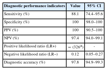
Diagnostic performance of FNAC in MEC
Individual cytomorphological features were evaluated against the histopathology to assess concordance using the McNemar test as detailed in Table 4. Mucinocytes and mucinous background showed high agreement, whereas perfect concordance was observed for nuclear pleomorphism and necrosis. However, a statistically significant discordance (p = .008) was obtained for lymphocytic infiltrate, reflecting its comparative underrepresentation in cytology smears.
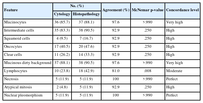
Concordance between the paired categorical data comprising various cytomorphological features on cytology and histopathology
DISCUSSION
FNAC of salivary gland lesions is a simple, cost-effective, easy, and minimally invasive tool for preoperative evaluation [8]. In the present study, 42 cases were studied irrespective of age group and either gender. The mean age in the present study was 42.2 years, which is concordant with the study done by Iftikhar et al. [9], and Shafique et al. [18]. The parotid gland (66.7%) was the most commonly involved salivary gland, which is similar to the findings of studies conducted by Wakely [17], and Iftikhar et al. [9]
The diagnostic accuracy of FNAC in salivary gland lesions varies and several authors have reported a wide range from 80.4% to 98.0%. Similarly, sensitivity varies from 70%–90% and specificity has been reported between 90% and 100% in several studies [19,20]. The sensitivity and specificity depend on many factors such as type of lesion, sampling technique, expertise needed to perform the fine-needle aspiration (FNA) procedure and expertise of the reporting cytopathologist [15-17,19,20]. Most of the earlier studies have reported performance metrics for salivary gland lesions as a whole rather than for individual tumor type [4,12]. Very few studies have specifically assessed the diagnostic accuracy of MEC. Several authors like Klijanienko and Vielh [4] (50 cases), Wakely [17] (76 cases), case series by Vasudevan et al. [7] (14 cases) and Joseph et al. [8] (6 cases) reported the diagnostic accuracy of FNAC in diagnosing MEC and observed a low accuracy range between 38%–52% (Supplementary Table S4). Following this, a study conducted by Iftikhar et al. [9] had concluded FNAC was not a reliable tool for diagnosing MEC due to a high number of FN cases. This lower sensitivity and diagnostic accuracy in the diagnosis of MEC at cytology has been attributed to several reasons like the inherent nature of the lesion, marked heterogeneity among and within the tumors, and experience of the reporting cytopathologists [2,8]. Most of the time, the presence of a cystic component, especially in low-grade MEC, leads to low cellular yield, responsible for a high rate of FN diagnosis [7,9]. Some studies have reported a very low sensitivity of 39% mainly due to the cystic nature of the lesion [4]. Similarly, accurate diagnoses of MEC require identification of three characteristic cell types, i.e., clear mucinous, intermediate, and squamous cells, which may not always be represented in the cytology smears. Significant overlap of cytomorphologic features between MEC and benign tumors like Warthin’s and pleomorphic adenoma further adds to the diagnostic dilemma [8,21].
In the present study, we report a high diagnostic accuracy of 97.8%. The reason for high diagnostic accuracy may be due to the fact that the study included histopathologically confirmed cases of MEC, which might have reduced misclassification. Also, the individual cytomorphologic features were evaluated and scored, which along with the consistent sampling technique, resulted in high diagnostic accuracy in the present study.
In the present study, grade-based stratification was performed on cytology. A high sensitivity of 87.9% for low-grade MEC and 88.9% for high-grade MEC was noted. This reflected a comparable diagnostic performance between low- and high-grade MEC for FNAC. Although low-grade MEC usually is challenging due to the cystic nature and scanty cellularity, diligent examination of cytomorphological features permitted high diagnostic accuracy in both groups. For grade-wise diagnostic performance analysis, intermediate-grade and high-grade MEC were grouped together (n = 9) because of the limited number of cases.
In the present study, low-grade MEC was most commonly encountered that is in 33/42 cases, which were classified further based on MSRSGC at cytology as one case in category II (non-neoplastic), one in category III (AUS), two cases as Benign (category IV A), and three cases in category V (suspicious) and 26 cases in category VI that were classified as malignant on cytology. A total of 88.1% of cases were categorized as malignant and suspicious for malignancy according to MSRSGC. In the present study, about 2.4% of cases were misdiagnosed as mucus retention cyst, which could be resolved by a diligent search for the presence of one of the three cell types i.e., mucus cells, squamoid cells, and intermediate cells. Also, in case of cystic swellings, repeat FNA from solid areas under image guidance and close follow-up is advocated in cystic lesions. One case was reported as AUS as the smears showed only mucin and occasional mucinocytes. Among two cases (4.76%) of the benign category, one case was wrongly reported as pleomorphic adenoma on cytology. This was due to the misinterpretation of clusters of epithelial cells and squamoid cells for squamous metaplastic cells and the mucin, which was wrongly interpreted as chondromyxoid matrix due to drying artifact in the smear. Careful examination of Giemsa-stained smears would avoid this pitfall, as chondromyxoid fibrillary stroma shows fibrillar filaments at the periphery of the stromal fragments. Another case of MEC was wrongly interpreted as Warthin’s tumor, which is the most common neoplasm confused with MEC, as it may show foam cells, mucinous metaplastic cells and due to precipitated material, which might appear like mucin. In the present study, three cases of low-grade MEC at histopathology were placed in the suspicious category of MSRSGC as they showed features of malignancy but lacked significant atypia. A similar pitfall was also reported by Wade et al. [2]. At histopathology, four cases of intermediate-grade MEC were classified into categories IVB (one case) and VI (three cases) on FNAC as per MSRSGC. One case diagnosed as SUMP showed features of pleomorphic adenoma with atypia, which turned out to be intermediate-grade MEC on histopathology. Atypia in pleomorphic adenoma is a known pitfall and careful observation of all available smears is essential to rule out malignancy. None were categorized into the benign or non-neoplastic category.
In the present study, five cases were confirmed as high-grade MEC on histopathology. These cases were placed in category V (one case) and category VI (four cases) of MSRSGC. In one case, which was reported as suspicious on cytology, clear features of malignancy; however, the material was insufficient to make a confident diagnosis of malignancy. Correct diagnosis was made in the remaining four cases; however, one among these four cases was wrongly subtyped as squamous cell carcinoma (SCC). Differentiating SCC from high-grade MEC is challenging. Keratinization is usually focal in MEC, whereas in SCC, keratin pearl formation, extensive keratinization, and intercellular bridges should allow accurate diagnosis. Multiple sampling and clinical correlation are required to avoid mistyping these lesions at cytology. Similar findings have been reported by Wakely [17].
In the present study, the most common morphological features observed were mucinocytes, mucinous background, and intermediate cells, which is concordant with observations made by Wade et al. [2].
In the present study, high concordance was observed for mucinocytes and mucinous background at cytology, highlighting their significant role in diagnosing low-grade MEC (Table 1). Earlier literature has also emphasized the role of extracellular mucin as an important feature aiding diagnosis, especially in low-grade MEC where cellular atypia may be minimal [12,22]. Necrosis and nuclear pleomorphism were consistently identified in both cytology and histopathology, depicting perfect concordance and better reproducibility. The presence of these two features at cytology should raise the possibility of malignancy, especially high-grade MEC [23]. However, lymphocytic infiltrate exhibited statistically significant discordance (p = .008), as cytology smears did not show its presence as compared to the histopathology sections. Literature reveals similar findings wherein stromal and inflammatory infiltrate are underrepresented in cytology smears or are diluted in mucin, making them less reliable in diagnosing MEC at cytology [4,11].
In the present study, an extremely high LR+ was obtained, which is a reflection of the absence of FP cases, which portends a strong rule-in capability of FNAC. The 0.12 LR− affirms the ability of FNAC to reduce the postsurgical probability of MEC when FNAC was negative for MEC. Despite this, the occurrence of cystic lesions and low-grade MEC with mucin predominance may account for a few FN cases [4,12].
FN cases accounted for 11.9% in the present study, which was comparable to that reported by Klijanienko and Vielh [4] (12%). In contrast, Wakely [17] reported a slightly lower FN rate of 8%, whereas Wade et al. [2] and Iftikhar et al. [9] reported a very high FN rate of 22% and 50%, respectively.
In MEC, the presence of the triad of mucin-secreting cells, intermediate cells, and squamous cells is crucial for making an accurate diagnosis of MEC at cytology. The maximum number of cases showed the presence of mucinocytes at both cytology and histopathology in the present study, highlighting the crucial role of these cell types, which also aids in differentiating MEC from other salivary gland neoplasms like adenoma with cystic change and acinic cell carcinoma. Similarly, another diagnostic feature noted in the present cohort was the mucinous dirty background, which is a well-established feature seen in benign mucous retention cyst and low-grade MEC. In benign mucous retention cyst, the mucin and the background are clean, whereas in low-grade MEC, the background is usually dirty with sprinkling of inflammatory cells in between; also, a diligent search for the accompanying cells is important, as in low-grade MEC intermediate cells can be seen in small clusters. These cells are small, basal-like cells with scant cytoplasm or large polygonal cells with pale cytoplasm and bland nuclei. These cells are usually underrecognized but crucial for diagnosis and hence careful search for them should be made in mucin-rich smears. Wade et al. [2] similarly emphasized the importance of intermediate cells as a diagnostic clue, particularly in aspirates where mucinocytes are sparse.
Most studies reported a low diagnostic accuracy for MEC at cytology mainly due to scant cellularity, absence of mucin in the background, and predominance of smear by one type of cell, as summarized in Supplementary Table S4 [2,4,9,17]. In the present study, the importance of multiple characteristic features allowed for accurate interpretation and improved diagnostic accuracy.
Correlation with clinicoradiological findings and repeat FNA under imaging guidance helps to avoid misinterpretation in indeterminate cases. Further ancillary techniques like immunocytochemistry and molecular tests would serve as an adjunct tool in refining the diagnosis and guiding clinicians in planning appropriate management.
FNAC is an important preoperative diagnostic investigation for MEC as reflected by high specificity, sensitivity, and overall diagnostic accuracy in the present study. Low cellularity, inadequate sampling and cystic lesions are major causes of FN diagnosis. Careful attention to individual cytomorphological features and interpretation of FNAC findings in an appropriate contextual manner with adequate clinicoradiological correlation can significantly improve the diagnostic accuracy, directing the clinicians for further management.
Supplementary Information
The Data Supplement is available with this article at https://doi.org/10.4132/jptm.2026.07.08.
Notes
Ethics Statement
Ethical clearance was obtained from the Institutional Ethical Committee, AIMS IEC/AIMS-301-2024. A written informed consent was obtained from all the patients before FNA procedure and publication.
Availability of Data and Material
The datasets generated or analyzed during the study are available from the corresponding author on reasonable request.
Code Availability
Not applicable.
Author Contributions
Conceptualization: S, AK. Data curation: S, DRS. Formal analysis: S, DRS, AK. Investigation: S, DRS. Methodology: S, AK. Project administration: AK, DRS. Resources: S, AK, DRS. Software: S, DRS. Supervision: AK. Validation: S, AK. Visualization: S, AK. Writing—original draft: S, AK. Writing—review & editing: S, AK, DRS. Approval of final manuscript: all authors.
Conflicts of Interest
The authors declare that they have no potential conflicts of interest.
Funding Statement
No funding to declare.