T-lymphoblastic leukemia/lymphoma: a comprehensive review of pathology, molecular features, and differential diagnosis
Article information

Abstract
T-lymphoblastic leukemia/lymphoma (T-ALL/LBL) is an aggressive neoplasm of immature T-lymphoid precursors that is classified as T-cell acute lymphoblastic leukemia when the primary site of involvement is the bone marrow and peripheral blood and as T-cell lymphoblastic lymphoma when it presents as a solid mass, most commonly in the anterior mediastinum. The neoplastic cells are defined by expression of T-lineage antigens (cytoplasmic or surface CD3) together with one or more markers of immaturity (terminal deoxynucleotidyl transferase, CD1a, CD34, CD99, or CD117) and must not fulfill criteria for early T-cell precursor acute lymphoblastic leukemia (ALL). This entity shows a marked male predominance (male:female ≈ 2:1) and predominantly affects children, adolescents, and young adults, accounting for approximately 15% of childhood ALL and 20%–25% of adult ALL cases. Pathologic diagnosis increasingly relies on recurrent molecular alterations such as activating NOTCH1 mutations (>60% of cases), transcription factor rearrangements, and cell-cycle regulator inactivation. Although intensive multiagent chemotherapy regimens have substantially improved outcomes, particularly in pediatric patients, adults and high-risk molecular subgroups continue to experience inferior survival. This review provides a practical, pathology-oriented update on the epidemiology, pathogenesis, diagnostic criteria, differential diagnosis, prognostic factors, and current treatment approaches for T-ALL/LBL.
INTRODUCTION
T-lymphoblastic leukemia/lymphoma (T-ALL/LBL) is an aggressive neoplasm of precursor lymphoid cells committed to the T-cell lineage [1]. In the fourth and fifth editions of the World Health Organization (WHO) classification, T-cell lymphoblastic lymphoma (T-LBL) has traditionally been defined by predominant extramedullary disease (most often a mediastinal mass) with limited bone marrow involvement (conventionally defined as fewer than 25% bone marrow blasts), while cases with extensive marrow and peripheral blood disease are designated T-cell acute lymphoblastic leukemia (T-ALL) [1-3]. This 25% figure is a convention used to assign a leukemia or lymphoma designation in patients with a mass lesion and is distinct from the blast threshold classically used to define an acute leukemia. However, continuing advances in immunophenotyping and molecular genetics are rapidly refining the diagnostic criteria for this neoplasm. As large-scale genomic studies continue to delineate the heterogeneous molecular landscape of this disease, the entity is increasingly being defined by specific molecular aberrations, with the most recent International Consensus Classification (ICC) recognizing multiple new provisional molecular entities based on distinct transcription factor rearrangements and other genetic alterations [4]. In this evolving landscape, pathologists play a pivotal role in the accurate diagnosis, subclassification, and risk stratification of this entity through the integrated interpretation of morphologic, immunophenotypic, flow cytometric, and molecular genetic findings [5].
EPIDEMIOLOGY
T-ALL/LBL exhibits a male predominance with a male-to-female ratio of approximately 2:1 [6]. The disease primarily affects children, adolescents, and young adults, with a peak incidence during the second decade of life [6-8]. It accounts for approximately 15% of acute lymphoblastic leukemia cases in children and 20%–25% of cases in adults [9,10]. Lymphoblastic lymphoma accounts for 1%–2% of all non-Hodgkin lymphomas [11], and 85%–95% of these are T-LBL [11-13]. Most cases arise sporadically, with no known environmental or infectious risk factors.
PATHOGENESIS
The pathogenesis of T-ALL/LBL involves a multi-step process of genetic alterations that disrupt normal T-cell development and promote uncontrolled proliferation. Several major molecular pathways have been implicated in leukemogenesis (Table 1) [14-29].
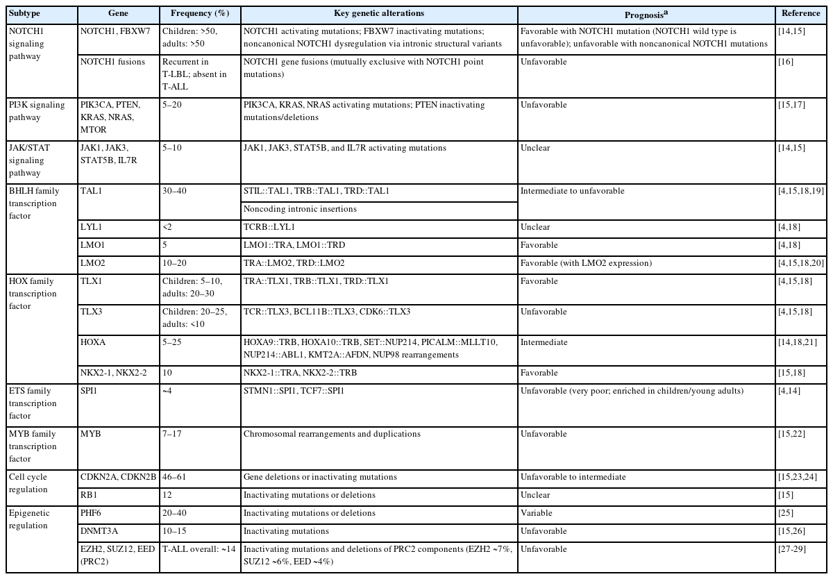
Key genetic alterations in T-ALL/LBL
Signaling pathway activation
Activating mutations in NOTCH1 represent the most common genetic alteration in T-ALL/LBL, occurring in approximately 60% of patients and leading to ligand-independent cellular signaling [30,31]. These mutations typically affect the heterodimerization or PEST domains, resulting in constitutive receptor activation and downstream transcriptional upregulation that inhibit T-cell differentiation while promoting proliferation and survival. Constitutive NOTCH1 signaling directly activates the MYC oncoprotein through a feed-forward transcriptional loop, which is thought to play a central role in leukemogenesis [32]. Inactivating mutations in FBXW7, the E3 ubiquitin ligase that targets NOTCH1 for degradation, are found in an additional 15%–20% of cases and further amplify this pathway. Gene fusions involving NOTCH1 have also been reported, almost exclusively in T-LBL, as well as non-canonical NOTCH1 intronic single nucleotide variations and intragenic deletions [14,16].
In parallel, hyperactivation of the phosphoinositide 3-kinase (PI3K)/Akt/mammalian target of rapamycin signaling pathway, most often driven by inactivation of the negative regulator PTEN (through deletion, mutation, or posttranslational mechanisms), is observed in a substantial proportion of T-ALL and cooperates with NOTCH1 to enhance cell growth, metabolism, and resistance to apoptosis [18,33].
Aberrant transcription factor expression
Greater than 50% of T-ALL cases exhibit enhanced expression of key transcription factors such as TAL1, TLX1, TLX3, LYL1, HOXA, LMO1, and LMO2, which define distinct molecular subgroups of the disease [34]. These alterations frequently result from chromosomal translocations that juxtapose the transcription factor genes with T-cell receptor (TCR) loci (TRA/D at 14q11 or TRB at 7q34), placing them under the control of strong TCR enhancers and causing their aberrant overexpression; this in turn drives leukemic cell proliferation and blocks normal T-cell differentiation [34]. In addition, recurrent gene fusions such as SET::NUP214, PICALM::MLLT10, NUP214::ABL1, and KMT2A::AFDN are present in roughly 25% of cases and result in dysregulation of the HOXA gene cluster, further reinforcing the oncogenic transcriptional program [35]. These transcription factor-driven subgroups carry distinct prognostic implications and are increasingly incorporated into refined molecular risk stratification schemes [18].
Cell cycle regulator inactivation
Inactivation of the tumor suppressor genes CDKN2A, CDKN2B, and/or RB1 is observed in the majority of T-ALL cases and represents one of the most frequent mechanisms of cell-cycle deregulation in this disease [23,24,34,36]. Biallelic deletions of the CDKN2A locus, which encodes both p16INK4A (a CDK4/6 inhibitor) and p14ARF (an MDM2 antagonist that stabilizes p53), occur in approximately 70% of cases [34]. Loss of these proteins simultaneously derepresses CDK4/6–RB signaling and impairs p53-mediated apoptosis, resulting in unchecked G1-to-S phase progression and enhanced proliferative capacity. Although deletions or inactivating events involving RB1 itself are less common, they further reinforce cell-cycle deregulation when present, typically in cooperation with NOTCH1 and transcription-factor alterations [34].
Cell adhesion and tissue tropism
T-ALL cells often express leukocyte function-associated antigen 1 in its active conformation, which mediates firm adhesion and extravasation through interactions with intercellular adhesion molecule 1, which facilitates leukemic infiltration into tissues [37]. Gene expression profiling and next-generation sequencing (NGS) studies have also revealed clear transcriptomic differences between T-LBL and T-ALL, with T-LBL demonstrating increased expression of cell adhesion proteins and upregulation of genes related to angiogenesis, potentially explaining its predilection for solid-mass formation rather than leukemic dissemination [3,18].
Epigenetic deregulation and cooperative mutations
Recurrent mutations in epigenetic regulators (e.g., PHF6, DNMT3A, KDM6A, EZH2) are observed in a substantial proportion of cases and contribute to altered chromatin accessibility and gene expression programs that cooperate with the above pathways. In a similar manner, mutations in specific components of the epigenetic writer complex PRC2, notably EZH2 and SUZ12, lead to loss of regulation of HOXA-driven developmental genes in T-ALL [27-29]. These multi-hit genetic events, NOTCH1/FBXW7 activation, transcription-factor deregulation, cell-cycle loss, and epigenetic changes, underlie the full leukemogenic transformation and influence distinct genomic subtypes of T-ALL/LBL [18,34].
CLINICAL, LABORATORY, AND RADIOLOGIC FEATURES
Clinical presentation
The clinical presentation of T-ALL/LBL varies depending on whether the disease primarily involves the bone marrow and peripheral blood (T-ALL) or presents as a mass lesion (T-LBL) [3]. Constitutional (B) symptoms, including fever, weight loss, and night sweats, are frequently observed [38]. A mediastinal mass is present in the majority of T-LBL cases (70%–90%), often producing respiratory symptoms such as cough, dyspnea, or chest pain, and superior vena cava syndrome is a well-recognized complication [3,39]. Mediastinal masses may also occur in T-ALL, although they are typically smaller than those seen in T-LBL [40]. Lymphadenopathy is present in the majority of cases, and hepatosplenomegaly is observed in a smaller subset [38]. Extranodal involvement can occur at sites including skin, testes, and central nervous system (CNS) [38]. Compared with B-lymphoblastic leukemia/lymphoma (B-ALL/LBL), mediastinal masses and CNS involvement are more common in T-ALL/LBL [5]. Bone marrow involvement manifests bone pain and symptoms related to cytopenias, including fatigue and pallor from anemia, bleeding from thrombocytopenia, and recurrent infections due to granulocytopenia [41].
Laboratory findings
The white blood cell count is significantly elevated in T-ALL, commonly exceeding 50,000/µL, while it is frequently within the normal range in T-LBL [40]. Anemia and thrombocytopenia are more common in T-ALL due to extensive bone marrow involvement [40].
Radiologic findings
In T-LBL, imaging typically reveals mediastinal widening, often with a mediastinum-to-thoracic ratio exceeding 0.75 on chest radiography and computed tomography (CT) (Fig. 1) [40,42]. Systemic lymphadenopathy is frequently demonstrated on CT, and pleural effusions, pericardial effusions, and airway and vasculature compression may also be observed [40,42]. In T-ALL, bone lesions such as metaphyseal banding, periosteal reactions, osteolytic lesions, osteosclerosis, or osteopenia may be present [40,43]. Mediastinal widening can occur in T-ALL but is typically less pronounced than in T-LBL [40,43].
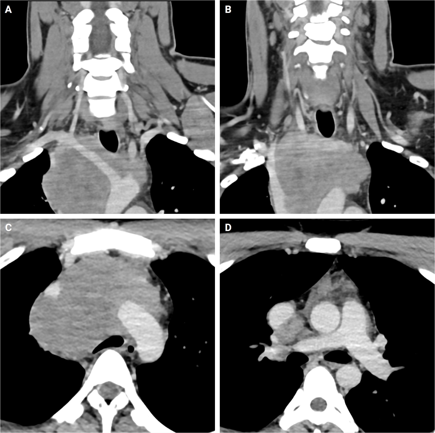
Representative contrast-enhanced computed tomography images of biopsy-proven T-cell lymphoblastic lymphoma in a 23-year-old male. (A) Posterior coronal view of mediastinal soft-tissue mass (70–80 Hounsfield units) encasing the right brachiocephalic artery, right common carotid, right subclavian, and left common carotid origin. (B) Anterior coronal view with mass laterally displacing, compressing, and invading the superior vena cava (SVC), concerning for SVC syndrome. (C) Axial view at the level of the aortic arch shows mass encasing the aortic arch and its branches as well as the SVC. (D) Axial view at the level of the pulmonary trunk shows mass abutting and partially compressing the right pulmonary artery.
PATHOLOGIC FEATURES
Morphology and cytology
T-ALL/LBL is characterized by a diffuse proliferation of blastoid, intermediate- to large-sized cells with high nuclear-to-cytoplasmic (N:C) ratios, fine chromatin, and nucleoli that range from inconspicuous to prominent [5]. The cells appear monotonous with back-to-back nuclei owing to their scant cytoplasm, and mitotic figures are readily appreciated [5].
In bone marrow biopsy specimens, the marrow is typically hypercellular with displacement of normal hematopoietic elements, although some residual trilineage hematopoiesis is usually present (Fig. 2A, B) [44]. Bone marrow necrosis has been reported in rare cases, and reticulin or collagen fibrosis may occasionally be seen [44,45]. Mediastinal mass biopsies show a similar diffuse proliferation of monotonous, intermediate-sized blastoid cells (Fig. 2C). In lymph nodes, T-LBL typically shows complete effacement of nodal architecture with capsular involvement. Less commonly, partial paracortical involvement sparing germinal centers may be observed. A “starry sky” pattern due to interspersed tingible-body macrophages is present in some cases, mimicking Burkitt lymphoma (Fig. 2D) [46].
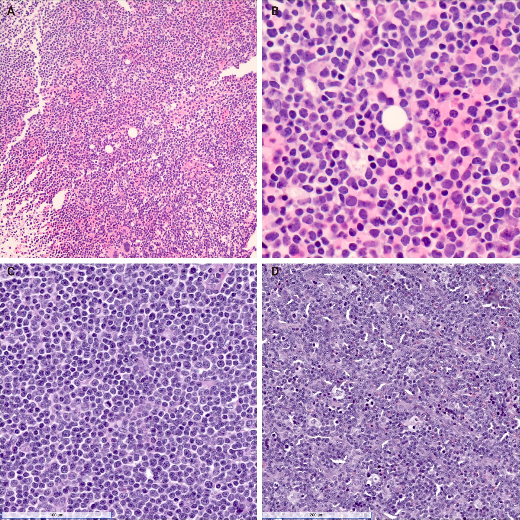
Representative microscopic images of T-lymphoblastic leukemia/lymphoma. (A, B) Bone marrow biopsy of T-cell acute lymphoblastic leukemia, with the low-power view demonstrating a diffuse, infiltrating pattern of blastoid cells with high nuclear-to-cytoplasmic ratios, and the high-power view more readily showing blasts with scant cytoplasm, fine chromatin, and appreciable mitoses. (C) Mediastinal mass biopsy of T-cell lymphoblastic lymphoma demonstrating a diffuse array of monotonous, intermediate-sized blastoid cells. (D) Additional mediastinal mass biopsy demonstrating an alternative “starry sky” pattern due to the presence of tingible body macrophages interspersed between blastoid cells.
Cytologic features on bone marrow aspirates or peripheral blood smears show intermediate- to large-sized blasts with a high N:C ratio, scant basophilic cytoplasm (occasionally vacuolated), round to oval nuclei that may be convoluted, fine dispersed chromatin, and nucleoli that range from indistinct to prominent (Fig. 3). Cytoplasmic azurophilic granules are uncommon. Blast lineage cannot be reliably determined by morphology alone, so flow cytometry and immunohistochemistry are essential for accurate diagnosis and management [47-49].
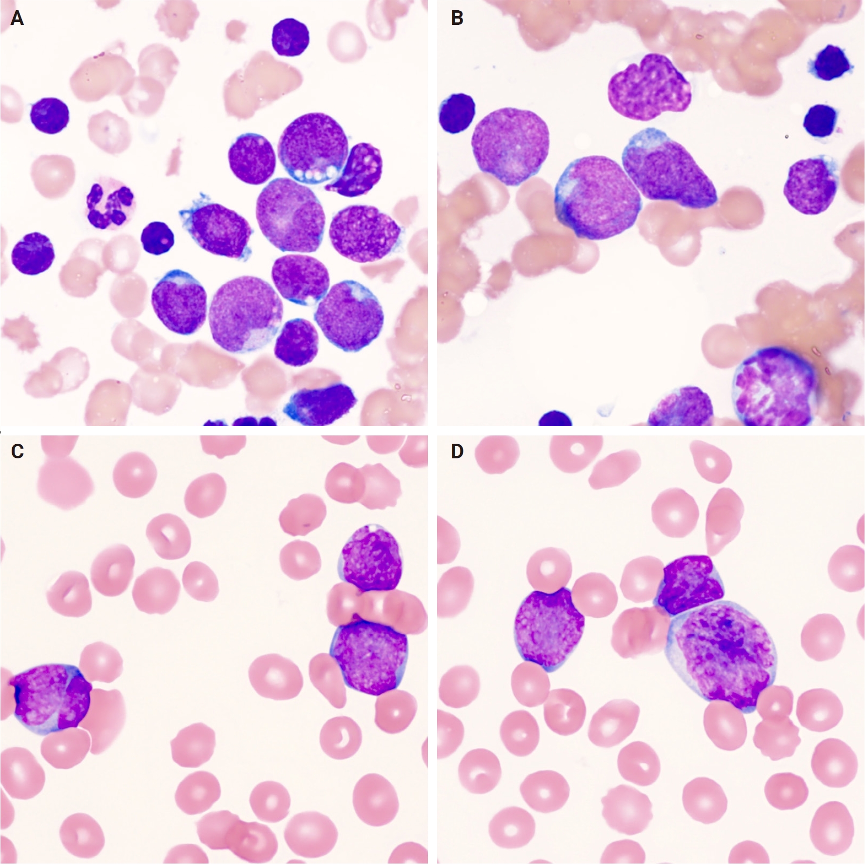
Representative cytologic images of T-lymphoblastic leukemia/lymphoma. (A, B) Bone marrow aspirates showing numerous T lymphoblasts with scant basophilic cytoplasm, fine chromatin, nuclear indentations, occasional prominent nucleoli, and cytoplasmic vacuoles (Wright-Giemsa). (C, D) Peripheral blood smears demonstrating similar blasts with scant cytoplasm, nuclear irregularities, and fine chromatin (Wright-Giemsa).
Immunophenotype
The neoplastic cells are positive for T-lineage markers, most consistently cytoplasmic CD3 (cCD3) and CD7, with CD2 and CD5 expressed in the majority of cases [50,51]. Markers of immaturity such as terminal deoxynucleotidyl transferase (TdT), CD1a, CD34, CD99, and CD117 are typically positive (Fig. 4) [1]. Dual CD4/CD8 co-expression is common in cortical-type tumors. Flow cytometry is essential for precise characterization of the immature T-cell phenotype (Figs. 5, 6) [47]. Aberrant expression of myeloid markers (CD13 or CD33) may occur but should prompt exclusion of early T-cell precursor lymphoblastic leukemia/lymphoma (ETP-ALL/LBL) [52].
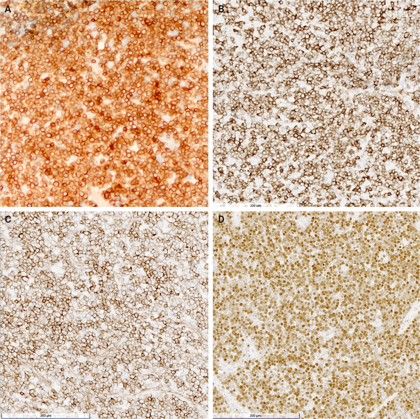
Representative high-power views of the immunohistochemical staining of T-lymphoblastic leukemia/lymphoma. Lymphoblasts with strong positive membranous CD3 (A) and positive membranous CD7 (B), indicating their T lineage. They also express positive membranous CD99 (C) and positive nuclear terminal deoxynucleotidyl transferase staining (D), indicating immaturity.
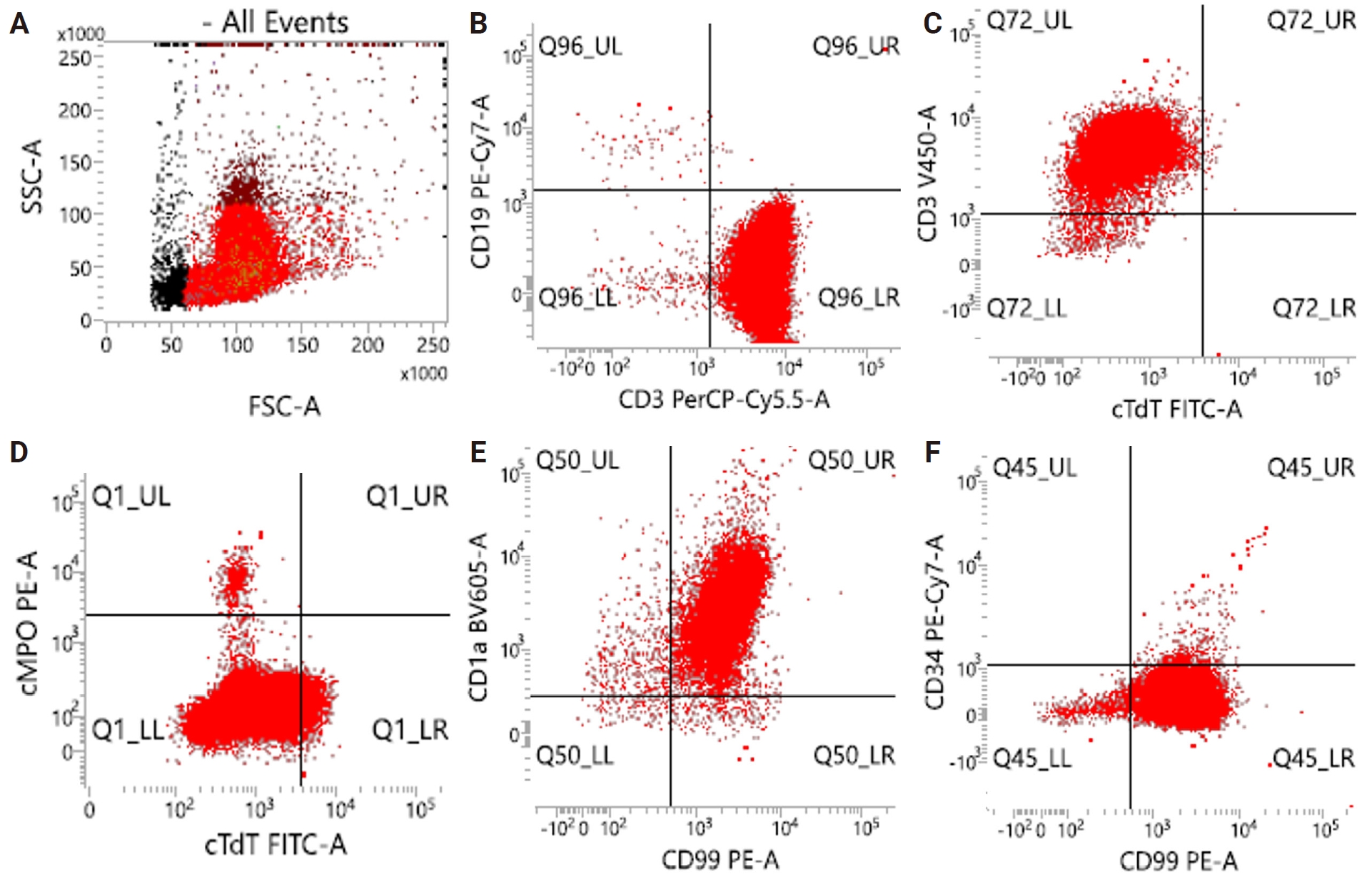
Representative flow cytometry from a mediastinal mass aspirate, diagnosed as T-cell lymphoblastic lymphoma (T-LBL). (A) The sample is composed almost entirely of blasts. (B–F) The blast population demonstrates positive expression of CD3, CD1a, and CD99, with little to no expression of CD19, myeloperoxidase (MPO), and CD34. Interestingly, cytoplasmic terminal deoxynucleotidyl transferase (TdT) is not expressed, which is a finding that is not typical of T-LBL.
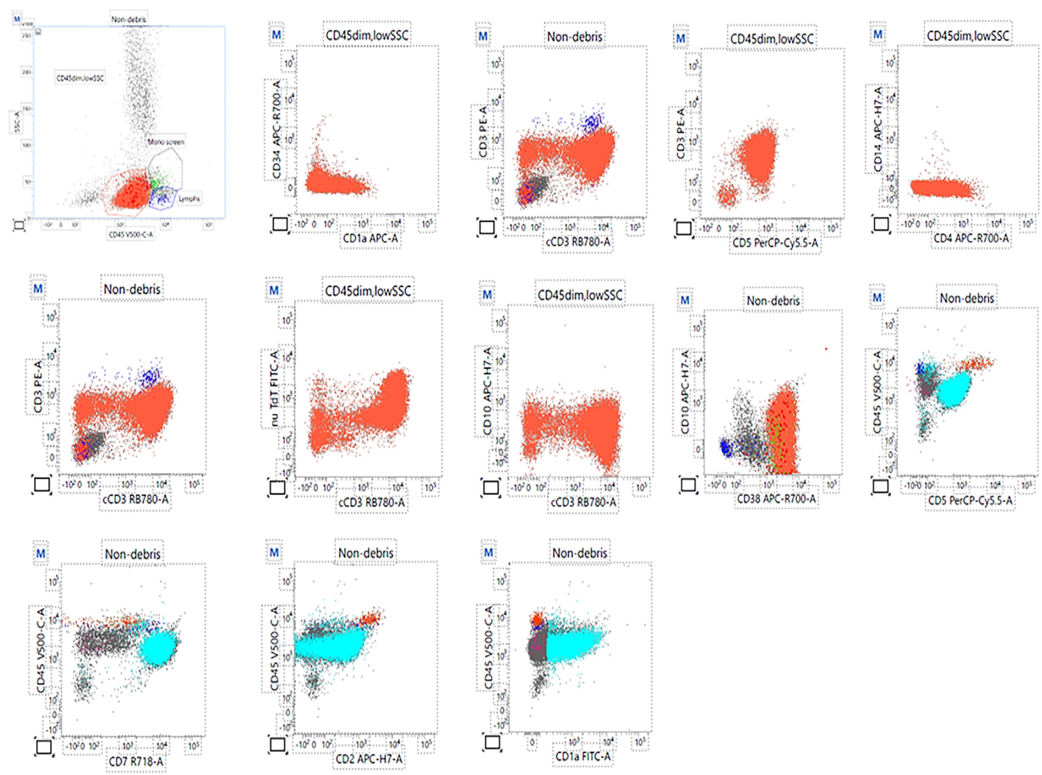
Representative flow cytometry from a bone marrow biopsy of a 44-year-old male. T-lymphoblasts comprise 90% of total events in the aspirate. They are positive for CD45 (dim), CD10 (partial dim), CD38, nuclear terminal deoxynucleotidyl transferase (TdT) (partial), CD1a (partial), CD7 (bright), CD5 (dim), CD2 (variable dim), sCD3 (variable dim), CD4 (subset dim), and cCD3 (variable). They are negative for CD34. CD117, CD13, CD33, HLA-DR, CD11b, CD16, CD64, CD14, CD300e, and CD123 are not shown, but are also negative in this case.
Molecular/cytogenetic features
Key genetic alterations in T-ALL/LBL include activating NOTCH1 mutations (seen in approximately 60% of cases), transcription factor rearrangements involving TAL1, TLX1, TLX3, LYL1, HOXA, and LMO1/2, and inactivation of CDKN2A/B [19,21,30,34,36]. These alterations, discussed in detail in the Pathogenesis section and summarized in Table 1, are diagnostically useful, inform prognosis, and are typically detected by cytogenetic analysis, fluorescence in situ hybridization, or NGS [35].
Pathologic diagnosis and classification
The fifth edition of the WHO Classification of Haematolymphoid Tumors recognizes two categories of precursor T-cell neoplasms: ETP-ALL and T-ALL/LBL, not otherwise specified (NOS) [2]. The ICC recognizes three main categories, T-ALL/LBL, ETP-ALL with BCL11B rearrangement, and ETP-ALL, NOS, along with eight provisional molecular entities defined by their transcription factor rearrangements or expression profiles (TAL1/2-R, TLX-R, TLX3-R, HOXA, LMO1/2-R, NKX2-R, SPI1-R, and BHLH, other) [4]. Additionally, recent integrative genomic studies have described an “ETP-like” molecular group that is defined by transcriptional features rather than immunophenotype, which may capture a broader set of cases than the immunophenotypic ETP-ALL definition and help inform future classification schemes [14].
The WHO and ICC share essentially the same core diagnostic criteria for T-ALL/LBL: the identification of hematopoietic progenitors expressing T-lineage–specific markers (surface CD3 [sCD3] or cCD3) with an aberrant immunophenotype that does not meet the criteria for the diagnosis of ETP-ALL [2,4]. Two distinct blast thresholds apply to these neoplasms and should not be confused. The threshold of 20% blasts in bone marrow or peripheral blood that conventionally defines an acute leukemia or involvement of an extramedullary site is desirable for the diagnosis of T-ALL/LBL. However, neither the WHO nor the ICC specifies a minimum blast percentage for lymphoblastic neoplasms and the diagnosis may be established at a lower blast burden, or in the absence of blood and marrow involvement altogether. The second is the cutoff of 25% bone marrow blasts applied to patients who present with a mass lesion, as mentioned in the introduction. This is used to designate cases as T-ALL when bone marrow blasts reach or exceed 25% and as T-LBL when they fall below that level. This 25% cutoff was adopted from cooperative group treatment protocols rather than from a demonstrated biologic boundary [2].
DIFFERENTIAL DIAGNOSIS
B-lymphoblastic leukemia/lymphoma
B-ALL/LBL expresses B-cell markers including CD19, cytoplasmic CD22, CD79a, and often CD10, while lacking T-cell markers such as CD3, CD5, and CD7; mediastinal masses are rare, unlike in T-ALL/LBL, and it represents the most common acute leukemia in children with a peak incidence in early childhood [4,53].
T-cell prolymphocytic leukemia
T-cell prolymphocytic leukemia is a rare aggressive leukemia of mature T cells often presenting with high white blood cell counts, splenomegaly, and skin involvement; it is characterized by small- to medium-sized cells with irregular nuclei that contrast with the immature blasts of T-ALL/LBL, overexpression or rearrangement of TCL1, and a lack of TdT expression [54].
Acute myeloid leukemia
Acute myeloid leukemias express myeloid markers such as CD13, CD33, CD117, and myeloperoxidase (MPO) with typical negativity for lymphoid markers like CD3 and TdT and may show Auer rods or other signs of myeloid differentiation on bone marrow smears [55].
Early T-cell precursor lymphoblastic leukemia/lymphoma
ETP-ALL/LBL is a subtype of T-ALL with a distinct immunophenotype that includes negativity for CD1a and CD8, weak or negative CD5, and positivity for one or more immature markers, including CD34 and HLA-DR, or myeloid markers, including CD117, CD13, or CD33; it is associated with poorer prognosis [56].
Natural killer–lymphoblastic leukemia/lymphoma
Natural killer (NK)–lymphoblastic leukemia/lymphoma is a rare entity characterized by expression of NK-cell markers like CD56 and cCD3 with negativity for sCD3 and B-cell and myeloid markers; it shares some mutations such as NOTCH1 and DNMT3A with T-ALL/LBL, but clinical presentation and immunophenotype aid differentiation [57].
Mixed-phenotype acute leukemia, T/myeloid
Mixed-phenotype acute leukemia, T/myeloid meets diagnostic criteria for both T-ALL and acute myeloid leukemia by immunophenotypic characterization [58]. The cells demonstrate markers of both T-cell lineage, denoted by cCD3 or sCD3 expression, and myeloid lineage, generally by MPO expression. Criteria for myeloid lineage may also be met by monocytic differentiation via the co-expression of two or more monocytic markers including CD11c, CD14, CD36, or CD64. Flow cytometry is typically required to confirm dual lineage expression [58,59].
Burkitt lymphoma
Burkitt lymphoma is a high-grade B-cell lymphoma with a characteristic histologic starry-sky pattern due to tingible-body macrophages. The cells are intermediate to large with basophilic cytoplasm and vacuoles, express mature B-cell markers including CD20 and surface immunoglobulin, are negative for TdT and CD34, and are molecularly defined by a MYC rearrangement typically via t(8;14) [60,61].
Thymoma
Thymoma is a tumor of thymic epithelial cells staining positive for cytokeratins and often accompanied by a lymphoid component of immature T cells that can resemble T-LBL with CD4/CD8 co-expression and CD1a or TdT positivity. The lymphoid component in thymoma demonstrates features of maturation on flow cytometry. Thymoma can readily be differentiated from T-LBL on histology and has a common clinical association with myasthenia gravis [62,63].
Myeloid sarcoma
Myeloid sarcoma is an extramedullary tumor of myeloid blasts positive for myeloid markers such as CD33, CD68, and MPO and is distinguished by the absence of T-lineage markers [64].
PROGNOSTIC FACTORS AND TREATMENT
Prognostic factors
Favorable prognostic factors include mutated NOTCH1 or FBXW7, TLX1 rearrangements, LMO rearrangements, NKX2-1 rearrangements, and low end-induction minimal residual disease (MRD) [17,18,65,66]. HOXA rearrangements confer intermediate risk [18]. Unfavorable factors include wild-type NOTCH1 or FBXW7, NOTCH1 fusions and intronic single nucleotide variants, PI3K pathway mutations, TAL1 rearrangement, and CNS involvement [4,14-18,66]. End-of-induction bone marrow MRD ≥0.1% is a powerful independent predictor of inferior event-free survival, particularly in pediatric T-LBL [67]. Advanced NGS-based genomic classifiers, when combined with MRD assessment and presenting white blood cell count, further refine risk stratification and identify very-high-risk subsets [68].
Prognosis and treatment
In pediatric patients treated with modern intensive protocols, 5-year event-free survival rates reach 78%–90% with relapse rates of approximately 12%–25% [17,69,70]. Adults continue to have inferior outcomes, with 5-year event-free survival ranging from 40%–70% [71,72]. The ETP phenotype, once considered uniformly high-risk, now demonstrates improved outcomes with contemporary pediatric regimens, although allogeneic transplantation is still frequently performed in this subgroup [71]. In relapsed or refractory disease, outcomes remain poor, particularly among infants [69,71].
Treatment consists of intensive multiagent chemotherapy regimens modeled after acute lymphoblastic leukemia protocols (e.g., hyperfractionated cyclophosphamide, vincristine, doxorubicin, and dexamethasone, augmented Berlin-Frankfurt-Münster, or Children’s Oncology Group protocols), frequently incorporating nelarabine and pegylated asparaginase for improved outcomes in T-ALL [71,73]. Intrathecal chemotherapy (methotrexate or cytarabine) is needed for prophylaxis against CNS relapse [74]. Allogeneic hematopoietic stem cell transplantation is another important arm of curative treatment for adult T-LBL and may be more effective than autologous hematopoietic stem cell transplant in high-risk adult patients [75].
Many emerging targeted and immunotherapeutic approaches for T-ALL/LBL are being developed. Inhibitors targeting NOTCH1, the JAK-STAT pathway, and multiple tyrosine kinases are currently being studied and take advantage of the wide array of genetic alterations seen in T-ALL/LBL [76]. Incorporation of BCL2 inhibitors, such as venetoclax, into T-ALL treatment is being investigated given improved survival rates seen with venetoclax utilization in ETP-ALL [71]. In cases of relapsed or refractory T-ALL/LBL, which face many therapeutic challenges, immunotherapies such as daratumumab and bispecific antibodies including blinatumomab are being studied in ongoing clinical trials [77]. Chimeric antigen receptor T-cell therapies targeting highly expressed surface antigens such as CD5 and CD7 represent a large area of investigation in this same population and show early promise [78].
CONCLUSION
Accurate diagnosis of T-ALL/LBL relies on integration of clinical presentation, radiologic findings, morphologic features, immunophenotypic profile, and molecular genetic data. Pathologists play a central and indispensable role in this process by ensuring precise subclassification, exclusion of morphologic mimics, identification of prognostically relevant alterations, and communication of actionable molecular information to the clinical team. Although T-ALL/LBL is an aggressive neoplasm, contemporary risk-adapted multiagent chemotherapy regimens combined with CNS prophylaxis and, in selected high-risk cases, allogeneic stem cell transplantation have rendered it potentially curable, particularly in pediatric cohorts. Continued refinement of molecular subclassification and the emergence of targeted and immunotherapeutic strategies hold considerable promise for further improving long-term outcomes, especially among adult patients and those with high-risk molecular features.
Notes
Ethics Statement
Not applicable.
Availability of Data and Material
Data sharing not applicable to this article as no datasets were generated or analyzed during the study.
Code Availability
Not applicable.
Author Contributions
Conceptualization: DD, ITL, AA. Supervision: AA. Writing—original draft: DD, ITL, MR, KS. Writing—review & editing: all authors. Approval of final manuscript: all authors.
Conflicts of Interest
The authors declare that they have no potential conflicts of interest.
Funding Statement
No funding to declare.
Disclaimer
The views expressed in this manuscript are those of the authors and do not necessarily reflect the official policy or position of Walter Reed National Military Medical Center, the Joint Pathology Center, the Defense Health Agency, the Department of the Army, the Department of the Navy, the Department of War, or the U.S. Government. The identification of specific products, scientific instrumentation, or organizations is considered an integral part of the scientific endeavor and does not constitute endorsement or implied endorsement on the part of the authors, the Department of War, or any component agency. Several of the authors are military service members or employees of the U.S. Government. This work was prepared as part of their official duties.