What’s new in neoplastic pulmonary pathology 2026: updates on grading, staging and molecular testing
Article information

Abstract
Neoplastic pulmonary pathology has continued to advance in recent years, including the introduction of histologic grading systems for both adenocarcinoma and squamous cell carcinoma, implementation of the UICC TNM 9th Edition classification, expanded molecular testing requirements, refinements in neuroendocrine tumor classification, and improvements in mesothelioma diagnostics. This newsletter summarizes the most important developments relevant to practicing pathologists.
LUNG ADENOCARCINOMA PATTERNS
• Accurate quantification of histologic patterns in resected adenocarcinomas is essential for grading and should be reported in 5% increments per WHO 5th edition recommendations.
• Cribriform and fused glandular patterns are classified as complex glandular (high-grade) patterns [1].
• Micropapillary and solid patterns remain the strongest individual adverse prognostic indicators. The recently described filigree pattern (Fig. 1) is considered a variant of the micropapillary pattern with equivalent adverse prognosis and should be classified accordingly [2]. Any amount of micropapillary pattern warrants documentation.
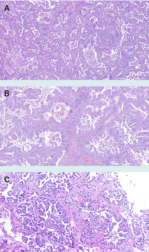
Classical and filigree micropapillary patterns in lung adenocarcinoma. (A) Classical micropapillary pattern with small papillary tufts lacking fibrovascular cores, floating within alveolar spaces. (B) and (C) Filigree pattern, characterized by delicate, elongated stacks of at least three cells without fibrovascular cores.
LUNG ADENOCARCINOMA GRADING
• The International Association for the Study of Lung Cancer (IASLC) Pathology Committee proposed a three-tier grading system for resected invasive non-mucinous lung adenocarcinoma in 2020, incorporating both the predominant histologic pattern and the proportion of high-grade patterns [1].
o Grade 1: lepidic predominant with <20% high-grade patterns (solid, micropapillary, complex glandular including cribriform and fused glands).
o Grade 2: acinar or papillary predominant with <20% high-grade patterns.
o Grade 3: any tumor with ≥20% high-grade patterns, regardless of the predominant pattern.
• Invasive mucinous adenocarcinoma is excluded from the IASLC grading system and represents a separate entity with distinct molecular features (frequent KRAS mutations and a subset with NRG1 and other fusions) and variable clinical behavior.
• The IASLC grading system has been validated in multiple international cohorts for overall survival, recurrence-free survival, and lung cancer-specific survival [3,4].
• The grading system is practical, reproducible (substantial interobserver agreement), and recommended for routine application in surgical pathology reports of resected lung adenocarcinomas.
• Importantly, grading applies only to resected specimens; no validated grading system exists for small biopsies of adenocarcinoma, in which accurate estimation of pattern proportions is not possible.
LUNG SQUAMOUS CELL CARCINOMA GRADING
• The IASLC Pathology Committee recently proposed a two-tier grading system for resected invasive lung squamous cell carcinoma based on tumor budding [5].
o Low-grade: 0-9 tumor buds per 0.785 mm2 (i.e., a 20× objective with field number 20 mm eyepiece).
o High-grade: ≥10 tumor buds per 0.785 mm2.
• Tumor budding was the only histologic feature independently associated with both recurrence-free survival and overall survival in multivariable analyses across two training sets.
• The cutoff of 10 buds aligns with the threshold recommended by the International Tumor Budding Consensus Conference (ITBCC 2016) for colorectal cancer.
LUNG CARCINOMA TNM STAGING: 9TH EDITION
• The UICC TNM 9th Edition classification for lung carcinoma (corresponds to AJCC Version 9), effective January 1, 2025, introduces changes to the N and M descriptors [6,7].
• T descriptors: no changes from the 8th edition. The existing size-based subcategories (T1a–T1c, T2a–T2b) are retained and validated.
• N descriptors: the N2 category is subdivided into N2a (metastasis confined to a single ipsilateral mediastinal or subcarinal station) and N2b (metastases in multiple mediastinal or subcarinal stations), reflecting significantly different prognoses.
• M descriptors: M1c is split into M1c1 (multiple extrathoracic metastases within a single organ system) and M1c2 (metastases in multiple organ systems).
• Stage groups IIA, IIB, IIIA, and IIIB are reordered based on the new N and M subdivisions.
• Spread through air spaces (STAS) is recommended as a histologic descriptor in the 9th edition. Although not yet incorporated into T classification, STAS has been validated as an independent adverse prognostic factor for stage I non-small cell lung cancer (NSCLC) [8].
• Pathologists should also continue to evaluate visceral pleural invasion (VPI), as it remains critical for assigning an accurate T category. In the 9th edition, VPI continues to change a tumor from T1 to T2a, irrespective of tumor size. The use of elastic stains (e.g., Verhoeff–van Gieson, orcein) is strongly recommended whenever VPI is suspected but not certain on H&E examination alone [9].
PREDICTIVE TESTING
• Reflex molecular testing of NSCLC is increasingly recommended at all stages, not just limited to advanced disease, reflecting the expanding role of targeted therapies in adjuvant and neoadjuvant settings. Testing should be initiated, where possible, by the pathologist without waiting for a clinician’s request (“reflex testing”) to minimize turnaround time [10,11].
• The current panel of actionable targets includes EGFR mutations; ALK, ROS1, and RET rearrangements; MET exon 14 skipping mutations and MET amplification; BRAF V600E, KRAS G12C, and ERBB2 (HER2) mutations; and NTRK fusions [12].
• STK11 and KEAP1 mutations have emerged as negative predictive biomarkers for immune checkpoint inhibitor response, particularly in KRAS-mutant adenocarcinomas. Their inclusion in molecular reports is increasingly advocated to inform immunotherapy decisions [13].
• PD-L1 immunohistochemistry remains a standard predictive biomarker for immunotherapy in NSCLC. Currently, only the Tumor Proportion Score (TPS, using cutoffs of 1% and 50%) is used for treatment decisions, assessed with approved assays (clones 22C3, 28-8, SP263). Whether alternative scoring algorithms incorporating immune cell expression, such as immune cell (IC) score, combined positive score (CPS), or the emerging tumor area positivity (TAP) score, may improve prediction in NSCLC remains under investigation.
• Broad-panel next-generation sequencing (NGS) is preferred over sequential single-gene testing to maximize tissue utilization and turnaround time, particularly in small biopsies with limited tissue.
• Liquid biopsy (circulating tumor DNA) serves as a complementary approach when tissue is insufficient, but it should not be used as a screening or primary diagnostic test for NSCLC.
MULTIPLE PRIMARY LUNG TUMORS
• The IASLC Pathology Committee published a combined four-step histologic and molecular classification algorithm for differentiating separate primary lung adenocarcinomas from intrapulmonary metastases, moving beyond the traditional Martini-Melamed criteria [14].
• Different histologic types (e.g., adenocarcinoma versus squamous cell carcinoma) remain sufficient to establish separate primaries.
• Multiple ground-glass or part-solid nodules corresponding to adenocarcinoma in situ (AIS), minimally invasive adenocarcinoma (MIA), or lepidic-predominant adenocarcinoma are classified as separate primaries with the single highest T category reported across all lesions.
• Multifocal mucinous adenocarcinomas are generally considered intrapulmonary metastases unless distinct histologic features support classification as separate primaries (in which case molecular studies are warranted).
• For multiple non-mucinous adenocarcinomas, molecular profiling using NGS has superior discriminatory power over histologic pattern comparison alone and should ideally be performed in all cases when feasible.
• Different oncogenic driver mutations between tumors strongly support separate primaries. However, a shared common hotspot driver mutation (e.g., EGFR L858R, KRAS G12C) is classified as indeterminate, because these high-prevalence mutations can arise independently in ~10% of paired tumors which share a hotspot driver by chance.
• To reduce the number of indeterminate cases, broader NGS profiling is recommended. One or more shared somatic mutations beyond the driver supports intrapulmonary metastasis. If no additional shared mutations are found, different TP53 mutation status favors separate primaries, while cases in which both tumors are TP53 wild-type remain inconclusive.
NEUROENDOCRINE TUMORS
• In small biopsies and metastasectomy specimens, classification into typical versus atypical carcinoid is not possible, as reliable assessment of mitotic count and necrosis requires completely resected and thoroughly sampled specimens. The designation "carcinoid, NOS" is accepted under the WHO 5th edition for these cases.
• Ki67 labeling index is increasingly used in pulmonary carcinoid tumors. Although not yet formally integrated into the WHO classification, Ki67 may identify a subset of high-grade neuroendocrine tumors with carcinoid morphology (“grade 3 NET”), analogous to the gastroenteropancreatic system [15,16].
• In resected carcinoids, the immunohistochemical panel (OTP, CD44, Ki67) can identify patients with a low risk of relapse over time [17].
• INSM1 (insulinoma-associated protein 1) has been adopted as a new immunohistochemical marker for neuroendocrine differentiation (Fig. 2), showing high sensitivity and specificity across the spectrum of pulmonary neuroendocrine neoplasms.
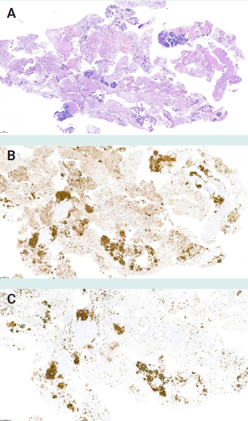
INSM1 immunohistochemistry in small cell lung carcinoma, EBUS-TBNA lymph node metastasis. (A) H&E showing small cell carcinoma with crush artifact. (B) Synaptophysin with strong diffuse cytoplasmic positivity. (C) INSM1 showing strong nuclear staining, confirming neuroendocrine differentiation.
• POU2F3 immunostaining identifies a distinct non-neuroendocrine (tuft cell-like) subtype of small cell lung cancer (SCLC) that demonstrates low expression or absence of classical neuroendocrine markers. POU2F3 can also be expressed in basaloid squamous cell carcinoma and other tumors, necessitating careful interpretation in context [18].
• Recent molecular studies have defined four transcriptional SCLC subtypes based on predominant expression of ASCL1, NEUROD1, POU2F3, or none of these markers [19]. While routine subtyping is not yet required for treatment decisions, pathologists should be aware that companion diagnostic testing for these markers may enter clinical practice in the near future.
• Large cell neuroendocrine carcinoma (LCNEC) comprises two molecular subgroups, an NSCLC-like subgroup with STK11, KEAP1, and KRAS alterations, and an SCLC-like subgroup with concurrent TP53 and RB1 inactivation. Although this distinction has not yet led to subgroup-specific treatment protocols, molecular testing of LCNEC is recommended, as tumors in the NSCLC-like subgroup may harbor actionable driver mutations, such as KRAS G12C or ALK fusions, amenable to targeted therapy [20].
• A novel "atypical SCLC" entity has been described in never/light smokers, characterized by proficient RB1 and TP53, chromothripsis-mediated oncogene amplification, and pathogenetic links to pulmonary carcinoid, suggesting transformation from a lower-grade neuroendocrine tumors as a distinct pathway of SCLC pathogenesis [21].
RARE AND REDEFINED THORACIC TUMORS
• Sarcomatoid carcinomas of the lung encompass pleomorphic carcinoma, spindle cell carcinoma, giant cell carcinoma, carcinosarcoma, and pulmonary blastoma. These tumors typically show aggressive behavior and may require broad immunohistochemical panels (cytokeratins, TTF1, p40) and molecular testing to confirm epithelial lineage.
• Thoracic SMARCA4-deficient undifferentiated tumor (Fig. 3) is recognized as a distinct entity in the WHO 5th edition under “other epithelial tumors”. It is characterized by undifferentiated or rhabdoid morphology, loss of SMARCA4 (BRG1) and typically concurrent SMARCA2 loss, strong male predominance, smoking history, and has a very poor prognosis. It must be distinguished from SMARCA4-deficient conventional NSCLC, which retains SMARCA2 expression [22].
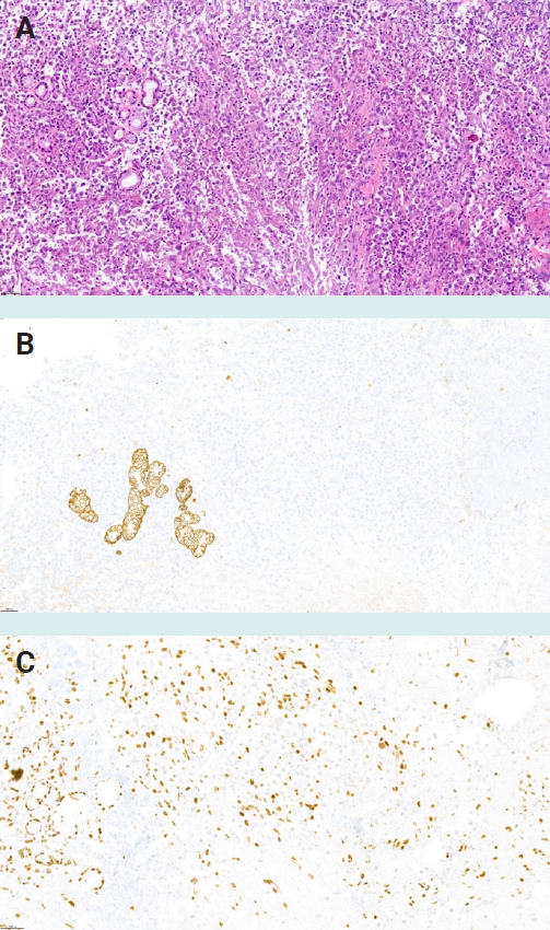
Thoracic SMARCA4-deficient undifferentiated tumor. (A) H&E showing epithelioid tumor cells with rhabdoid morphology. (B) Pan-cytokeratin (AE1/AE3) is negative in tumor cells. (C) SMARCA4 (BRG1) shows reduction/loss of nuclear expression in tumor cells, with retained staining in infiltrating inflammatory cells serving as an internal positive control.
• NUT carcinoma (Fig. 4) is a highly aggressive tumor defined by NUTM1 gene rearrangements (most commonly BRD4::NUTM1). It should be considered in the differential diagnosis of poorly differentiated (often p63 positive) carcinomas in the thorax, particularly in young patients. Immunohistochemistry for NUT protein (specific speckled nuclear staining) is the primary screening tool.
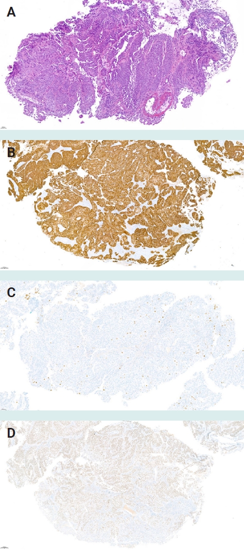
NUT carcinoma of the thorax. (A) H&E showing nests of monotonous undifferentiated tumor cells. (B) Cytokeratin 5/6 with strong diffuse positivity, confirming squamous lineage. (C) p40 shows only scattered single cells with nuclear positivity. (D) NUT immunohistochemistry with diffuse nuclear positivity in tumor cells.
• Hyalinizing clear cell carcinoma, a salivary gland-type tumor harboring EWSR1::ATF1 or EWSR1::CREM fusions, can rarely present as a primary lung tumor and should be recognized to avoid misdiagnosis as squamous cell carcinoma or mucoepidermoid carcinoma.
• PEComas (perivascular epithelioid cell tumors) of the lung include benign clear cell tumor and lymphangioleiomyomatosis. Diagnosis rests on coexpression of melanocytic (HMB45, Melan-A) and smooth muscle (SMA, desmin) markers. Rare malignant PEComas may occur and should be considered in the differential diagnosis of unusual lung tumors with clear cell morphology.
• Primary pulmonary mesenchymal tumors are rare. Solitary fibrous tumor shows characteristic STAT6 nuclear expression (reflecting NAB2::STAT6 fusion), inflammatory myofibroblastic tumor harbors ALK rearrangements in approximately 50% of cases, and synovial sarcoma carries the SS18::SSX fusion. Recognition of these entities requires immunohistochemical and molecular confirmation.
PLEURAL MESOTHELIOMA
• Diagnosis of mesothelioma in small biopsies and cytology remains challenging. The combination of clinical context, imaging, immunohistochemistry, and CDKN2A FISH can support a malignant diagnosis even in limited samples.
• Mesothelial origin of the tumor cells must always be confirmed with at least two positive mesothelial markers.
• A combined immunohistochemical panel of BAP1, MTAP, Merlin, and p53 has been validated for distinguishing mesothelioma from benign mesothelial proliferations, with increasing sensitivity when markers are combined [23].
o BAP1 loss by IHC has moderate sensitivity and near 100% specificity for mesothelioma.
o MTAP loss (surrogate for homozygous CDKN2A deletion) has moderate sensitivity and near 100% specificity.
o Loss of NF2 (Merlin) by IHC has been shown to have moderate sensitivity and 100% specificity for mesothelioma when complete loss of expression is used as the diagnostic criterion.
o Diffuse mutant-pattern p53 staining is infrequent in mesothelioma (approximately 7%) but may represent the only immunohistochemical abnormality in rare cases. Null-pattern p53 is not specific for malignancy and should not be used as a diagnostic criterion.
o CDKN2A FISH remains useful, particularly when IHC results are equivocal or in sarcomatoid mesothelioma, where BAP1 loss is less frequent.
• GATA3 expression has been reported in approximately 70% of sarcomatoid mesotheliomas and may assist in distinguishing them from lung sarcomatoid carcinoma, particularly when combined with calretinin and WT1 [24].
• Biphasic mesothelioma should be diagnosed in a small biopsy if both sarcomatoid and epithelioid components are present, regardless of the amount of each component.
• Grading of epithelioid mesothelioma is based on a two-tier system incorporating nuclear grade (low versus high, based on nuclear atypia and chromatin pattern) and mitotic count, with the presence of necrosis as an additional adverse feature.
o High nuclear grade and elevated mitotic count are associated with significantly worse survival.
o Grading is recommended for routine reporting in epithelioid mesothelioma and can be assessed in resection specimens and sufficiently large biopsies [25].
• The TNM staging system for pleural mesothelioma has also been updated in the 9th edition. Pathologists reporting resection specimens should apply the current staging criteria, paying attention to the depth and extent of invasion.
• Low-grade germline-mutant BAP1-associated mesothelioma (L-BAM) is a newly proposed entity describing mesotheliomas in carriers of germline BAP1 mutations [26].
o L-BAM is typically of epithelioid subtype with tubulopapillary and/or trabecular architecture, superficial stromal invasion, minimal nuclear atypia, and low mitotic activity. It is not distinguishable from sporadic mesothelioma by morphology or immunohistochemistry alone.
o Multiserosal involvement (pleura and peritoneum) is common and represents multiple primary tumors rather than metastatic spread. Staging should account for this distinction.
o Despite a more indolent course than sporadic mesothelioma, L-BAM ultimately progresses to lethal disease.
• Mesothelioma in situ (MIS) is recognized in the WHO 5th edition as a preinvasive mesothelial neoplasm, although the entity remains subject to further data accrual [27–29].
o MIS is characterized by a single layer of relatively bland mesothelial cells on the serosal surface without evidence of stromal invasion. It can also present as a well-differentiated papillary mesothelial tumor-like proliferation.
o The diagnosis cannot be made on morphology alone and requires demonstration of BAP1 loss or MTAP loss by immunohistochemistry, or CDKN2A homozygous deletion by FISH, in the mesothelial cells.
o Clinically, MIS is associated with recurrent pleural effusions without radiologic evidence of a tumor mass. Reported cases remain rare (<50), and MIS carries a high risk of progression to invasive mesothelioma, although the time course is variable and unpredictable.
Meet the Authors
Dr. Tiefenbacher joined PathologyOutlines. com as an author in 2025. He works as a pathologist at the Vienna General Hospital / Medical University of Vienna, where he primarily practices pulmonary pathology.
Dr. Brčić has been an author for PathologyOutlines.com since 2023, a member of the Board of Reviewers since 2025, and part of the Editorial Board for Thoracic Pathology since 2026. He practices pulmonary pathology at the Vienna General Hospital / Medical University of Vienna, Austria.