Targeted next-generation sequencing–based mutational signature analysis in endometrial cancer: utility and limitations across molecular subtypes
Article information

Abstract
Background
Molecular subtype classification is essential for staging endometrial cancer, planning adjuvant treatments, and stratifying prognosis. However, the correlation between mutational signature analysis using tumor-only targeted next-generation sequencing (NGS) data and molecular subtypes, as well as the interpretive value at the individual case level, remains inadequately characterized.
Methods
We performed mutational signature analysis on tumor-only targeted NGS data from 72 endometrial cancer cases using the SATS R package. Molecular classification into four subtypes (POLE-mutated, mismatch repair deficient [MMRd], TP53-mutant, and no specific molecular profile [NSMP]) was established through a standard stepwise algorithm that integrated NGS and immunohistochemistry.
Results
Out of the 72 endometrial cancers, 12 were classified as POLE-mutated, 10 as MMRd, six as TP53-mutant, and 44 as NSMP. All POLE-mutated tumors exhibited POLE-associated signatures (SBS10a/SBS10b), including cases with non-hotspot POLE exonuclease variants and TMBs below the conventional ultramutation threshold. MMRd-associated signatures showed a broader distribution across molecular subtypes; while the summed MMRd-associated signatures were typically higher in MMRd tumors, low-level contributions were also noted in subsets of NSMP and TP53-mutant tumors. Within the POLE-mutated group, MMRd-associated signatures were more pronounced in cases displaying subclonal MMR protein loss as determined by immunohistochemistry.
Conclusions
In this small, clinically selected cohort, targeted NGS-based mutational signatures provided exploratory, complementary information regarding POLE-associated mutagenesis and mixed MMR-related patterns. Overlapping signature distributions, dependence on the analytical pipeline, and the use of tumor-only targeted sequencing limit case-level interpretation. Mutational signature analysis should be interpreted in conjunction with established molecular tests and should not be used as a stand-alone classifier.
INTRODUCTION
Endometrial cancer is a heterogeneous disease, and recent advancements in molecular classification have enhanced risk stratification and clinical decision-making. In 2013, The Cancer Genome Atlas identified four molecular groups of endometrial cancer: POLE-ultramutated, microsatellite instability (MSI) hypermutated, copy-number low, and copy-number high [1]. This classification has been adapted for clinical practice using surrogate markers—POLE mutation testing and immunohistochemistry (IHC) for mismatch repair (MMR) proteins and p53—resulting in four clinically applicable groups: POLE-mutated, MMR-deficient (MMRd), p53-abnormal, and no specific molecular profile (NSMP) [2-4].
POLE-mutated endometrial cancers are characterized by pathogenic somatic mutations in the exonuclease domain of the POLE gene, which disrupt its essential 3'-5' proofreading activity during DNA replication. This deficiency in proofreading leads to an accumulation of somatic mutations, resulting in an ultramutated phenotype, typically defined as a tumor mutation burden (TMB) of ≥100 mutations per megabase (mut/Mb). Consequently, this has led to the widely used term “POLE-ultramutated cancer” [1]. These tumors exhibit an excellent prognosis regardless of histologic grade or stage. The pathogenic POLE mutations are primarily found within exons 9–14 of the exonuclease domain. The five most common and well-characterized hotspot mutations are P286R, V411L, S297F, A456P, and S459F [5-8].
MMRd endometrial cancers result from defective MMR function, leading to MSI and a hypermutated phenotype (10–100 mut/Mb range) [9]. The underlying deficiency can result from germline mutations in MMR genes (MLH1, MSH2, MSH6, and PMS2) associated with Lynch syndrome, or from somatic alterations such as MLH1 promoter hypermethylation. MMRd endometrial cancers typically have an intermediate prognosis among the four molecular groups.
Despite standardized algorithms for molecular classification, interpretation challenges persist in certain cases. First, non-hotspot POLE variants of unknown significance exist beyond the established hotspot POLE exonuclease mutations, complicating the distinction between pathogenic and non-pathogenic variants. To address this issue, a scoring system that incorporates base change frequencies, indels, and TMB has been proposed [10]. Second, some POLE-mutated tumors present with TMB below the ultramutation threshold of 100 mut/Mb [11,12]. Third, some POLE-mutated tumors display subclonal or heterogeneous loss of MMR protein expression on IHC, indicating secondary MMR deficiency. In these instances, additional evidence beyond conventional testing would be beneficial for confident interpretation.
Mutational signature analysis in cancer genomics identifies characteristic patterns of somatic mutations linked to specific mutagenic processes [13]. This method reflects the underlying mutational mechanisms active in tumors and was initially characterized using whole-exome or whole-genome sequencing data in research settings.
With the clinical adoption of molecular classification in endometrial cancer, the use of targeted next-generation sequencing (NGS) panels is increasing in real-world clinical practice. However, applying mutational signature analysis to targeted NGS data presents several challenges, including limited genomic coverage, formalin-induced artifacts [14], and the difficulty of reliably distinguishing germline from somatic variants in tumor-only sequencing. Despite these limitations, recent studies have shown that reliable extraction of mutational signatures is feasible from targeted NGS panels, particularly for POLE-associated signatures [15,16].
This study evaluated mutational signatures derived from tumor-only targeted NGS across molecular subtypes of endometrial cancer, focusing on their interpretive utility and limitations in diagnostically challenging scenarios. These challenges include non-hotspot POLE variants, POLE-mutated tumors with non-ultramutated TMB, and tumors exhibiting subclonal MMR protein loss.
MATERIALS AND METHODS
Case selection
This retrospective study included 72 endometrial cancer cases that underwent tumor-only targeted NGS using the Oncomine Comprehensive Assay Plus (OCA Plus, Thermo Fisher Scientific, Waltham, MA, USA) panel for clinical purposes at Seoul St. Mary's Hospital between August 9, 2023, and February 27, 2024. Of the 74 cases initially sequenced, two were excluded due to poor DNA quality from older formalin-fixed paraffin-embedded (FFPE) blocks.
All cases were reviewed by two pathologists (M.L. and J.K.). Histologic type was classified according to the 2020 WHO Classification of Tumours [17]. Tumor grade and stage were assigned using International Federation of Gynecology and Obstetrics (FIGO) grading criteria and the FIGO 2023 staging system [18], respectively.
Immunohistochemistry
IHC staining for MMR proteins (MLH1, MSH2, MSH6, and PMS2) was performed as part of the routine clinical workup and used for molecular classification. The antibody clones used were MLH1 (M1), MSH2 (G219-1129), MSH6 (SP93), and PMS2 (A16-4), all sourced from Roche/Ventana (Tucson, AZ, USA). Interpretation of the IHC results followed the guidelines outlined in the College of American Pathologists Protocol for Endometrial Carcinoma ver. 5.1.0.0 [19].
MMR protein expression was categorized into three groups: intact, complete loss, and subclonal loss. Complete loss was defined as the absence of nuclear staining in tumor cells, while internal controls retained staining. Subclonal loss was characterized by distinct tumor regions that displayed complete absence of nuclear staining adjacent to areas with retained expression [19].
Targeted NGS
All FFPE tissue sections were reviewed by a pathologist to confirm that tumor content was at least 10% on hematoxylin and eosin–stained slides. Tumor-rich areas were isolated through manual microdissection of these slides.
Genomic DNA was extracted from FFPE tissue blocks using the RecoverAll Total Nucleic Acid Isolation Kit (Invitrogen, Thermo Fisher Scientific, Waltham, MA, USA). The quantity and quality of the nucleic acids were evaluated using the NanoDrop 2000 spectrophotometer and the Qubit fluorometer (both from Thermo Fisher Scientific). Library preparation was performed with the OCA Plus panel (Thermo Fisher Scientific), and sequencing was conducted on the Ion S5 XL platform using Ion 550 Chips loaded via the Ion Chef System (Thermo Fisher Scientific).
Reads were aligned to the hg19 reference genome, and variants were called and annotated using Ion Reporter (ver. 5.20, Thermo Fisher Scientific). Variants with a variant allele frequency below 1.5% were excluded from downstream analysis. MSI status was assessed using the NGS-derived MSI score from Ion Reporter, with MSI-high defined as a score ≥25, microsatellite stable (MSS) as <20, and indeterminate as 20–24. TMB was automatically calculated by Ion Reporter as the number of somatic nonsynonymous variants per megabase of the interrogated genomic region.
Mutational signature analysis from targeted NGS data
Single base substitution (SBS) mutational catalogs were created from filtered somatic variants by assigning each variant to its trinucleotide context, resulting in a 96-channel SBS profile for each sample. In the tumor-only sequencing approach, post-calling filtering focused on eliminating likely germline variants and technical artifacts, which included database-based filtering using Ion Reporter. Signature attribution was conducted with Signature Analyzer for Targeted Sequencing (SATS, ver. 1.0.4) [20], implemented in R ver. 4.4.2, utilizing signeR ver. 2.6.0 [21] as the foundational framework. A panel-specific opportunity matrix was used to express signature exposures on a per-megabase scale. SBS profiles were then refit against the COSMIC SBS reference set (ver. 3.2) [13]. The fidelity of the reconstruction was evaluated by calculating the cosine similarity between the observed and reconstructed 96-channel SBS profiles. Signature burdens were normalized by the callable panel size and expressed as mutations per megabase. The total burden associated with MMRd signatures was determined by aggregating the burdens of SBS6, SBS15, SBS21, SBS26, and SBS44.
MMRd-associated signature proportions were compared between POLE-mutated tumors with intact and subclonal MMR protein expression using a two-sided Wilcoxon rank-sum test. A p-value <.05 was considered statistically significant.
Molecular classification and POLE mutation pathogenicity
Molecular classification into four groups (POLE-mutated, MMRd, TP53-mutant, and NSMP) followed a stepwise algorithm integrating NGS and IHC results [2-4].
Tumors were initially assessed for pathogenic POLE variants. Cases containing one of the five established hotspot mutations (P286R, V411L, S297F, A456P, or S459F) were classified as POLE-mutated [5-8]. For non-hotspot variants, pathogenicity was assessed by literature review [10,22].
Tumors without a pathogenic POLE mutation were evaluated for MMR status. Those exhibiting complete loss of nuclear expression in one or more MMR proteins by IHC were classified as MMRd. Among the remaining POLE-wild-type and MMR-proficient tumors, those harboring pathogenic or likely pathogenic TP53 variants were designated as TP53-mutant; variants of uncertain significance were excluded. The results of p53 IHC were available for all cases and were reviewed as supportive information but were not used to override the sequencing-based classification. When TP53 sequencing and p53 IHC findings were discordant, the sequencing-based classification was retained. Accordingly, we refer to this sequencing-defined study group as “TP53-mutant” rather than “p53-abnormal.” Cases that did not meet the criteria for any of the three categories were classified as NSMP.
RESULTS
Clinicopathologic features and molecular classification
A total of 72 endometrial cancer cases were included. Molecular subtype distribution was as follows: POLE-mutated (n = 12, 16.7%), MMRd (n = 10, 13.9%), TP53-mutant (n = 6, 8.3%), and NSMP (n = 44, 61.1%).
Among 12 POLE-mutated cases, 11 (91.7%) were endometrioid histologic type. Most were high-grade (FIGO grade 3 or high grade, n = 9, 75.0%), with half presenting at stage I (n = 6, 50.0%) and half at stage III (n = 6, 50.0%). Nine tumors carried canonical hotspot mutations within the exonuclease domain of POLE (Table 1).
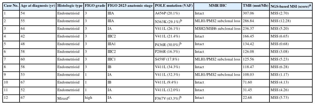
Clinicopathologic and molecular characteristics of POLE-mutated endometrial cancers
All 10 MMRd tumors were endometrioid histologic type. The majority were FIGO grade 2 (n = 8, 80.0%) and presented at stage I (n = 7, 70.0%). NGS-based MSI analysis classified four cases as MSI-high, five as MSS, and one as MSI-indeterminate (Table 2).
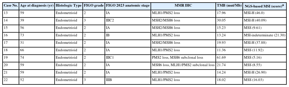
Clinicopathologic and molecular characteristics of endometrial cancers with MMRd by IHC
Mutational signature profiles across molecular subtypes
Mutational signature profiles across molecular subtypes are summarized in Supplementary Fig. S1. POLE-associated signatures were consistently enriched in the POLE-mutated subtype, while MMRd-associated signatures were observed variably in the MMRd subtype. NSMP and TP53-mutant tumors generally exhibited lower TMB and less subtype-specific signature profiles.
POLE-associated signatures in POLE-mutated tumors
TMB in POLE-mutated tumors ranged from 22.68 to 307.06 mut/Mb. Nine cases (75.0%) were classified as ultramutated (TMB ≥100 mut/Mb), while the remaining three were hypermutated but below the ultramutation threshold (TMB values of 22.68, 31.45, and 71.60 mut/Mb). SBS10a and SBS10b were identified as the predominant signature contributors across all 12 POLE-mutated cases (Fig. 1A, B). Among POLE-mutated tumors, stage III cases exhibited a higher median POLE-associated signature burden and a broader distribution compared to stage I cases, although the number of cases was limited (Fig. 2A).
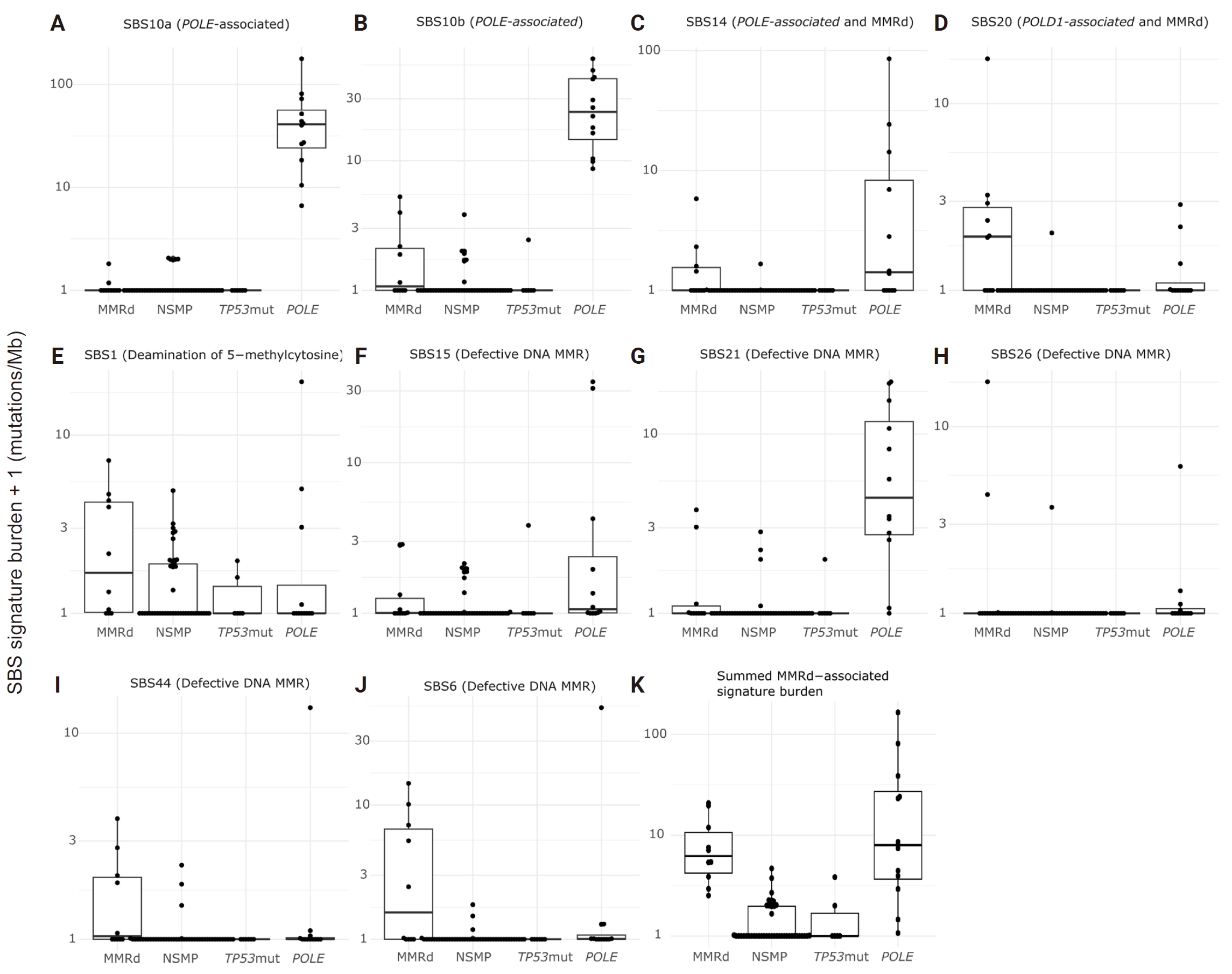
Distribution of selected mutational signatures across molecular subtypes. Panels show SBS10a (A), SBS10b (B), SBS14 (C), SBS20 (D), SBS1 (E), SBS15 (F), SBS21 (G), SBS26 (H), SBS44 (I), SBS6 (J), and the summed MMRd-associated signature burden (K) across MMRd, NSMP, TP53-mutant, and POLE-mutated tumors. Signature burden + 1 (mut/Mb) is shown on a logarithmic scale; therefore, a plotted value of 1 represents an estimated burden of 0 mut/Mb. The summed burden comprises SBS6, SBS15, SBS21, SBS26, and SBS44. SBS, single base substitution; MMRd, mismatch repair deficient; NSMP, no specific molecular profile; Mut, mutations; Mb, megabase.
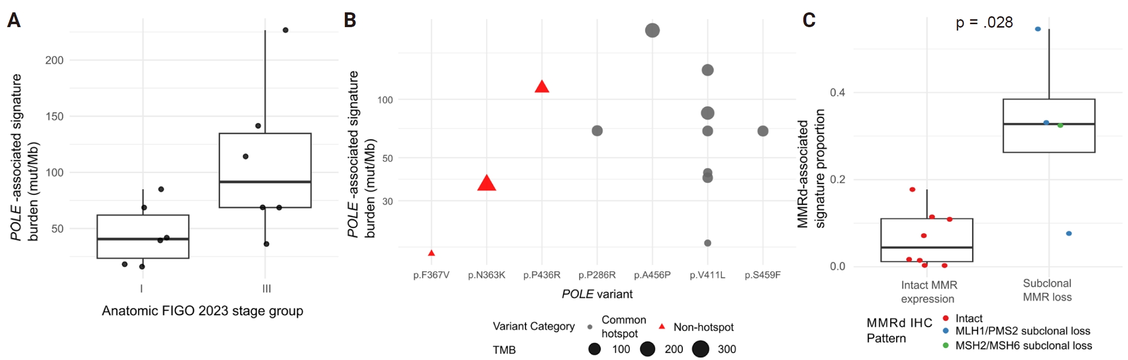
Distribution of POLE-associated and MMRd-associated mutational signatures within POLE-mutated endometrial cancers. (A) POLE-associated signature burden according to anatomic stage. Stage III tumors show a descriptively higher median burden and a wider distribution than stage I tumors; no formal group comparison was performed. (B) POLE-associated signature burden according to the POLE variant. Point size indicates tumor mutational burden, and point shape indicates common hotspot versus non-hotspot variants. The y-axis is shown on a logarithmic scale. (C) Proportion of the total attributed SBS burden assigned to MMRd-associated signatures according to MMR IHC status. Tumors with subclonal MMR protein loss have higher proportions than tumors with intact MMR protein expression (two-sided Wilcoxon rank-sum test, p = .028). Point color indicates the specific MMR IHC pattern. MMRd, mismatch repair deficient; SBS, single base substitution; MMR, mismatch repair; IHC, immunohistochemistry; FIGO, International Federation of Gynecology and Obstetrics; TMB, tumor mutational burden.
Signature profiles in tumors with non-hotspot POLE variants
Three tumors carried non-hotspot exonuclease domain variants: p.N363K (case 2) [22] and p.P436R (case 5) [10], both of which have been reported as pathogenic, and p.F367V (case 12), which is interpreted as likely pathogenic based on published evidence for other substitutions affecting the same residue. All three exhibited contributions from the POLE-associated signature (Fig. 2B). These signature findings were interpreted as supportive evidence of POLE-associated mutagenesis rather than independent confirmation of variant pathogenicity.
Case 2 (N363K) displayed a mixed signature pattern, with contributions from the combined POLE-associated and mismatch repair deficiency signature (28.1%), the defective DNA mismatch repair signature (23.5%), and the POLE-associated signature (8.9%) (Supplementary Fig. S2A). Case 5 (P436R) exhibited a profile dominated by the POLE-associated signature (79.2%) (Supplementary Fig. S2B). Case 12 (F367V) also showed a predominant POLE-associated signature (62.5%) (Supplementary Fig. S2C).
Distribution of MMRd-associated signatures across molecular subtypes
Individual MMRd-associated signatures exhibited variable distributions across molecular subtypes and did not distinctly separate MMRd tumors from NSMP or TP53-mutant tumors (Fig. 1C–J). The summed MMRd-associated signature burden, calculated from SBS6, SBS15, SBS21, SBS26, and SBS44, tended to be higher in MMRd tumors compared to NSMP and TP53-mutant tumors. However, the distributions overlapped across molecular subtypes. Low-level summed burdens were observed in subsets of NSMP and TP53-mutant tumors, while some MMRd tumors displayed relatively low summed values. Additionally, POLE-mutated tumors demonstrated variable summed MMRd-associated signature burdens, particularly in cases with subclonal MMR protein loss (Fig. 1K).
Four POLE-mutated tumors exhibited subclonal MMR protein loss on IHC. These tumors presented mixed mutational signature profiles, characterized by predominant POLE-associated signatures and variable contributions from MMRd-associated signatures (Supplementary Fig. S3). The proportions of MMRd-associated signatures varied according to MMR IHC status within POLE-mutated tumors (Fig. 2C). Tumors with subclonal MMR protein loss exhibited higher proportions of MMRd-associated signatures compared to tumors with intact MMR protein expression (p = .028). In contrast, tumors with intact MMR protein expression generally showed low proportions of MMRd-associated signatures.
DISCUSSION
In this study, tumor-only targeted NGS-based mutational signature analysis revealed varying levels of interpretability depending on the molecular context. POLE-associated signatures were consistently detected in POLE-mutated tumors and may provide supportive evidence in selected diagnostically challenging cases, including tumors with non-hotspot POLE variants or tumor mutation burdens below the conventional ultramutation threshold. In contrast, MMRd-associated signatures exhibited overlapping distributions across molecular subtypes. Although summed MMRd-associated signatures tended to be higher in MMRd tumors, low-level contributions were also observed in subsets of NSMP and TP53-mutant tumors, with some MMRd tumors displaying low values. These findings suggest that MMRd-associated signatures derived from targeted NGS should not be used as a stand-alone classifier for molecular subtype assignment but may be interpreted as complementary information alongside MMR IHC, MSI status, TMB, and institutional or platform-specific analytical experience.
These results indicate that targeted NGS-based signature analysis requires careful interpretation that takes into account the specific platform and pipeline used. Since signature attribution may vary based on panel design, genomic coverage, sequencing chemistry, variant filtering, tumor-only workflow, and refitting methods, the same numerical signature burden may not carry identical diagnostic implications across different assays. Therefore, targeted-panel signature results should be interpreted in the context of assay-specific validation data, accumulated institutional experience, and concordance with established molecular classification methods. Universal cutoffs should be applied cautiously unless supported by data generated from the same or comparable analytical workflows.
The 2023 FIGO staging update now integrates molecular classification directly into tumor staging for endometrial carcinoma, recommending its routine application where feasible. Under the 2023 FIGO staging system, endometrial carcinomas otherwise classified as stage I or II are assigned to stage IAmPOLEmut when a pathogenic POLE mutation is identified, regardless of histologic type or other uterine factors [18]. The National Comprehensive Cancer Network treatment guidelines also recommend POLE mutation testing as part of the molecular subtype classification algorithm [23]. The clinical relevance of identifying the POLE-mutated subtype has increased considerably. Updated European guidelines classify stage I–II POLE-mutated endometrial carcinomas as low risk and recommend no adjuvant therapy [24]. Consistent with this approach, PORTEC-4a recently demonstrated that molecular profile-guided adjuvant treatment maintained locoregional control while allowing 46% of patients with a favorable molecular profile to avoid adjuvant therapy [25]. Ongoing trials, including RAINBO POLEmut-BLUE and TAPER, are further evaluating molecularly guided treatment de-escalation [26,27].
A recognized challenge in the molecular classification of endometrial cancer is interpreting non-hotspot POLE variants of uncertain pathogenicity. All three cases in our study exhibited detectable POLE-associated signature contributions, providing supportive evidence for POLE-associated mutagenesis in these tumors. This finding is notable because standard diagnostic approaches primarily rely on established hotspot mutations, leaving non-hotspot variants without a definitive classification framework. Mutational signature analysis may help address this gap by offering supportive evidence of POLE-associated mutagenesis, independent of the variant hotspot status.
The POLE-score was not formally assessed in these cases because it may not be directly transferable to the present data [10]. The scoring system was developed using whole-exome sequencing and incorporates TMB, the proportions of C>A, T>G, and C>G substitutions, the indel proportion, and variant recurrence. In our tumor-only targeted-panel workflow, restricted genomic coverage, panel-specific trinucleotide opportunities, and post-calling filtering of likely germline variants and technical artifacts could influence the observed substitution proportions and panel-derived TMB. Moreover, the filtered dataset used for SBS analysis did not provide an indel proportion directly comparable with the whole-exome sequencing–derived measure. Applying the original thresholds could therefore result in inaccurate score assignments. We consequently interpreted the available TMB and POLE-associated signature profiles descriptively and did not use a formal POLE-score to determine variant pathogenicity.
Beyond variant classification, another challenge arises when POLE-mutated tumors exhibit a TMB below the ultramutation threshold. In our cohort, three POLE-mutated cases had a TMB below 100 mut/Mb (specifically, 22.68, 31.45, and 71.60 mut/Mb), yet POLE-associated signatures remained the predominant contributor in all three. Pathogenic POLE variants impair exonuclease domain proofreading activity, resulting in progressive mutational accumulation. Non-ultramutated cases may therefore represent an earlier stage of tumor development, occurring before enough mutations have accumulated to exceed the ultramutation threshold. The reasons for the lower panel-derived TMB in these tumors remain uncertain and may include biological heterogeneity and assay-related factors. Nevertheless, the predominance of POLE-associated signatures provided complementary support for POLE-associated mutagenesis despite TMB values below 100 mut/Mb. The detection of POLE-associated signatures, regardless of TMB level, provides supportive evidence for the classification of these tumors within the POLE-mutated subtype.
Approximately 3% of endometrial cancers harbor more than one molecular classifier feature [11]. The coexistence of POLE mutations with MMR deficiency can occur as a secondary event, in which POLE-induced mutagenesis disrupts MMR genes or pathways [10,11]. In this context, the presence of subclonal MMR protein loss on IHC presents an interpretive challenge, as it remains uncertain whether these findings indicate true MMR deficiency or are merely technical artifacts. In our cohort, four out of 12 POLE-mutated tumors (33.3%) exhibited subclonal MMR protein loss on IHC. Within POLE-mutated tumors, subclonal MMR protein loss was associated with higher MMRd-associated signature proportions at the group level. However, the distributions overlapped, and the small subgroup size precludes use of a case-level cutoff.
The higher substitution counts in POLE-mutated and some MMRd tumors may improve signature attribution, whereas estimates in tumors with low mutation burdens are likely to be less stable. Clinically, this suggests that mutational signature analysis may serve as valuable molecular evidence when standard diagnostic criteria are ambiguous, particularly in challenging scenarios such as non-hotspot POLE variants of uncertain significance, POLE mutations with non-ultramutated TMB, and subclonal MMR protein loss that is difficult to interpret using IHC alone.
This study has several limitations. First, the cohort was retrospective and relatively small, with key subsets—such as tumors with non-hotspot POLE variants or mixed molecular features—represented by few cases. Second, signature profiling was conducted using a tumor-only targeted panel with a limited genomic footprint, which can lead to unstable attribution when substitution counts are low; small contributions from overlapping SBS processes may not be reliably distinguishable. Consequently, we treated signature results as supportive descriptors rather than definitive evidence, especially in low mutation-burden tumors. Third, matched normal sequencing was not available. Despite filtering through databases, residual germline variation cannot be entirely ruled out, and platform- or pipeline-specific artifacts, including those related to FFPE changes, may influence SBS catalogs. Thus, the observed signature patterns may vary when different sequencing platforms or attribution pipelines are utilized.
The present analysis defined the TP53-mutant group by TP53 mutation status to maintain consistency with the sequencing-based analytical framework. The sequencing-defined TP53-mutant group is not fully interchangeable with the conventional p53-abnormal subgroup defined by IHC. This difference may have affected group composition and limits direct comparison with studies using IHC-based molecular classification.
Only four of the 10 IHC-defined MMRd tumors were classified as MSI-high by NGS, whereas five were MSS and one was MSI-indeterminate. This finding indicates a notable degree of discordance between the two testing modalities. Although such discordance was pronounced in our cohort, MMRd/MSS tumors are recognized in endometrial carcinoma. MMR IHC assesses loss of protein expression, whereas MSI analysis measures a downstream genomic phenotype; therefore, the two assays are not interchangeable. Endometrial carcinomas may show subtle or locus-specific microsatellite shifts, particularly in tumors with MSH6 loss, which may remain below assay-specific thresholds for MSI-high classification. Differences in tumor sampling, intratumoral heterogeneity, tumor purity, microsatellite loci included in the panel, and bioinformatic thresholds may further contribute. Accordingly, MSS by NGS does not exclude MMRd defined by IHC. The relatively high discordance in this small, clinically selected cohort also supports cautious, platform-specific interpretation of both results [28,29].
In this small, clinically selected cohort, mutational signatures derived from tumor-only targeted NGS showed potential complementary value for characterizing POLE-associated mutagenesis and mixed MMR-related patterns. However, overlapping distributions, combined polymerase–MMR signatures, limited mutation counts, and assay-specific attribution constrain case-level interpretation. Mutational signature analysis should be interpreted in conjunction with established molecular tests and should not be used as a stand-alone classifier. These exploratory findings require validation in larger, independently characterized cohorts.
Supplementary Information
The Data Supplement is available with this article at https://doi.org/10.4132/jptm.2026.09.07.
Notes
Ethics Statement
The Institutional Review Board of Seoul St. Mary's Hospital (KC24RISI0312) approved this study and waived the requirement for written informed consent due to its retrospective nature. All procedures were conducted in accordance with the 1964 Declaration of Helsinki and its later amendments, or comparable ethical standards.
Availability of Data and Material
The datasets used and/or analyzed during the current study are available from the corresponding author on reasonable request.
Code Availability
Not applicable.
Author Contributions
Conceptualization: JK. Formal analysis: UK, AL, KHL, SJL, SHH, DL, JK, ML. Investigation: UK, AL, KHL, SJL, SHH, DL, JK, ML. Methodology: JK, ML. Writing—original draft: UK, JK, ML. Writing—review and editing: UK, AL, KHL, SJL, SHH, DL, JK, ML. Approval of final manuscript: all authors.
Conflicts of Interest
The authors declare that they have no potential conflicts of interest.
Funding Statement
This study was supported by Research Fund of Seoul St. Mary’s Hospital, The Catholic University of Korea (J.K. No. 5-2024-B0001-00076).