Detection of BRAFV600E Mutations in Papillary Thyroid Carcinomas by Peptide Nucleic Acid Clamp Real-Time PCR: A Comparison with Direct Sequencing
Article information
Abstract
Background
Papillary thyroid carcinoma (PTC) of the thyroid is the most common endocrine malignancy. High prevalence of an activating point mutation of BRAF gene, BRAFV600E, has been reported in PTC. We assessed the efficiency of peptide nucleic acid clamp real-time polymerase chain reaction (PNAcqPCR) for the detection of BRAFV600E mutation in PTC in comparison with direct sequencing (DS).
Methods
A total of 265 thyroid lesions including 200 PTCs, 5 follicular carcinomas, 60 benign lesions and 10 normal thyroid tissues were tested for BRAFV600E mutation by PNAcqPCR and DS.
Results
The sensitivity and accuracy of the PNAcqPCR method were both higher than those of DS for the detection of the BRAFV600E mutation. In clinical samples, 89% of PTCs harbored the BRAFV600E mutation, whereas 5 follicular carcinomas, 50 benign lesions and 10 normal thyroid tissues lacked the mutation. The mutation was associated with aggressive clinical behaviors as extrathyroid invasion (p=0.015), lymph node metastasis (p=0.002) and multiple tumor numbers (p=0.016) with statistical significance.
Conclusions
The PNAcqPCR method is efficiently applicable for the detection of the BRAFV600E mutation in PTCs in a clinical setting.
Various oncogenes and tumor suppressor genes regulate the development and progression of papillary thyroid carcinoma (PTC), but only a few of these have been identified to date.1 PTC is the most common endocrine malignancy in the thyroid, accounting for 85-90% of all thyroid cancers.2 The most frequent genetic alterations in BRAF are the BRAF activating point mutations, BRAFV600E and T1796A,3,4 which are important events in the development of PTC. Three Raf families of serine/threonine kinases have been identified as c-RAF, A-raf and BRAF in human.5-8 Mitogenic signals are transduced from the cell membrane to the nucleus by these proteins and they mediate cell differentiation and proliferation. The mutational hot spot codon and nucleotide incorrectly reported in earlier reports as 599 and 1,796 should be correctly located at codon 600 and nucleotide 1,799.9 Codon 600 is in the domain of BRAF, and the V600E mutation makes the enzyme more constitutively active than the wild-type BRAF. This suggests that therapy with RAF kinase inhibitors may be useful for PTC with the BRAFV600E mutation. It will be necessary to evaluate the presence or absence of BRAFV600E mutations in PTC to maximize successful treatment of the disease. There are various methods to detect BRAFV600E mutations, including direct Sanger's sequencing (DS),4,9-12 single strand conformation polymorphism/denaturant gradient gel electrophoresis with direct sequencing,13 allele-specific fluorescent probe melting curve analysis,14 mutant allele-specific polymerase chain reaction (PCR),15 restriction fragment length polymorphism,16 pyrosequencing17 and allele-specific PCR using a dual priming oligonucleotide system,18 although the detection rates vary between methods. The DS method has been a gold standard for detecting mutations even though it has two important disadvantages: low sensitivity (20-50%) and a risk of contamination when handling PCR reaction components.19 It is necessary to apply a more sensitive and specific method for the detection of BRAFV600E mutations in PTC for diagnosis and treatment. The peptide nucleic acid clamp real-time PCR (PNAcqPCR) procedure has been developed for enhanced amplification,20 and has successively detected KRAS mutations in colorectal cancers with specificity and sensitivity.21 In this study, we analyzed 200 PTCs for the presence of the BRAFV600E mutation by the two methods to evaluate their efficiency for BRAFV600E mutant detection, and the results of BRAFV600E in PTC were analyzed with clincopathologic parameters.
MATERIALS AND METHODS
Cell culture
To validate the sensitivity and specificity of the commercial PNA Clamp BRAF mutation detection kit (Panagene Ltd., Daejeon, Korea), two human cancer cell lines (HeLa, Korea cell line bank No. 10002; BRAF wild type and COLO 205, Korea cell line bank No. 10222; BRAFV600E, T1799A, mutant type) were used as PNA clamping standards for real-time PCR. HeLa cells (BRAF wild type) were grown in Dulbecco's modified Eagle's medium (Hyclone, Thermo, Logan, UT, USA), while COLO 205 cells were grown in RPMI 1640, supplemented with 10% fetal bovine serum (Sigma, St. Louis, MO, USA). The cells were cultivated under standard conditions (humidified atmosphere, 5% CO2 in air, 37℃).
Tumor samples
A total of 205 cases of surgically resected thyroid malignant lesions (200 PTCs and 5 follicular carcinomas), 50 benign lesions (15 follicular adenomas, 10 cases of nodular hyperplasia and 10 cases of Hashimoto thyroiditis) and 10 normal thyroid tissues adjacent to nodular hyperplasia were included in this study. Eighty cases of papillary microcarcinoma (≤1 cm) were included to see if the BRAFV600E mutation occurs from early carcinogenesis. Informed consent was obtained from the patients for the collection of the tumor specimens, and the study protocol was approved by the Soonchunhyang University, College of Medicine Ethics Committee. The tissues were fixed in 10% neutral buffered formalin for 8-14 hours at room temperature for paraffin block.
Preparation of genomic DNA
Genomic DNAs were extracted from the cell lines using the High Pure PCR Template Preparation kit (Roche, Mannheim, Germany), according to the manufacturer's instructions, and the DNA was diluted to a concentration of 50 ng/µL for evaluation of the test. DNA from the COLO 205 cells, T1799A (V600E), was diluted with the DNA from the HeLa cells to give mutation/wild-type ratios of 1%, 5%, 10%, 20%, 50%, and 100%. The DNAs extracted from the cell lines were stored at -20℃ until use. The genomic DNAs from the formalin-fixed paraffin-embedded (FFPE) sections were extracted from 5×10 µm sections using the QIAmp DNA FFPE tissue kit (Qiagen, Valencia, CA, USA) as per the instructions.
DNA sequencing
The PCR reactions were performed in a final volume of 50 µL containing 1×PCR buffer, 200 µmol/L dNTPs, 200 nmol/L of each primer and 2.5 U of Taq polymerase (Solgent, Daejon, Korea). The forward primer was 5'-AAACTCTTCATAATGCTTGCTCTG-3'. The reverse primer was 5'-GGCCAAAAATTTAATCAGTGGA-3'. PCR was performed in a C1000 Thermal Cycler system (Bio-Rad, Hercules, CA, USA) with an initial denaturation temperature at 95℃ for 5 minutes followed by 40 cycles of 30 seconds at 95℃, 30 seconds at 62℃, and 1 minute at 72℃, and a final extension at 72℃ for 5 minutes. Amplicons of 231 bp were purified using the PCR purify kit according to the manufacturer's instructions (COSMO Co. Ltd., Seoul, Korea). Briefly, the amplicons were mixed with 5 volumes of PB buffer and the sample was loaded onto a column. After washing with NW buffer, the DNA was eluted with Tris buffer (10 mM Tris-Cl [pH 8.5]). DNA sequencing was performed with the PRISM 3100 DNA analyzer (Applied Biosystems, Carlsbad, CA, USA) using the ABI Prism BigDye Terminator ver. 3.1 (Applied Biosystems) with the forward primer.
PNAcqPCR
The BRAFV600E mutation was tested using the PNA Clamp BRAF mutation detection kit according to the manufacturer's instructions. Briefly, PCR was performed in a total volume of 20 µL that contained 50 ng of DNA, 13 µL of real-time SYBR Green PCR master mix and each of the primers and PNA probes for codon 600. The PCR control lacked a PNA probe and contained the wild type template. The PCR cycling conditions were at 94℃ for 5 minutes. followed by 40 cycles of four temperature steps (94℃ for 30 seconds, 70℃ for 20 seconds, 63℃ for 30 seconds and 72℃ for 30 seconds), and a final extension at 72℃ for 5 minutes. The PNA probe designed to hybridize completely to the wild-type BRAF allele securely inhibited the amplification of the wild-type BRAF allele, while the PNA/mutant-type allele hybrid was unstable due to base pair mismatch, which is why the extension by polymerase occurred. The threshold cycle (Ct) was automatically calculated from the PCR amplification plots where fluorescence was plotted against the number of cycles. Delta-Ct values were calculated as the Ct values of the samples minus those of the controls. The higher delta-Ct value showed that the mutant was efficiently amplified. A cut-off value of 2.0 was used for determining the presence of mutant DNA.
Interpretation of the mutation data and statistical analysis
For calculating sensitivity, specificity and accuracy of each detection method, true-positive (TP), true-negative (TN), false-positive (FP), and false-negative (FN) results were defined as follows. Detection of a BRAFV600E mutation in a tissue pathologically confirmed as PTC, in 200 cases, was considered as TP. Detection of a BRAFV600E mutation in a specimen pathologically confirmed as a normal, benign lesion or follicular carcinoma, in 75 cases, was categorized as FP. Failure to detect a BRAFV600E mutation in a normal, benign lesion or follicular carcinoma in 75 cases, was considered as TN, and failure to detect a BRAFV600E mutation in a PTC in 200 cases, was categorized as FN. Diagnostic values were calculated as follows: sensitivity=TP/(TP+FN)×100; specificity=TN/(TN+FP)×100; positive predictive value (PPV)=TP/(TP+FP)×100; negative predictive value=TN/(TN+FN)×100; accuracy=(TP+TN)/(TP+TN+FP+FN)×100. Analysis by the κ statistic was performed to evaluate the agreement between each method and histopathologic diagnosis (κ≥0.75, excellent; κ=0.40-0.75, fair or good; κ<0.40, poor). The χ2 test and Student's t-test were used to analyze the relationship between clinicopathologic features and BRAFV600E mutation in patients with PTC. A p-value of less than 0.05 was considered statistically significant.
RESULTS
Sensitivity and specificity of the PNAcqPCR compared to direct sequencing using cell lines for the detection of the BRAFV600E mutation
The serially diluted mutant cells (COLO 205) in the wild type cell (HeLa) background were tested by PNAcqPCR in comparison to DS. The delta-Cts of the 1%, 5%, 10%, 20%, 50%, and 100% mutant were 3.63, 5.5, 6.34, 6.96, 8.62, and 9.96, respectively. The delta-Ct of the 1% mutant was larger than the cutoff value of 2.0 and the kit sensitively detected mutation in 1% of the mutants in the wild type background. For sequencing, the mutation was detected barely by manual reading in 50% of the mutants in the wild type background (Fig. 1).

Comparison of efficiency in detection of the BRAFV600E mutation between peptide nucleic acid (PNA) clamp real-time polymerase chain reaction (PCR) and direct sequencing. (A) The delta Ct of PNA clamp real-time PCR in 1% mutant is 3.63, which is larger than the cutoff value of 2.0 to detect mutants. (B) The sequencing barely detects mutation in 50% of mutants by manual reading.
Detection of the BRAFV600E mutation in thyroid samples by PNAcqPCR compared to Direct Sequencing
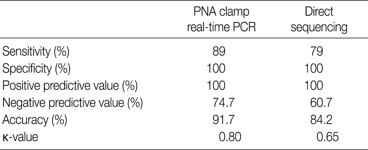
Of the 200 PTC samples, 178 were positive (89%) by PNAcqPCR and 158 (79%) were positive by DS. All of the positive cases by DS were positive by PNAcqPCR as well. We failed to detect the BRAFV600E mutation either by PNAcqPCR or by DS in follicular carcinomas and benign thyroid lesions including follicular adenoma, nodular hyperplasia, Hashimoto thyroiditis and normal thyroid tissue. In 80 cases of papillary microcarcinoma, 71 cases (89%) were positive for BRAFV600E. The sensitivity to detect BRAFV600E by PNAcqPCR was 89% whereas it was 79% by DS. The specificity and PPV was 100% by the two methods. The accuracy to detect BRAFV600E was 91.7% by PNAcqPCR whereas it was 84.2% by DS. The Cohen's kappa coefficient of PNAcqPCR was 0.80, which is excellent, whereas that of DS was 0.65, which is fair or good (Table 1).
Correlation between BRAFV600E mutation and clinicopathologic parameters in papillary thyroid carcinomas
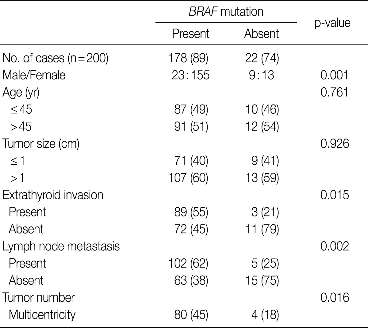
We analyzed the association between the presence of the BRAFV600E mutation and various clinicopathologic parameters in patients with PTC, including sex, age, tumor size, extrathyroid invasion, lymph node metastasis and tumor multicentricity. As shown in Table 2, age and tumor size did not differ significantly in patients with or without the BRAFV600E mutation. The female patients had a higher prevalence of BRAFV600E which was statistically significant (p=0.001), compared to the male patients. The tumors with BRAFV600E had higher probabilities of extrathyroid extension (p=0.015). The tumors with nodal metastasis (p=0.002) and with multicentricity (p=0.016) had a statistically higher prevalence of BRAFV600E than that of non-metastasis and of single location (Table 2).
DISCUSSION
PTC occurs due to oncogenic constitutive activation of the mitogen-activated protein kinase pathway by the BRAFV600E mutation.3 The prevalence of the BRAFV600E mutation in PTC is different according to the detection methods, ranging from 32% to 90%.3,4,10-18 In this study, the BRAFV600E mutation was detected in 89% (178/200) of PTC samples by the PNAcqPCR method, which is in concordance with the highest prevalence reported by Lee et al.18 In Korea, the BRAFV600E mutation has been reported to be present in 83-90% of PTCs,12,17,18 which is higher than what is reported (32-69%) in other countries.3,4,10,11,13-16 The reason behind higher prevalence of BRAFV600E in PTC in Korea compared to other countries is not clear. However, a recent epidemiologic study reported that high iodine intake may be a significant risk factor for the presence of the BRAFV600E mutation.22 Koreans usually consume sufficient iodine through the ingestion of seaweed products.23 Korean patients with thyroid diseases ingest considerably more iodine than healthy subjects, especially patients with thyroid cancer.24
The PNAcqPCR detects the BRAFV600E mutation with high sensitivity. The COLO 205 cell line is a heterozygous mutant cell, and the sequencing chromatogram of 100% mutant cells demonstrated low wild type A peak. In order to test sensitivity to detect the BRAFV600E mutation, heterozygous mutant cells proportionally mixed with wild type cells may be used.25 As shown in this study with cell lines, the PNAcqPCR detected a 1% BRAF mutant in the background of the wild type with high sensitivity, though the gold standard DS detected a 50% mutant in the background of the wild type. The PNAcqPCR is a sensitive and specific method to detect point mutations.26,27 Several chemical features make PNA a superior PCR clamp for specific alleles. PNA cannot be used as a primer for polymerization, nor can it be used as a substrate for the exonuclease activities of Taq polymerase. In addition, the melting temperature (Tm) of a perfectly matched PNA-DNA duplex is higher than that of a DNA-DNA duplex of the same length or nucleotides, and a single mismatch destabilizes the PNA-DNA hybrids, causing a Tm shift.26 Therefore, a PNA probe can specifically block chain elongation on a perfectly matched wild template without interfering with these reactions on the mutant templates with mismatched bases.26,27 This enables the detection of mutants by real-time PCR rapidly and sensitively.
DS is the "gold standard" which has been used to detect point mutations. This method, however, takes 2-3 days and has relatively low sensitivity, since samples must contain at least 20% mutant cells for successful mutant detection.19 In this study, DS failed to detect the BRAFV600E mutation in 20 samples that were positive for this mutation by PNAcqPCR. These samples were microcarcinomas that had diameters less than 1 mm with a few tumor cells.
Comparing the efficiency between PNAcqPCR and DS for the detection of the BRAFV600E mutation, the sensitivity and accuracy of PNAcqPCR method were both higher than those of DS. However, the specificity and PPV of the two methods were both 100%. The PNAcqPCR method has several advantages over DS under clinical settings. First, it is more sensitive for the detection of the BRAFV600E mutation than DS. Second, it requires only simple real-time PCR equipment rather than an expensive sequencer. Third, the result can be obtained rapidly by this method. Fourth, as it uses a single tube reaction, cross contamination of the samples can be minimized.
Although the BRAFV600E mutation is one of the most common genetic alterations in PTC, the exact biological characteristics, pathological features and clinical behaviors of tumors harboring BRAF mutations are still unclear. Lee et al.28 reported by meta-analysis including 1,168 patients from 12 studies that the BRAF mutation significantly correlated with histologic subtype (tall cell variant and conventional type), presence of extrathyroidal extension, and advanced clinical stage, but not with age, sex, race or tumor size. The association between the BRAFV600E mutation and lymph node metastasis was found to be of borderline significance (odds ratio, 1.5; 95% confidence interval, 0.992 to 2.268; p=0.055).
In this study, the BRAFV600E mutation was associated with aggressive clinical behaviors including extrathyroid invasion (p=0.015), lymph node metastasis (p=0.002) and multiple tumor numbers (p=0.016) with statistical significance. This result is concordant with that of meta-analysis by Lee et al.28 It is noticeable that the BRAFV600E mutation occurs in 89% of papillary microcarcinomas. This shows that the BRAFV600E mutation plays a role in early carcinogenesis.
Because of its high prevalence, and its association with aggressive clinical behaviors, BRAFV600E in PTC has become an attractive target for small molecular kinase inhibitors, which are currently in development. BAY43-9006 (sorafenib) and PLX4032 are novel signal transduction inhibitors that are intended to prevent tumor growth by blocking the RAS signaling pathway, and stop tumor angiogenesis by inhibiting the vascular endothelial growth factor receptor-2/platelet derived growth factor receptor-beta signaling cascade.29,30 Identification of the BRAFV600E activating point mutation by a rapid laboratory method could have significant clinical value, and maximize therapeutic efficacy by drugs used in PTC. The PNAcqPCR method could be a method of choice for the detection of BRAFV600E with sensitivity and rapidity. Also, it appears feasible to use both routine FFPE tissue samples and minute amount of cells obtained by fine needle aspiration.
In summary, in this study, the BRAFV600E mutation was detected in 89% of PTCs, which is one of the highest levels of prevalence reported. This suggests that BRAFV600E provides a genetic marker differentiating papillary carcinomas from other types of thyroid carcinomas, and suggests that the BRAFV600E mutation can play a significant role in the carcinogenesis of papillary carcinoma. The BRAFV600E mutation was associated with aggressive tumor behaviors including extrathyroid invasion, multiplicity and regional lymph nodal metastasis. The PNAcqPCR method for the detection of BRAFV600E is sensitive, and is applicable in a clinical setting to maximize the therapeutic efficacy of small molecular kinase inhibitors.
Acknowledgments
We thank the Soonchunhyang College of Medicine Gene Bank for providing us with the materials used in this study.
Notes
No potential conflict of interest relevant to this article was reported.

