Adenocarcinoma Arising in a Colonic Duplication Cyst: A Case Report and Review of the Literature
Article information
Duplications in the alimentary tract are uncommon congenital abnormalities that may occur anywhere from the oral cavity to the anus, with the ileum being the most common site.1 Malignant change in a duplication is extremely rare.
Here, we report a case of adenocarcinoma arising in a colonic duplication cyst in a 23-year-old female and provide a review of the relevant literature.
CASE REPORT
A 23-year-old female patient was admitted with a palpable abdominal mass. The mass had been noted three years previously. On physical examination, a nontender and movable mass measuring approximately 7 cm was observed at the right lower quadrant.
The patient's serum carcinoembryonic antigen (CEA) level was 6.51 ng/mL (normal range, <5.0 ng/mL), and that of cancer antigen 19-9 was 47.71 U/mL (normal range, <37 U/mL).
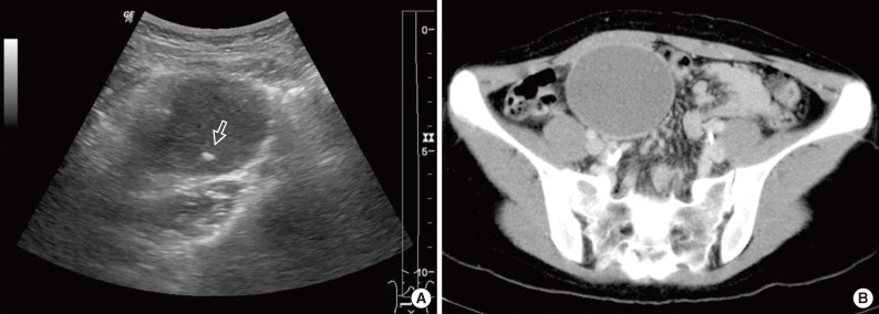
Ultrasonography disclosed a hypoechoic cystic mass with an internal echogenic dot in the right lower quadrant. The echogenic dot was thought to be either an intraluminal secretion or necrotic debris (Fig. 1A). Computed tomography (CT) revealed a nonenhancing cystic mass measuring 7 cm, with linear calcification located adjacent to the medial side of the ascending colon (Fig. 1B). No enlarged regional lymph nodes, ascites, or other abnormalities were observed.

Radiologic findings. (A) A hypoechoic mass with an echogenic dot (arrow) in the right lower quadrant is observed on ultrasonography. (B) A cystic mass with linear calcification in the mesentery adjacent to the ascending colon is observed on computed tomography.
A laparoscopic excision of the mass was performed. During the operation, the mass was observed to be attached to the ascending colon mesentery, having the blood supply by the right colic artery.
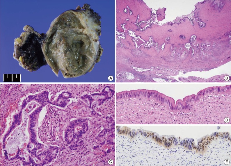
On gross examination, the cystic mass measured 8.6×6.4×2.9 cm, and its outer surface was smooth; mesenteric fat adhered to the outer surface. The maximum thickness of the cystic wall was 1.5 cm, and focal calcification was noted within the wall. The cyst contained brownish-colored mucoid material (Fig. 2A).

Gross and microscopic findings. (A) The cystic mass measured 8.6×6.4×2.9 cm and contained brownish-colored mucoid material. (B) Full-thickness of two smooth muscle layers and adjacent mesenteric soft tissue are invaded by tumor cells. (C) The tumor is moderately differentiated adenocarcinoma. (D) A focal area of atypical columnar epithelium, compatible with dysplasia, is noted on the surface. (E) The dysplastic epithelial cells are positive for p53.
Microscopically, the cyst was found to have two well-organized layers of smooth muscle with an infiltrating adenocarcinoma forming irregular tubules (Fig. 2B, C). The tumor had invaded the pericystic mesenteric soft tissue and metastasized to eight of 16 mesenteric lymph nodes. Perineural and lymphovascular invasions were noted. Non-neoplastic glandular epithelium was not found; instead, a focal area of atypical columnar epithelium, which was compatible with low-grade dysplasia, was observed (Fig. 2D). On immunohistochemical study, tumor cells were positive for cytokeratin 7 (1:100, OV-TL 12/30, Dako, Glostrup, Denmark), cytokeratin 20 (1:100, KS20.8, Dako), CEA (prediluted, II-7, Dako), CDX2 (1:50, AMT 28, Novocastra, Newcastle upon Tyne, UK), and p53 (1:100, DO-7, Dako). The dysplastic epithelium was also positive for p53 (Fig. 2E).
Positron emission tomography-CT was performed after surgery and revealed no hypermetabolic areas other than the previous operation site. Two months after surgery, the patient started adjuvant chemotherapy.
DISCUSSION
Duplications are rare congenital anomalies that may occur anywhere along the alimentary tract.1 The most common site of involvement is the ileum. Colonic duplications comprise 4% to 18% of all duplications, and occur most often in the cecum. Strict morphologic criteria for the diagnosis of duplication have been established: 1) attachment to the alimentary tract, 2) presence of smooth muscle layers, and 3) presence of lining epithelium resembling that of the alimentary tract. Our case met all of these criteria.
Malignant change in a duplication is very rare, and adenocarcinoma is the most common histologic type of malignancy found in these unusual cases.1-10 However, squamous cell carcinoma, carcinoid tumor, gastrointestinal stromal tumor, and leiomyosarcoma have also been reported.
Thirteen cases of adenocarcinoma arising in a duplication of the colon have been reported to date in the English literature (Table 1).1-10 Among the 14 cases, including the current case, four patients were male and 10 patients were female. The mean age at diagnosis was 48.8 years (range, 23 to 72 years) and our patient was the youngest. Some of the patients, including our patient, were relatively young, suggesting that the epithelium of the duplication has a high risk of carcinogenesis. The most common symptoms were abdominal pain and a palpable mass, and the mean size was 10.2 cm (range, 3 to 20 cm). Calcifications in the cystic wall or calculi in the lumen were common findings and were present in six of the eight cases where relevant data were available. Peripheral calcifications were present in three cases and calculi were found in four of six cases. Calcification in a duplication can be a worrisome finding because calcification is extremely rare in benign duplications.6 The preoperative serum CEA level was available for five cases and was found to be elevated in four cases in which patients presented with advanced stage disease at the time of initial diagnosis. According to our survey, except for one case without relevant data, all cases had invasion beyond the muscular wall. Metastasis to a regional lymph node or to a distant organ was reported in three cases.3,9 Although there was insufficient data on the duration of follow-up, five of six cases involved uneventful clinical courses. One patient died 41 months after diagnosis due to extensive metastasis.9 No follow-up data was provided for the remaining eight patients. Due to its rarity and nonspecific symptoms, we thought that, in the present case, the diagnosis was made at an advanced stage. Calcification within the mass and elevated serum CEA level appear to be useful for preoperative prediction of malignant change in duplications.
In conclusion, intestinal duplication is an uncommon congenital abnormality, and malignant change in a duplication is extremely rare. Differential diagnosis of a cystic mass located in or adjacent to the gastrointestinal tract should include duplication. When serum CEA level is high, and/or calcification within the mass is observed, the possibility of adenocarcinoma arising in a duplication cyst should be considered.
Notes
No potential conflict of interest relevant to this article was reported.
