Adenocarcinoma Arising in Gastric Duplication Cyst
Article information
Alimentary tract duplications may occur from the oral cavity to the rectum. The ileocecal valve area is the most commonly affected site.1 Gastric duplication cysts are relatively rare congenital anomalies that constitute only 2% to 8% of alimentary tract duplications.2 These gastric duplication cysts are mostly discovered in children, with most patients presenting during the first year of their life. Associated symptoms often include nausea, vomiting, hematemesis, and abdominal pain.3 Ulceration, bleeding, rupture, and fistula formation are known complications. There have been a few case reports on gastric duplication cysts associated with malignancy.1,2,4,5 Previously reported malignant lesions have included adenocarcinoma, mixed adenocarcinoma and squamous cell carcinoma, and neuroendocrine carcinoma. Here, we report a case of adenocarcinoma arising in a gastric duplication cyst, the first such occurrence to be reported in Korean patients.
CASE REPORT
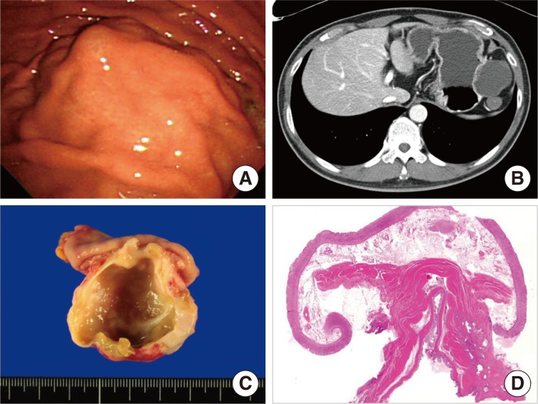
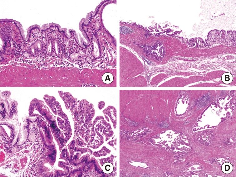
A 56-year-old man was admitted to Asan Medical Center with a gastric submucosal lesion that was detected by esophagogastroduodenoscopy during a regular check-up (Fig. 1A). The overlying gastric mucosa was normal except for a slight elevation, and no associated symptoms were present. His medical and family histories were unremarkable. A complete blood count and serum chemistry tests were normal. A computerized tomography (CT) scan revealed a 5.5-cm low attenuating cystic mass located in the submucosal layer along the greater curvature of the body (Fig. 1B). The radiologic differential diagnoses were cystic degeneration of the neurogenic tumor, gastrointestinal stromal tumor (GIST), and gastric duplication cyst. The patient underwent a wedge resection to remove the lesion, and an unilocular cystic mass measuring 5.5×3.5×2.5 cm was identified in the proper muscle layer with smooth and intact overlying gastric mucosa. Thick, yellowish fluid filled the cyst and there were no connections between the cyst and the lumen of the stomach. The cystic wall had a regionally thickened portion (0.6 cm) and its cut surface was firm and granular (Fig. 1C). Microscopically, the cystic wall was comprised of all of the layers of the stomach wall (from the mucosa to the muscularis propria). The proper muscle layer of the cyst merged into the proper muscle layer of the stomach, sharing a common wall (Fig. 1D). The lining epithelial cells of the cyst were columnar and mucus-secreting cells resembling foveolar cells (Fig. 2A). There were multiple foci of high grade dysplasia (Fig. 2B, C) and adenocarcinoma showing invasion to the proper muscle layer of the cyst was observed (Fig. 2D). The histological features of the carcinoma resembled conventional adenocarcinomas commonly observed in the gastrointestinal tract. The carcinoma did not extend beyond the cyst wall, and neither the proper muscle nor the subserosal layer of the surrounding normal stomach harbored any tumor cells. The overlying gastric mucosa was normal and the resection margins were clear. Three regional lymph nodes were harvested during the wedge resection, which showed no metastasis.

(A) Esophagogastroduodenoscopy reveals gastric submucosal lesion with normal overlying mucosa. (B) Abdominal computed tomography shows a 5.5-cm cystic lesion, located along the greater curvature side of the stomach, during the arterial phase. (C) An unilocular cyst is readily seen below the mucosa when the lesion is bisected. The inner surface of the cyst is smooth but the cut surface of the cyst wall shows focal irregularity. (D) The cyst wall shares a common wall with the gastric proper muscle layer.
DISCUSSION
Gastric duplication is an extremely rare congenital anomaly in adults. It usually presents as a saccular or elongated cystic mass attached to the stomach. This is surrounded by at least one coat of smooth muscle, usually fusing the muscularis propria of the cyst and the stomach. The cyst is lined by typical gastric mucosa, often accompanied by patches of ectopic intestinal, colonic, respiratory epithelium, or pancreatic tissue. The presence of pancreatic tissue or gastric mucosa leads to complications such as peptic ulcers or acute pancreatitis. Most of the gastric duplications are located in the greater curvature, because gastric duplications usually originate dorsal to the primitive intestine.6
Although gastric duplication is usually found as an asymptomatic mass, there can be symptoms associated with complications, such as pain, obstruction, ulceration, and hemorrhage. The malignant transformation of gastric duplications is very rare and only nine cases have been reported in the literature. Eight out of these nine cases presented with clinical symptoms such as abdominal pain, vomiting, and weight loss. However, our patient was asymptomatic and the lesion was detected during a regular check-up. The most common histologic type of malignancy arising in gastric duplications was adenocarcinoma (seven cases), but squamous cell carcinoma (one case) and neuroendocrine carcinoma (one case) were also reported.1,2,4,5 Six cases showed invasion to the adjacent stomach wall, but three cases were confined within the wall of the duplication cyst, as in the present case.
Differential diagnoses based on radiologic or endoscopic findings are cystic neoplasms or tumors with cystic degeneration, such as a GIST, neuroendocrine tumor, neurogenic tumor, or heterotopic pancreas. An abdominal CT or magnetic resonance imaging can be a useful tool in differentiating duplication cysts from other cystic lesions. However, depending on the series, up to 70% of the cases can be misdiagnosed as solid lesions due to the variable amount of highly concentrated proteins in cystic contents.6 Therefore, histological confirmation through endoscopic ultrasonography-guided aspiration or biopsy is generally recommended to rule out the possibility of malignant changes in gastric duplication cysts.6,7
The present case of adenocarcinoma arising in a gastric duplication cyst presented as a submucosal tumor during endoscopic examination. Adenocarcinoma arising in gastritis cystica profunda (GCP)8,9,10 is an important differential diagnosis, both histologically and endoscopically. GCP is a benign lesion characterized by herniated or displaced normal gastric glands in the submucosa, which sometimes show cystic dilatations. GCPs can also present as polypoid lesions and are thus sometimes called gastritis cystica polyposa. Although most GCPs are found in a previously operated stomach, they can also be found in an unoperated stomach. The major difference between a gastric duplication cyst and a GCP is that a GCP does not have a smooth muscle coat surrounding the lesion. Additionally, a gastric duplication cyst is usually located in the proper muscle layer, but GCP does not go deeper than the submucosa. Finally, a gastric duplication cyst is a congenital lesion, whereas a GCP is thought to be a reactive lesion due to chronic gastritis.
The location of malignancy is also different in the cases of adenocarcinomas associated with GCPs versus the cases associated with gastric duplication cysts. In GCPs, the focus of the carcinoma is located in the superficial portion of the GCP and invades below the submucosal layer,10 but the focus of a carcinoma arising in a duplication cyst lies in the mucosal layer of the cyst wall and invades beyond the cyst wall and further into the gastric wall layers.1,2,4,5 Therefore, an adenocarcinoma associated with GCP can be diagnosed by endoscopic biopsies, but an adenocarcinoma arising in a duplication cyst cannot be confirmed without wedge resection.
In summary, a gastric duplication cyst is a rare congenital anomaly with a potential for malignant transformation. It should be considered as a differential diagnosis of cystic lesions in the stomach, and wedge resection is the most effective way to confirm the diagnosis.
Notes
No potential conflict of interest relevant to this article was reported.
