Mediastinal Glomus Tumor: A Case Report and Literature Review
Article information
Abstract
A glomus tumor in the mediastinum is very uncommon, and only five cases have been reported in the English literature. We recently encountered a 21-year-old woman with an asymptomatic mediastinal mass that measured 5.3 × 4.0 cm. Surgical excision was performed, and the tumor was finally diagnosed as mediastinal glomus tumor with an uncertain malignant potential. After reviewing this case and previous reports, we analyzed the clinicopathologic features associated with progression of such a tumor.
Glomus tumors are rare mesenchymal neoplasms that originate from modified smooth muscle cells (glomus body), which help regulate body temperature and control blood pressure [1,2]. Although these tumors typically develop in the dermis or subcutis in the acral area, they can occur in deep soft tissue or visceral organs including the esophagus, stomach, rectum, kidney, lung, mediastinum, and urinary bladder [1,3-6].
Most glomus tumors are benign. Occasionally, these tumors exhibit atypical histologic features such as large size, infiltrative growth pattern, high nuclear grade, increased mitotic activity and atypical mitosis. Such atypical tumors have been known to be associated with local recurrence or distant metastasis [4,7]. Pathologic classification of glomus tumor has recently been suggested based on various clinicopathologic parameters [7].
Glomus tumors located in the mediastinum are extremely rare, and only five cases have been reported in the English literature [1-3,8,9]. We encountered a case of mediastinal glomus tumor in a 21-year-old woman, and reviewed the clinicopathologic characteristics of this case and previous mediastinal glomus tumor cases.
CASE REPORT
A 21-year-old woman was referred to our hospital due to a mediastinal mass that was incidentally detected on a chest X-ray performed during health screening. The patient did not complain of any notable symptoms. Chest computed tomography scan revealed a densely-enhanced, round to oval-shaped mass in the right cardiophrenic angle of the anterior inferior mediastinum. Small tortuous vascular structures were noted near the tumor (Fig. 1A). The patient underwent thoracoscopic mediastinal mass excision.
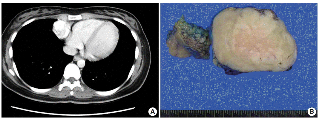
Radiologic and macroscopic findings. (A) Chest computed tomography reveals a well-demarcated enhanced mass in the anterior inferior mediastinum. (B) Mediastinal mass shows a gray-white homogeneous cut surface with no necrotic or hemorrhagic focus.
On gross examination, the specimen was a relatively well-demarcated solid mass with a rubbery consistency, measuring 5.3 ×4.0 cm. The cut surface of the tumor was tan yellow and had a nodular appearance without a necrotic or hemorrhagic area (Fig. 1B).
Microscopically, the tumor was enveloped by a variable, thickened fibrous capsule with no evidence of infiltration into adjacent tissue. A solid growth pattern was evident, with prominent vascular structures composed of small- to medium-sized blood vessels. Medium-sized vessels occasionally appeared with staghorn features and myxoid changes in their walls (Fig. 2A). The tumor displayed both low and high cellular areas. The high cellular areas appeared in vague nodular configuration, and the low cellular area had edematous and myxoid change and was present between high cellular areas (Fig. 2A, B). High-powered examination revealed that the tumor was composed of round epithelioid cells, which had clear to eosinophilic cytoplasm with sharply defined borders. The centrally-located vesicular nuclei were round to polygonal, with a mild convoluted contour of nuclear membranes. One or two small inconspicuous nucleoli were noted. Nuclear pleomorphism was not identified (Fig. 2C, D). Mitotic activity was infrequent, observed in fewer than 1/50 high power fields. Atypical mitosis was not found. Lymphovascular or perineural invasion was not observed. Immunohistochemical staining for smooth muscle actin and vimentin showed diffuse and focal positivity, respectively. Tumor cells were not immunoreactive for CD34, calretinin, cytokeratin, desmin, or neuroendocrine markers, including synaptophysin and chromogranin (Fig. 2E, F). Based on the histology and immunohistochemical results, a pathologic diagnosis of glomus tumor with uncertain malignant potential was made. There was no clinicoradiologic evidence of recurrence or metastasis for seven months after surgery.

Macroscopic finding. (A) The tumor displays a sheet-like growth pattern with increased vascularity, and some medium-sized vessels within the tumor has a staghorn appearance. (B) Edematous and myxoid changes are prominent in hypocellular areas. (C, D) Tumor cells have a well-defined cell border and pale to eosinophilic cytoplasm. The nuclei are round and concentrically located with one or two inconspicuous nucleoli. (E) Immunohistochemical staining for smooth muscle actin is positive in tumor cells. (F) CD34 immunohistochemistry is nonreactive in tumor cells.
DISCUSSION
Glomus tumors are unusual benign smooth muscle neoplasms that comprise fewer than 2% of all soft tissue tumors [10,11]. These tumors are preferentially located in the subungal area of extremities and have a characteristic clinical triad that includes pain, pinpoint tenderness and hypersensitivity to cold temperatures [12]. Clinically, this type of tumor is usually benign, and malignant cases have rarely been reported [13]. Additionally, mediastinal glomus tumors, first described in 1949, are extremely rare [2]; only five cases have been described in the English literature [1-3,8,9]. This is the sixth case of a glomus tumor in the mediastinum.
The pathologic differential diagnoses of mediastinal glomus tumor include carcinoid tumor, hemangiopericytoma, epithelioid leiomyoma, primitive neuroectodermal tumor, and paraganglioma. Characteristic morphologic features that are helpful for distinguishing these tumors include uniform, round tumor cells with centrally-located nuclei, a well-defined cell membrane and immunohistochemial results that are positive for actin and equivocal or negative for CD34, neuroendocrine markers (chromogranin A, synaptophysin, neuron specific enolase) and CD99 [1].
A previous article reported that mediastinal glomus tumors tend to have atypical histologic appearance with cytologic pleomorphism and infiltrate into surrounding tissue [1]. However, clinicopathologic features or distinct classification of glomus tumors in the mediastinum or visceral organs have not been established due to their infrequency. We comprehensively reviewed the clinicopathologic data of mediastinal glomus tumors; this data and the current case are presented in Table 1.
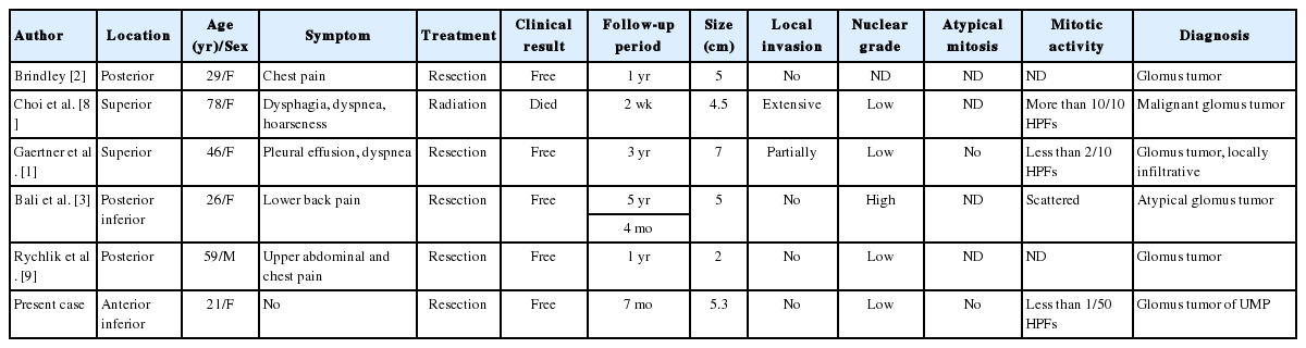
Clinicopathologic data of reported glomus tumor cases of mediastinum
Compared with previous cases, our patient was young and did not present with any symptoms. In addition, although previous mediastinal glomus tumors were frequently found in the posterior superior area, this case was noted in an unusual location, the anterior inferior mediastinum. To investigate the clinicopathologic characteristics in malignant mediastinal glomus tumors, we searched for histologic descriptions in former reports. A previous report indicated that, in a fatal mediastinal glomus tumor case [8], advanced age, extensive local invasion, and brisk mitotic activity were significant and correlated with poor prognosis in mediastinal glomus tumors.
Glomus tumors with atypical features should be evaluated for variable clinicopathologic features, based on recently-defined classifications. These classifications include tumor location and size, nuclear atypia, and mitosis including atypical ones. The tumors can be categorized into four groups (Table 2) [4,7]. Since glomus tumors that arise in visceral organs, including the mediastinum, can be considered deeply-located tumors, they should be classified as malignant glomus tumor or glomus tumor of uncertain malignant potential. However, only one of six such reported patients died, and the remainder had no evidence of recurrence or metastasis during follow-up. Therefore, we collected and analyzed glomus tumor cases in visceral organs to define and outline characteristics for increased diagnostic accuracy, based on prognosis and adequate treatment.

We described an extremely rare case of mediastinal glomus tumor with no subject symptoms and reviewed the clinicopathologic features of previously reported cases. Considering the unpredictable prognosis of mediastinal glomus tumor, we suggest the need for histopathologic assessment parameters through collection and observation of glomus tumors that originate in the visceral organs.
Notes
Conflicts of Interest
No potential conflict of interest relevant to this article was reported.
Acknowledgements
This work was supported in part by the Soonchunhyang University Research Fund. We appreciate the expert counsel of Jae Ro, MD, PhD, Houston Methodist Hospital.