E-submission
Articles
- Page Path
- HOME > J Pathol Transl Med > Volume 46(2); 2012 > Article
-
Original Article
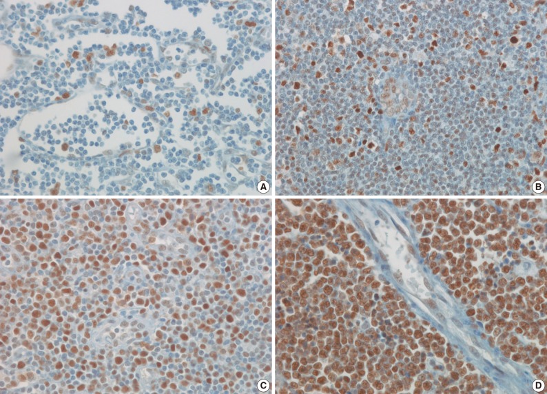
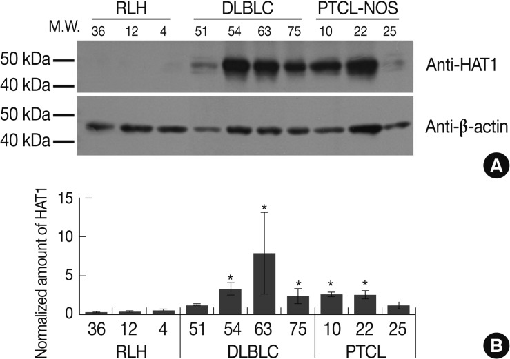
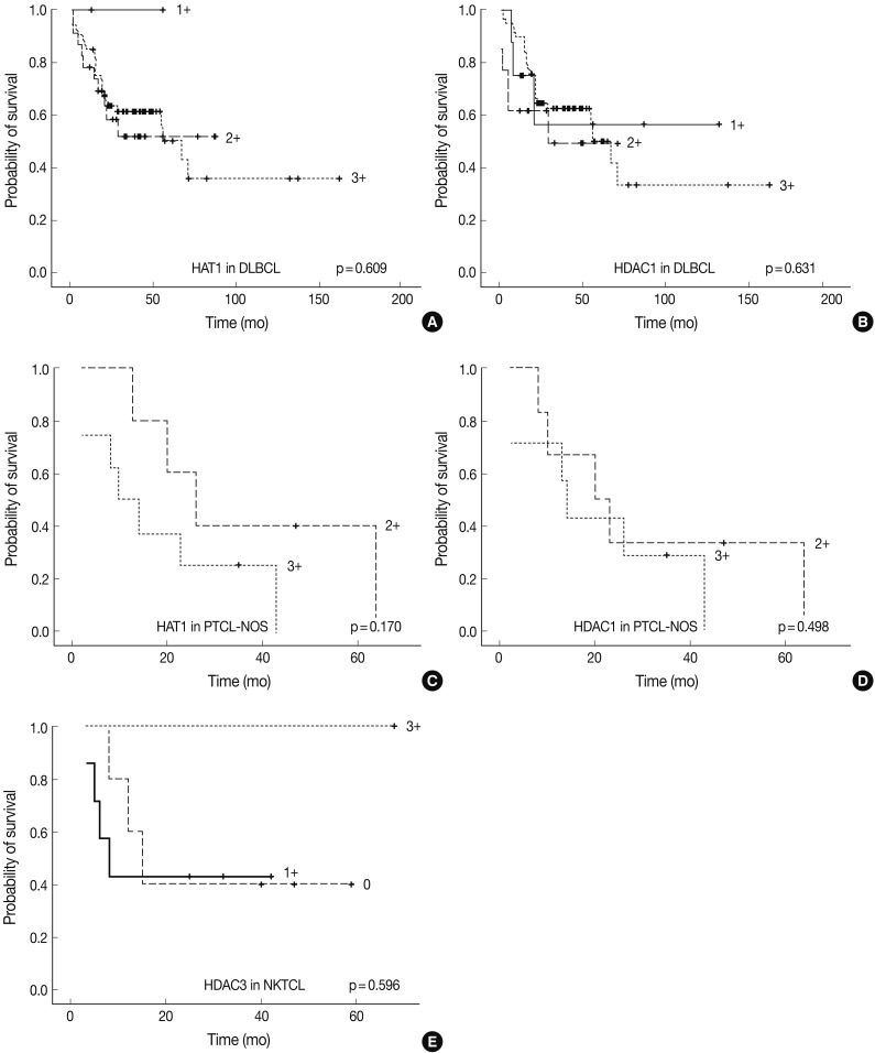
Expression of HAT1 and HDAC1, 2, 3 in Diffuse Large B-Cell Lymphomas, Peripheral T-Cell Lymphomas, and NK/T-Cell Lymphomas - Soo Kee Min, Young Ho Koh1, Yunwoong Park1, Hyo Jung Kim2, Jinwon Seo, Hye-Rim Park, Seong Jin Cho3, In Sun Kim4
-
Korean Journal of Pathology 2012;46(2):142-150.
DOI: https://doi.org/10.4132/KoreanJPathol.2012.46.2.142
Published online: April 25, 2012
Department of Pathology, Hallym University Sacred Heart Hospital, Hallym University College of Medicine, Anyang, Korea.
1Ilsong Institute of Life Science, Hallym University, Anyang, Korea.
2Department of Hemato-Oncology, Hallym University Sacred Heart Hospital, Hallym University College of Medicine, Anyang, Korea.
3Department of Pathology, Hallym University Kangdong Sacred Heart Hospital, Hallym University College of Medicine, Seoul, Korea.
4Department of Pathology, Korea University College of Medicine, Seoul, Korea.
- Corresponding Author: Seong Jin Cho, M.D. Department of Pathology, Hallym University Kangdong Sacred Heart Hospital, 150 Seongan-ro, Gangdong-gu, Seoul 134-701, Korea. Tel: +82-2-2224-2557, Fax: +82-2-2224-2214, 'apilas@hanmail.net'
© 2012 The Korean Society of Pathologists/The Korean Society for Cytopathology
This is an Open Access article distributed under the terms of the Creative Commons Attribution Non-Commercial License (http://creativecommons.org/licenses/by-nc/3.0) which permits unrestricted non-commercial use, distribution, and reproduction in any medium, provided the original work is properly cited.
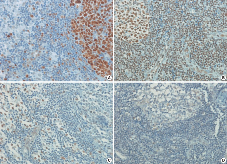

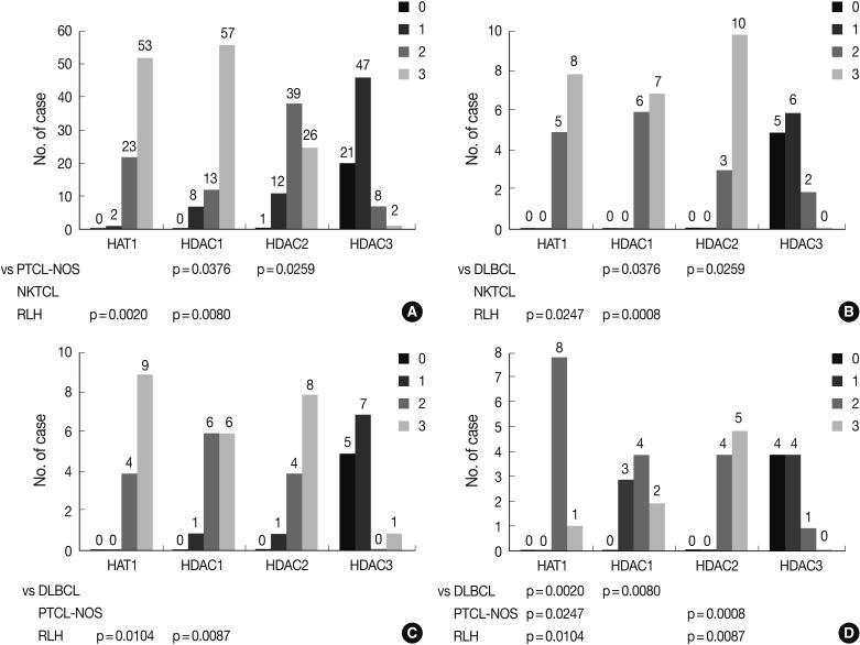


Figure & Data
References
Citations
- Chidamide and orelabrutinib synergistically induce cell cycle arrest and apoptosis in diffuse large B-cell lymphoma by regulating the PI3K/AKT/mTOR pathway
Chunyan Wu, Shilv Chen, Zhimin Wu, Jiao Xue, Wen Zhang, Shan Wang, Xindong Zhao, Shaoling Wu
Journal of Cancer Research and Clinical Oncology.2024;[Epub] CrossRef - Targeting HDACs for diffuse large B-cell lymphoma therapy
Chunyan Wu, Qiao Song, Sophie Gao, Shaoling Wu
Scientific Reports.2024;[Epub] CrossRef - Epigenetic regulation in hematopoiesis and its implications in the targeted therapy of hematologic malignancies
Ailin Zhao, Hui Zhou, Jinrong Yang, Meng Li, Ting Niu
Signal Transduction and Targeted Therapy.2023;[Epub] CrossRef - Understanding HAT1: A Comprehensive Review of Noncanonical Roles and Connection with Disease
Miguel A. Ortega, Diego De Leon-Oliva, Cielo Garcia-Montero, Oscar Fraile-Martinez, Diego Liviu Boaru, María del Val Toledo Lobo, Ignacio García-Tuñón, Mar Royuela, Natalio García-Honduvilla, Julia Bujan, Luis G. Guijarro, Melchor Alvarez-Mon, Miguel Ánge
Genes.2023; 14(4): 915. CrossRef - HAT1: Landscape of Biological Function and Role in Cancer
Vincenza Capone, Laura Della Torre, Daniela Carannante, Mehrad Babaei, Lucia Altucci, Rosaria Benedetti, Vincenzo Carafa
Cells.2023; 12(7): 1075. CrossRef - Recent advancement of HDAC inhibitors against breast cancer
Syed Abdulla Mehmood, Kantrol Kumar Sahu, Sounok Sengupta, Sangh Partap, Rajshekhar Karpoormath, Brajesh Kumar, Deepak Kumar
Medical Oncology.2023;[Epub] CrossRef - Noncoding rules of survival: epigenetic regulation of normal and malignant hematopoiesis
LaShanale Wallace, Esther A. Obeng
Frontiers in Molecular Biosciences.2023;[Epub] CrossRef - Potential Therapeutic Use of Aptamers against HAT1 in Lung Cancer
José Ignacio Klett-Mingo, Celia Pinto-Díez, Julio Cambronero-Plaza, Rebeca Carrión-Marchante, Miriam Barragán-Usero, María Isabel Pérez-Morgado, Eulalia Rodríguez-Martín, Mª Val Toledo-Lobo, Víctor M González, M. Elena Martín
Cancers.2022; 15(1): 227. CrossRef - Modulation of serine/threonine-protein phosphatase 1 (PP1) complexes: A promising approach in cancer treatment
Bárbara Matos, John Howl, Carmen Jerónimo, Margarida Fardilha
Drug Discovery Today.2021; 26(11): 2680. CrossRef - Histone acetyltransferase 1 promotes gemcitabine resistance by regulating the PVT1/EZH2 complex in pancreatic cancer
Yan Sun, Dianyun Ren, Yingke Zhou, Jian Shen, Heshui Wu, Xin Jin
Cell Death & Disease.2021;[Epub] CrossRef - Deciphering genes associated with diffuse large B-cell lymphoma with lymphomatous effusions: A mutational accumulation scoring approach
Sina Abdollahi, Seyedeh Zahra Dehghanian, Liang-Yi Hung, Shiang-Jie Yang, Dao-Peng Chen, L. Jeffrey Medeiros, Jung-Hsien Chiang, Kung-Chao Chang
Biomarker Research.2021;[Epub] CrossRef - The contributory roles of histone deacetylases (HDACs) in hematopoiesis regulation and possibilities for pharmacologic interventions in hematologic malignancies
Mahdieh Mehrpouri, Atieh Pourbagheri-Sigaroodi, Davood Bashash
International Immunopharmacology.2021; 100: 108114. CrossRef - Emerging role of histone deacetylase inhibitors in the treatment of diffuse large B-cell lymphoma
Mingyang Wang, Xiaosheng Fang, Xin Wang
Leukemia & Lymphoma.2020; 61(4): 763. CrossRef Effective Treatment with PD-1 Antibody, Chidamide, Etoposide, and Thalidomide (PCET) for Relapsed/Refractory Natural Killer/T-Cell Lymphoma: A Report of Three Cases
Lijun Du, Lei Zhang, Ling Li, Xin Li, Jiaqin Yan, Xinhua Wang, Xiaorui Fu, Zhenchang Sun, Xudong Zhang, Zhaoming Li, Jingjing Wu, Hui Yu, Yu Chang, Zhiyuan Zhou, Feifei Nan, Xiaolong Wu, Li Tian, Mingzhi Zhang
OncoTargets and Therapy.2020; Volume 13: 7189. CrossRef- Overexpressed histone acetyltransferase 1 regulates cancer immunity by increasing programmed death-ligand 1 expression in pancreatic cancer
Ping Fan, Jingyuan Zhao, Zibo Meng, Heyu Wu, Bo Wang, Heshui Wu, Xin Jin
Journal of Experimental & Clinical Cancer Research.2019;[Epub] CrossRef - Histone modifications: A review about the presence of this epigenetic phenomenon in carcinogenesis
Emanuely Silva Chrun, Filipe Modolo, Filipe Ivan Daniel
Pathology - Research and Practice.2017; 213(11): 1329. CrossRef - Histone Acetyltransferase 1 Promotes Cell Proliferation and Induces Cisplatin Resistance in Hepatocellular Carcinoma
Xin Jin, Shenghua Tian, Pingping Li
Oncology Research Featuring Preclinical and Clinical Cancer Therapeutics.2017; 25(6): 939. CrossRef - HDACs and HDAC Inhibitors in Cancer Development and Therapy
Yixuan Li, Edward Seto
Cold Spring Harbor Perspectives in Medicine.2016; 6(10): a026831. CrossRef - Histone deacetylase inhibitors and epigenetic regulation in lymphoid malignancies
Diana Markozashvili, Vincent Ribrag, Yegor S. Vassetzky
Investigational New Drugs.2015; 33(6): 1280. CrossRef - Genome-Wide Association Study of Event-Free Survival in Diffuse Large B-Cell Lymphoma Treated With Immunochemotherapy
Hervé Ghesquieres, Susan L. Slager, Fabrice Jardin, Amelie S. Veron, Yan W. Asmann, Matthew J. Maurer, Thierry Fest, Thomas M. Habermann, Marie C. Bene, Anne J. Novak, Sylvain Mareschal, Corinne Haioun, Thierry Lamy, Stephen M. Ansell, Herve Tilly, Thomas
Journal of Clinical Oncology.2015; 33(33): 3930. CrossRef - Histone deacetylase 2 controls p53 and is a critical factor in tumorigenesis
Tobias Wagner, Peter Brand, Thorsten Heinzel, Oliver H. Krämer
Biochimica et Biophysica Acta (BBA) - Reviews on Cancer.2014; 1846(2): 524. CrossRef - Targetome profiling and functional genetics implicate miR-618 in lymphomagenesis
Alan Fu, Aaron E Hoffman, Ran Liu, Daniel I Jacobs, Tongzhang Zheng, Yong Zhu
Epigenetics.2014; 9(5): 730. CrossRef - Expression of Histone Deacetylases HDAC1, HDAC2, HDAC3, and HDAC6 in Invasive Ductal Carcinomas of the Breast
Jinwon Seo, Soo Kee Min, Hye-Rim Park, Dong Hoon Kim, Mi Jung Kwon, Lee Su Kim, Young-Su Ju
Journal of Breast Cancer.2014; 17(4): 323. CrossRef - Diffuse large B-cell lymphoma
Maurizio Martelli, Andrés J.M. Ferreri, Claudio Agostinelli, Alice Di Rocco, Michael Pfreundschuh, Stefano A. Pileri
Critical Reviews in Oncology/Hematology.2013; 87(2): 146. CrossRef - Histone deacetylase inhibitors activate CIITA and MHC class II antigen expression in diffuse large B‐cell lymphoma
Kelly A. Cycon, Kathleen Mulvaney, Lisa M. Rimsza, Daniel Persky, Shawn P. Murphy
Immunology.2013; 140(2): 259. CrossRef
 PubReader
 Cite this Article