E-submission
Articles
- Page Path
- HOME > J Pathol Transl Med > Volume 47(6); 2013 > Article
-
Original Article
A Different Perspective on Macroscopic Sampling of Cholecystectomy Specimens - Asuman Argon, Ayşe Yağcı, Funda Taşlı, Tulu Kebat, Senem Deniz, Nazif Erkan1, Gül Kitapçıoğlu2, Enver Vardar
-
Korean Journal of Pathology 2013;47(6):519-525.
DOI: https://doi.org/10.4132/KoreanJPathol.2013.47.6.519
Published online: December 24, 2013
Department of Pathology, Izmir Bozyaka Training and Research Hospital, Izmir, Turkey.
1Department of General Surgery, Izmir Bozyaka Training and Research Hospital, Izmir, Turkey.
2Department of Biostatistics and Medical Communication, Ege University Faculty of Medicine, Izmir, Turkey.
- Corresponding Author: Asuman Argon, M.D. Department of Pathology, Izmir Bozyaka Training and Research Hospital, Izmir 35100, Turkey. Tel: +90-2322505050, Fax: +90-2322502997, 'asumanargon@gmail.com'
• Received: September 3, 2013 • Revised: October 26, 2013 • Accepted: October 28, 2013
© 2013 The Korean Society of Pathologists/The Korean Society for Cytopathology
This is an Open Access article distributed under the terms of the Creative Commons Attribution Non-Commercial License (http://creativecommons.org/licenses/by-nc/3.0/) which permits unrestricted non-commercial use, distribution, and reproduction in any medium, provided the original work is properly cited.


Figure & Data
References
Citations
Citations to this article as recorded by 
- Ultrasonographic features of gallbladder wall thickening in dogs with hypoalbuminemia
Masahiro Murakami, Hock Gan Heng, Sarah Steinbach, Mario Sola
Veterinary Quarterly.2023; 43(1): 1. CrossRef - Can the sampling method affect the detection of incidental gallbladder carcinoma? Comparative analysis of two sampling methods
Ezgi Hacihasanoglu, Esra Pasaoglu, Merve Cin, Enver Yarikkaya, Nevra Dursun, Sevim Baykal Koca
Annals of Diagnostic Pathology.2023; 67: 152187. CrossRef - Current management of incidental gallbladder cancer: A review
Claudio F. Feo, Giorgio C. Ginesu, Alessandro Fancellu, Teresa Perra, Chiara Ninniri, Giulia Deiana, Antonio M. Scanu, Alberto Porcu
International Journal of Surgery.2022; 98: 106234. CrossRef - Accuracy of Right Upper Quadrant Ultrasound in Estimating Gallbladder Wall Thickness
Lindsay Cefalu, Robert McMurray, Grant Sizemore, Gerald Bieniek, Michael Lustik, Christopher Yheulon
Surgical Laparoscopy, Endoscopy & Percutaneous Techniques.2019; 29(1): 26. CrossRef - Optimal block sampling of routine, non‐tumorous gallbladders
Newton A C S Wong
Histopathology.2017; 71(1): 162. CrossRef - The Relationship Between Intracholecystic Papillary-Tubular Neoplasms and Invasive Carcinoma of the Gallbladder
Asuman Argon, Funda Yılmaz Barbet, Deniz Nart
International Journal of Surgical Pathology.2016; 24(6): 504. CrossRef
 PubReader
 ePub Link
-
 Cite this Article
- Cite this Article
-
- Close
- Download Citation
- Close
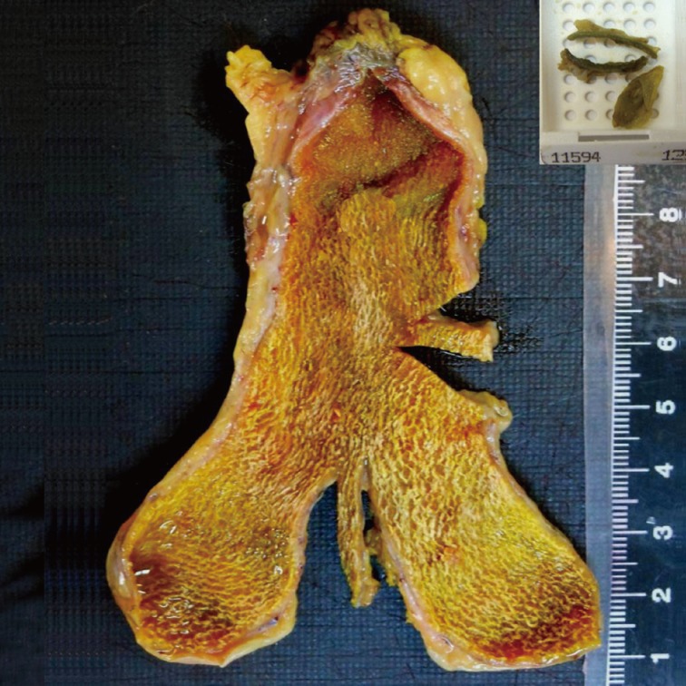
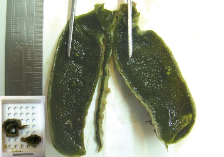
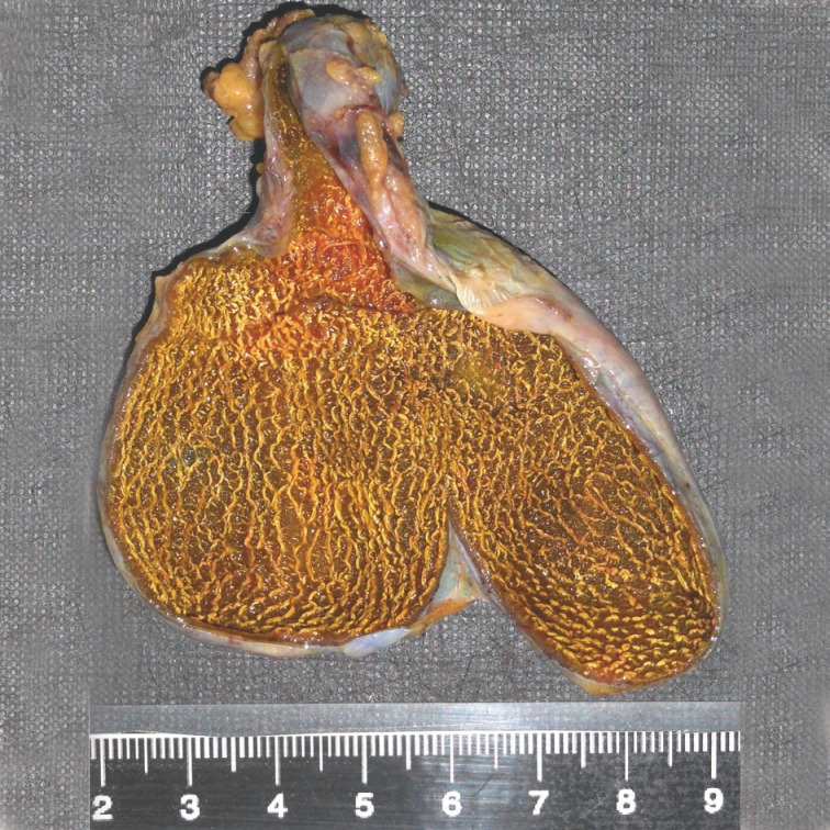
- Figure
-