Nuclear Expression of Hepatitis B Virus X Protein Is Associated with Recurrence of Early-Stage Hepatocellular Carcinomas: Role of Viral Protein in Tumor Recurrence
Article information
Abstract
Background:
Hepatitis B virus (HBV) plays well-known roles in tumorigenesis of hepatocellular carcinoma (HCC) in infected patients. However, HBV-associated protein status in tumor tissues and the relevance to tumor behavior has not been reported. Our study aimed to examine the expression of HBV-associated proteins in HCC and adjacent nontumorous tissue and their clinicopathologic implication in HCC patients.
Methods:
HBV surface antigen (HBsAg), HBV core antigen (HBcAg), and HBV X protein (HBx) were assessed in 328 HBV-associated HCCs and in 155 matched nontumorous tissues by immunohistochemistry staining.
Results:
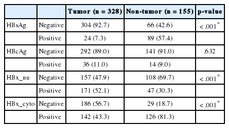
The positive rates of HBsAg and cytoplasmic HBx staining in tumor tissue were lower than those in nontumorous tissue (7.3% vs. 57.4%, p < .001; 43.4% vs. 81.3%, p < .001). Conversely, nuclear HBx was detected more frequently in tumors than in nontumorous tissue (52.1% vs. 30.3%, p < .001). HCCs expressing HBsAg, HBcAg, or cytoplasmic HBx had smaller size; lower Edmondson-Steiner (ES) nuclear grade, pT stage, and serum alpha-fetoprotein, and less angioinvasion than HCCs not expressing HBV-associated proteins. Exceptionally, nuclear HBx-positive HCCs showed higher ES nuclear grade and more frequent large-vessel invasion than did nuclear HBx-negative HCCs. In survival analysis, only nuclear HBx-positive HCCs had shorter disease-free survival than nuclear HBx-negative HCCs in pT1 and ES nuclear grade 1–2 HCC subgroup (median, 126 months vs. 35 months; p = .015).
Conclusions:
Our data confirmed that expression of normal HBV-associated proteins generally decreases in tumor cells in comparison to nontumorous hepatocytes, with the exception of nuclear HBx, which suggests that nuclear HBx plays a role in recurrence of well-differentiated and early-stage HCCs.
Hepatocellular carcinoma (HCC) is the third most fatal cancer worldwide and poses a major burden to the healthcare system [1]. More than 50% of HCC cases overall and 70%–80% of HCC cases in hepatitic B virus (HBV)–endemic regions are attributable to chronic HBV infection [2]. The mechanism of viral hepatitis-mediated induction of HCC involves the direct mutagenic effect of the virus on the host genome and the indirect effect of the inflammation-necrosis regeneration cycle in the setting of chronic hepatitis [3].
HBV is a member of the Hepadnaviridae family and has four important viral proteins: hepatitis B virus surface antigen (HBsAg), hepatitis B virus core antigen (HBcAg), hepatitis B virus X protein (HBx), and viral DNA polymerase. HBsAg and HBcAg are structural proteins that are the main components of the viral capsule and core. These proteins induce an immune response in infected hosts and can be used for the assessment of viral replication activity. Thus, they serve as clinical markers for diagnosis and follow-up of viral hepatitis patients via serum tests [4]. HBx is a key regulatory nonstructural protein of the virus that is at the intersection of HBV infection, replication, pathogenesis, and carcinogenesis [5].
Integration of viral genomes in the host genome is considered a possible mechanism of hepatocarcinogenesis, and this notion is supported by the observation that a large portion of HCC have integrated HBV sequence encoding HBx and a truncated pre-S2/S protein [6]. HBx protein regulates the cell cycle and DNA repair genes of host cells and induces cellular transformation via transactivation (protein interactions) in the nucleus and cytoplasm [5].
Clinically, a high viral load of HBV is associated with HCC recurrence, and early antiviral treatment can increase disease-free survival (DFS) and overall survival of HCC patients [7,8]. A specific mutated form of HBV known as genotype C was reported to be associated with HCC occurrence in cirrhotic patients [9]. HBsAg positivity in non-neoplastic liver tissue was reported as a risk factor for HCC recurrence [10].
HBsAg expression is generally lower in tumor cells compared with adjacent non-neoplastic hepatocytes, and the HBx protein expression in non-neoplastic hepatic parenchyma is associated with the development of HCC in patients with chronic viral hepatitis [11,12]. Previous studies of the expression of HBV genes and HBx protein in HCC and adjacent non-neoplastic hepatic parenchyma generally focused on the mechanisms underlying the HBV-related hepatocarcinogenesis.
The aim of this study was to assess the expression of the HBsAg, HBcAg, and HBx proteins in HCC and nontumorous liver tissue and to examine the histologic features of HCCs, possible correlations with hepatitis serum markers, and their influence on cancer prognosis.
MATERIALS AND METHODS
Patients and clinicopathologic parameters
We enrolled 328 HBV hepatitis patients who had been diagnosed with HCC based on a resected specimen and whose medical records and formalin-fixed paraffin blocks of tumor tissue were available from the archives of the Department of Pathology at Seoul National University Hospital (SNUH) from 1998 to 2004. We excluded co-infected hepatitis C virus hepatitis patients and patients who had neither serologic nor clinical evidence of HBV infection. The matched non-neoplastic hepatic parenchyma was available for 155 of the 328 patients. Clinical information, such as age, sex, surgical procedure, underlying etiology of liver disease, preoperative serum α-fetoprotein (AFP, μg/mL), preoperative treatment, and postoperative tumor recurrence, was collected from the medical records. Serological results of HBsAg, anti-HBs (HBsAb), IgG anti-HBc (HBcAb), hepatitis B virus e antigen (HBeAg), and anti-HBe (HBeAb) were based on the most recent preoperative tests from medical records. Depending on the state of the serum viral marker, liver function test, and clinical symptom of hepatic failure or portal hypertension (e.g., hypoalbuminemia, prolonged prothrombin time, ascites, hyperbilirubinemia, or hepatic encephalopathy), “asymptomatic carriers” had positive serum viral markers, but a normal liver function test and no clinical symptom of hepatic failure, while “noncirrhotic” patients had positive serum viral marker with abnormal liver function test, but no symptom of hepatic failure. Patients with symptom of hepatic failure were assigned to the cirrhotic patient group. Disease recurrence was defined as newly appearing lesions diagnosed by radiologic examinations such as ultrasonography and X-ray computed tomography, or based on serum tumor markers such as AFP, after an operation. Pathologic information, such as tumor size, number of tumors, gross type (vaguely nodular, expanding nodular, nodular with perinodal extension, and multinodular confluent), angioinvasion, large vessel invasion, Edmondson-Steiner (ES) nuclear grade, histologic pattern of the tumor, cellular type of tumor cells (hepatic vs. non-hepatic including giant, pleomorphic, spindle, and clear cell types), and extent of tumor invasion, was collected from pathology reports and review of the slides. Criteria for pathologic T stage (pT) followed the liver tumor staging of the American Joint Committee on Cancer, seventh edition [13]. Clinicopathologic parameters were assessed according to the general rules for examining primary liver cancer [14].
Of the 328 patients, 283 were male and 45 were female (M:F ratio of 6.3:1), with a median age of 55 years (range, 25 to 80 years). The mean size of a tumor was 5.44 cm (range, 0.8 to 24.0 cm). Most patients (92.1%, 302/328) had undergone partial hepatectomy, such as right or left lobectomy, caudate lobectomy, or segmentectomy, whereas the remaining patients (7.9%, 26/328) had undergone total hepatectomy for transplantation. Follow-up periods ranged from 0 to 161 months (median, 51 months). This study was approved by the Institutional Review Board of Seoul National University Hospital (H-1011-046-339).
Tissue microarray construction
Hematoxylin and eosin slides were reviewed, and one representative formalin-fixed paraffin-embedded (FFPE) archival block was selected for each case. Each core tissue biopsy (2 mm in diameter) was taken from individual FFPE blocks (donor blocks) and arranged in recipient paraffin blocks (tissue array blocks) using a trephine. Immunohistochemical studies were performed on 13 array blocks containing 328 HCC samples and on six array blocks containing 155 samples of HCC-adjacent non-neoplastic tissue as a healthy control (Superbiochips Laboratories, Seoul, Korea). Each tissue microarray had four cores of normal liver, normal bile duct, and normal gastrointestinal tract mucosa as a negative control.
Immunohistochemistry and interpretation
Sections (4 μm) were stained for HBcAg (1:800, hepatitis B core antigen rabbit polyclonal antibody, Cat. No. B0586, DAKO, Copenhagen, Denmark), HBsAg (1:200, HBsAg mouse monoclonal antibody, clone number S1-210, Cell Marque, Rocklin, CA, USA), and HBx (1:100, HBx mouse monoclonal antibody, clone number 3F6-G10, Thermo, Rockford, IL, USA) after antigen retrieval using a microwave and pH 6.0 citrate buffer. The slides were stained according to methods specified in the Ultravision LP kit (Lab Vision, Fremont, CA, USA) or the Envision kit (Dako, Glostrup, Denmark) using the Bond polymer Refine Detection kit (Leica, Wetzlar, Germany). Unequivocal cytoplasmic staining for HBsAg in more than 5% of tumor cells was considered positive, and unequivocal nuclear and cytoplasmic staining for HBcAg in more than 5% of tumor cells was considered positive as described in a previous study [4]. For HBx, both cytoplasmic and nuclear staining were observed in tumorous and nontumorous liver tissue; thus, we separately assessed the cytoplasmic and nuclear expression of HBx. The intensity of cytoplasmic staining was graded as weak, moderate, or strong by two pathologist (H.Y.Jung and K.-B.Lee), and a grade higher than moderate was considered as positive criteria. Nuclear staining was assessed by image analysis of digitally scanned slides (Nuclear V9 algorithm, ScanScope, Aperio Technologies, Vista, CA, USA). The intensity of nuclear staining was graded as 1+ (intensity range, 230 to 210), 2+ (intensity range, 210 to 188), or 3+ (intensity range, 188 to 162), and more than 5% of tumor cells with more than 2+ intensity were considered as positive criteria.
Statistical analysis
Comparative analysis of HBsAg, HBcAg, and HBx expression with clinicopathologic parameters was assessed using the chisqaure test. Survival analysis was performed using the Kaplan-Meier method. The results were considered statistically significant when p-values were <.05. All calculations were performed using the PASW statistics ver. 18.0 (SPSS Inc., Chicago, IL, USA).
RESULTS
Positive rates of HBsAg, HBcAg, and HBx expression in HCC
The representative immunohistochemical stainings and expression rates of HBsAg, HBcAg, and HBx in the nucleus and cytoplasm are summarized in Table 1 and Fig. 1, respectively. Among the 328 patients with HBV, the positive rate for HBsAg was significantly lower in HCC than in nontumorous tissue (7.3% vs 57.4%, respectively; p<.001), and the positive rate for nuclear HBx was higher in HCC than in nontumorous tissue (52.1% [171/328] vs 30.3% [47/155], respectively; p<.001). Cytoplasmic HBx expression was less frequent in HCC samples than in nontumorous tissue (43.3% [142/328] vs 81.3% [126/155], respectively; p<.001).

The percentage of patients positive for HBV proteins in the hepatocellular carcinoma and non-tumorous liver
Clinicopathologic characteristics of HBsAg-, HBcAg-, and HBx-expressing HCC
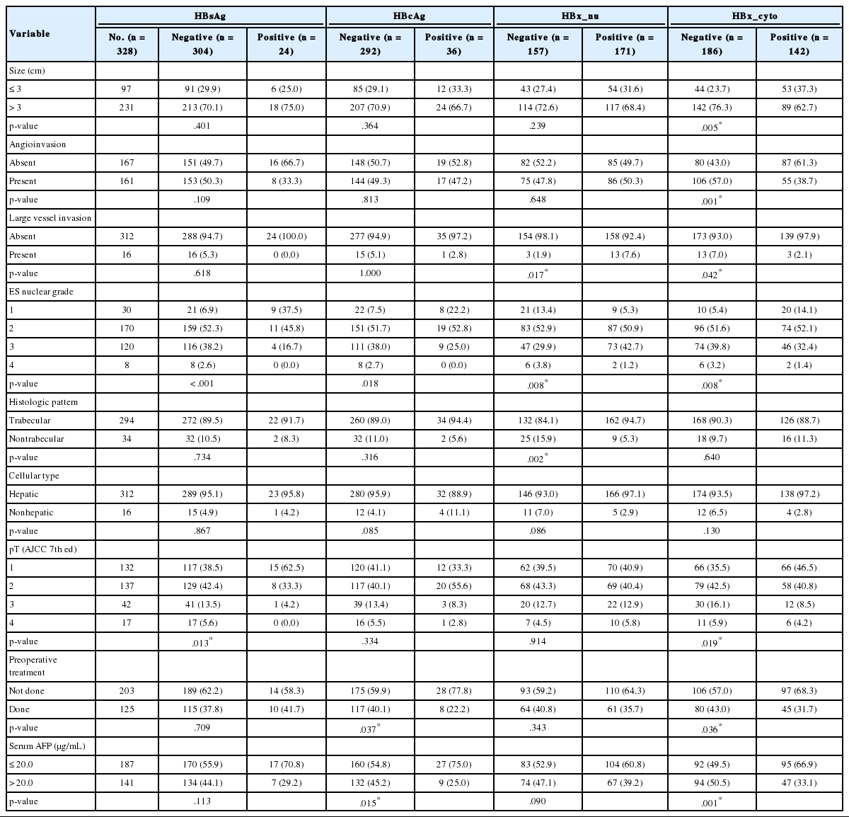
The clinicopathologic features and expression of HBsAg, HBcAg, and HBx are summarized in Table 2. HBsAg-expressing HCCs had lower ES nuclear grade and lower pT stage than HCCs without HBsAg expression (p<.001 and p=.013, respectively). HBcAg was more frequently expressed in well-differentiated HCCs (lower ES grade) and in HCCs without preoperative treatment, such as transarterial chemoembolization, radiofrequency ablation, or percutaneous ethanol injection, as well as in patients who had lower serum AFP (p=.018, p=.037, and p=.015, respectively). Cytoplasmic expression of HBx was associated with similar features: lower ES nuclear grade, smaller tumor size, less frequent angioinvasion, lower pT stage, and a lower serum AFP level compared to HCCs not expressing cytoplasmic HBx (p<.05) (Table 2). Compared to HBsAg and HBcAg, nuclear HBx was more frequently expressed in HCCs with higher ES grade and with large-vessel invasion (p=.008, and p=.017, respectively). Although nuclear HBx was more frequently expressed in HCCs with high ES nuclear grade, the histologic pattern of HCCs with nuclear HBx expression was mostly trabecular and retained the hepatic cell type (p=.002 and p=.086, respectively) (Table 2). Age and sex were not different between HBV protein positive groups and negative groups (data not shown).
Correlation between serum hepatitis B markers and expression of HBV proteins in HCC
We set out to test whether the serum level of hepatitis B markers or the expression profile of nontumorous liver tissue showed any correlation with the expression of HBV proteins in HCCs. All patients with HBsAg-positive HCCs were HBsAg positive in serum. Patients with positive serum HBeAb results showed lower positive rates of HBsAg in HCCs than did patients with negative serum HBeAb (10.8% [7/65] vs 1.8% [2/108], p=.027). Expression of HBcAg and expression of nuclear HBx in HCCs were not significantly concordant with the serum status of HBsAg, HBsAb, HBcAb, HBeAg, and HBeAb. As for the clinical stage of hepatitis, nuclear HBx was more frequently expressed in HCCs of noncirrhotic chronic hepatitis patients compared to asymptomatic carriers or cirrhotic patients (noncirrhotic vs asymptomatic vs cirrhosis; 75% [15/20] vs 35% [13/37] vs 43.9% [54/123], p=.023).
Progression of HBsAg-, HBcAg-, and HBx-expressing HCC
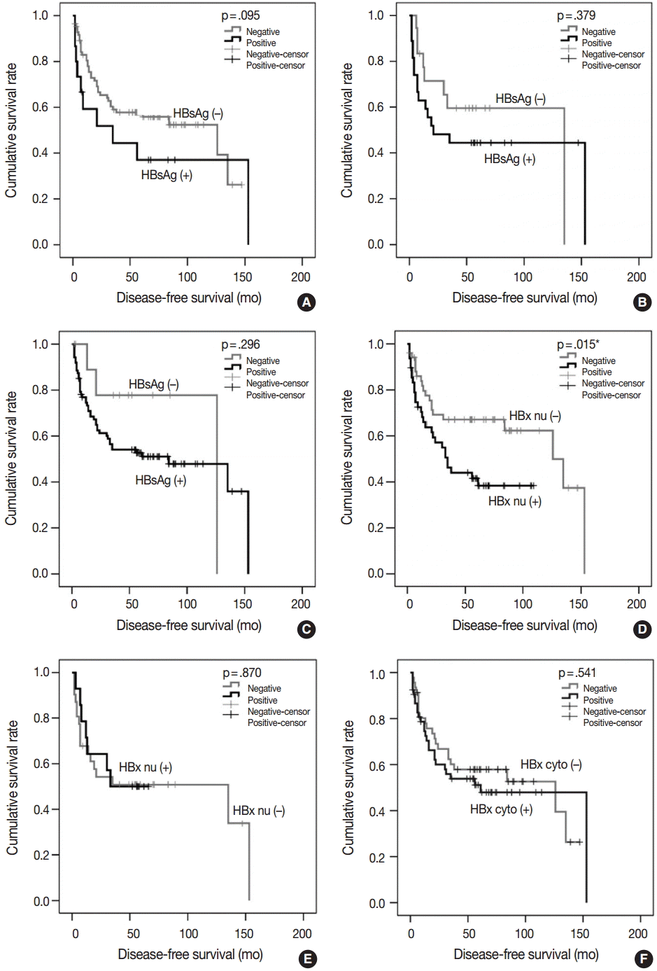
To test whether the HBsAg, HBcAg, or HBx status of tumors influences cancer progression, we analyzed DFS time after operation in the 328 patients with HBV-associated HCC using Kaplan-Meier analysis. We could not determine any correlation between DFS time and the expression status of HBsAg, HBcAg, and HBx in tumors and nontumorous tissue or the serum levels of hepatitis markers (data not shown). The size and multiplicity of the tumor, angioinvasion, ES nuclear grade, and the extent of tumor invasion were statistically significant prognostic factors for tumor recurrence in the entire sample of 328 patients (data not shown). The low frequency of positive HBsAg in HCCs and well-differentiated histology of HBsAg-expressing HCCs may be a confounder of the above-mentioned negative results in the entire group, so we stratified the study group and analyzed progression-free survival in 99 patients with early-stage (pT1) and well-differentiated HCC (ES nuclear grade 1 and 2); these patients were a relatively low-risk group for HCC recurrence. In 48.5% (48/99) of the patients, the tumor recurred and median DFS time was 84 months, ranging from 2 to 153 months. As shown in Table 3, nuclear HBx in tumors was a statistically significant prognostic factor (p=.015), whereas the presence of HBsAg in a tumor showed a tendency for poor prognosis of early-stage HCCs, but did not reach statistical significance (p=.095). Cytoplasmic expression of HBx in tumor and nontumorous tissue was not associated with tumor recurrence. In nontumorous liver tissue, HBsAg-, HBcAg-, and HBx-positive patients had a relatively lower median DFS, but this effect was statistically insignificant (log-rank p>.05) (Table 3). The presence of HBsAg in the tumor, serum, and nontumorous tissue was correlated with shorter DFS and early recurrence compared to HBsAg-negative patients, as shown in Fig. 2, but this effect was not statistically significant (Table 3). Survival analysis for the high-risk group (advanced stages, pT2–4) or higher nuclear grade (ES nuclear grade 3 and 4) showed no significant difference in DFS according to the status of HBV-associated proteins in serum, tumor, and nontumor tissues (data not shown).

Disease-free survival analysis of 99 pT1 and well-differentiated HBV-related HCCs in relation to the expression of HBV proteins

Cumulative disease-free survival curves of 99 patients with pT1 and well-differentiated hepatocellular carcinoma in relation to the presence of hepatitis B virus (HBV) surface antigen (HBsAg) and HBV X protein (HBx) in a tumor, nontumorous tissue, and serum. HBsAg in tumors (A), HBsAg in nontumorous tissue (B), HBsAg in serum (C), nuclear HBx (HBx nu) in tumors (D), HBX nu in nontumorous tissues (E), and cytoplasmic HBx (HBx cyto) (F) in tumors. *p<.05.
DISCUSSION
In this study, we found that the expression of HBsAg, representing normal viral replication in infected cells, is less frequent in tumors than nontumorous hepatocytes, and HBx, which is one of the key proteins in hepatocarcinogenesis, could be preferentially expressed in different subcellular compartments. The nuclear expression of HBx occurs more frequently in tumors than in nontumorous hepatocytes, whereas the cytoplasmic expression of HBx is less frequent in tumors, according to our results. HCCs with pronounced expression of HBsAg have well-differentiated histology, but HCCs with nuclear expression of HBx showed increased nuclear atypia and aggressive behavior. When the test group is restricted to well-differentiated and to early-stage tumors to minimize the influence of tumor-related prognostic factors, then HCCs expressing nuclear HBx proteins show a higher risk of early recurrence after operation.
Persistence of HBx is important for the pathogenesis of early HCC development, and HBx expression in the liver during chronic HBV infection may be an important prognostic marker for the development of HCC [5,12]. Even seronegative HBV patients have HBx gene and protein expression in HCC, which are consistent with the hepatocarcinogenic properties of HBx [15]. The subcellular location of HBx, as assessed by immunohistochemistry in previous studies, is generally in the cytoplasm of hepatocytes or tumor cells and is consistent with the biological functions of HBx, i.e., protein-protein interactions in cytoplasmic signaling pathways [5]. The biological role of HBx in the nucleus may involve direct interaction with DNA, RNA, or transcription factors, but the existing data are scarce. Recently, it was shown in an in vitro study using a human cell line that when HBx is targeted to the nucleus by a nuclear localization signal, it can restore HBx-deficient HBV replication, whereas HBx containing a nuclear export signal cannot, suggesting that nuclear localization of HBx is required for viral replication [16]. In addition, chip-based chromatin immunoprecipitation with expression microarray profiling for HCCs identified 184 gene targets that might be directly deregulated by HBx via targeting from indirect protein-DNA binding as well as transcriptional factors directly interacting with HBx [17].
Our results show that nuclear, not cytoplasmic, HBx expression correlates with aggressiveness of HCC tumors, such as nuclear grade or large vessel invasion, and with early recurrence after an operation. These data suggest that the interaction of HBx with nuclear proteins might be more important for tumor progression than for cytoplasmic interactions of HBx. Although the HBx protein may be involved in metastasis and tumor invasiveness by regulating proteins that control the extracellular matrix, angiogenesis, or epithelial mesenchymal transition, the reason for the paucity of clinical evidence regarding the HBx protein and HCC recurrence or prognosis could be the difficulties with interpretation of HBx immunohistochemistry [18-21]. In the majority of the studies on HBx, cytoplasmic staining was considered positive expression, and nuclear staining was not investigated [15,18,19,22]. Because we minimized the influence of well-known prognostic factors, such as vascular invasion, size, multiplicity, extent of tumor invasion, and differentiation, by selecting HCCs of pT1 stage and ES nuclear grade 1 and 2, we expected that HBx in nontumorous tissue would be a prognostic factor, but the results showed that it was not. We tried to find a link between the pattern of recurrence and the HBx expression, but the recurrence pattern (presented as a single intrahepatic mass versus multiple or disseminated intrahepatic or extrahepatic masses) was not different depending on the HBx expression in tumor and non-tumor tissues. However, nuclear HBx (–) HCCs both in tumor and nontumor were more frequently presented as a single intrahepatic mass at recurrence time after resection (rate of a single intrahepatic mass, HBx_nu[+] in tumor and non-tumor vs HBx_nu[+] in tumor or non-tumor 59.3% [16/27] vs 37.0% [27/73], p=.388), but this result was not statistically significant. Further research is needed to understand how nuclear expression of HBx promotes tumor recurrence before pathologic features of tumor aggressiveness have appeared.
The mechanism of HCC recurrence in HBV patients is regrowth of microscopically or macroscopically leftover tumor cells or de novo occurrence of HCC. The viral influence on HCC recurrence can be explained by the de novo tumor recurrence. Su et al. [23] reported that a higher serum HBV DNA load was an important risk factor associated with recurrence in patients with HBV-associated HCC without antiviral therapy after resection, but this observation was not applicable in advanced stage HCC. Tsai et al. [10] reported that a ground-glass hepatocyte pattern or HBsAg expression in nontumorous liver tissues was a prognostic marker for the recurrence of HBV-related HCC after hepatic resection; these data also support the viral influence on HCC recurrence. Our finding that HBsAg-expressing HCCs recur earlier than do HBsAg-negative HCCs in patients with early-stage and well-differentiated HCC is in line with the previous reports except for the source of HBsAg production, although this finding was not statistically significant.
Last, our results were limited due to the one core-construct of the tissue microarrays that were used. However, staining on full sections for preliminary study showed that staining pattern was relatively patched, but generally weak to moderate areas were alternatively mixed with strongly stained areas, not totally negative areas, and the size of the study groups (n=483) might be large enough to compensate this problem.
In summary, we report that nuclear expression of HBx in HBV-associated HCCs increases in tumors compared to nontumorous tissue, and HBsAg-expressing HCCs have a tendency to recur early, even if they are well-differentiated histologically and the surgical procedure is performed at an early stage. Tissue expression of HBsAg was correlated with serum HBsAg. Nuclear expression of HBx in tumor tissue assessed by immunohistochemistry can be a useful prognostic marker for prediction of tumor recurrence in early-stage HCC patients and may ultimately lead to novel therapeutic strategies for managing HBV-associated HCC and for researching HBV-associated hepatocarcinogenesis.
Notes
Conflicts of Interest
No potential conflict of interest relevant to this article was reported.
Acknowledgements
This research was supported by a grant of the Korea Health Technology R&D Project through the Korea Health Industry Development Institute (KHIDI), funded by the Ministry of Health & Welfare, Republic of Korea (grant number: HI14C3298).

