Correlation between myoferlin expression and lymph node metastasis in papillary thyroid carcinoma
Article information
Abstract
Background
Myoferlin is a multifunctional protein expressed in various normal and cancer cells, with novel oncogenic roles being newly discovered. Recently, correlations have been found between myoferlin expression and unfavorable prognosis in various carcinomas. This study investigated the prognostic role of myoferlin expression in papillary thyroid carcinoma (PTC), specifically that associated with nodal metastasis.
Methods
We collected clinicopathological data and PTC tissues from 116 patients who had been admitted to Gyeongsang National University Hospital in 2010. Immunohistochemical analysis was performed on surgical specimen-derived tissue microarray blocks. Myoferlin expression was graded, and the relationship between expression level and pathological features of tumors based on the American Joint Committee on Cancer staging system was evaluated.
Results
Of the 116 patient samples, 100 cases exhibited positive myoferlin expression. Higher grade of myoferlin expression was correlated with lower T category group (p = .010). Presence of lymph node metastasis was determined to be significantly correlated with low-grade myoferlin expression (p = .019), with no significant difference between pN1a and pN1b tumors.
Conclusions
Our study revealed an adverse correlation between myoferlin expression and pathological features of PTC, evidence of the potential prognostic role of myoferlin in PTC lymph node metastasis.
In general, papillary thyroid carcinoma (PTC) has an excellent prognosis, with survival rates of 96% at 5 years and 93% at 10 years [1]. Unlike most other malignant tumors, the presence of lymph node metastasis has not been considered an independent risk factor for survival and has traditionally not been included in the criteria for survival risk stratification of papillary and follicular thyroid carcinoma. The current tumor, nodes, and metastasis (TNM) classification by the American Joint Committee on Cancer (AJCC) staging system categorizes the N category of PTC into N0 or N1 based on the existence of regional lymph node metastasis [2]. In patients younger than 55 years, determination of the AJCC prognostic stage. group for patient survival is not affected by the N category. Recent studies have suggested that nodal metastases in differentiated thyroid cancer are prognostic of not only an increased risk of recurrence, but also of poorer survival, particularly among older patients [3–6]. Additional variable features are also considered prognostic, including the size, number, and existence of metastatic nodal extracapsular extensions [7–10].
Myoferlin is a membrane-anchored protein of the ferlin family and is expressed in various cells such as myoblasts and endothelial cells. Myoferlin is involved in many cell functions such as cell fusion, cell growth and repair, and vesicle trafficking [11,12]. It also plays a role in oncogenesis through the regulation of cell proliferation and homeostasis and induces the epithelial-mesenchymal transition, leading to tumor metastasis [13–15]. The expression of this protein is found in various cancers including non-small cell lung cancer, breast cancer, pancreatic adenocarcinoma, clear cell renal cell carcinoma, hepatocellular carcinoma, and endometrioid carcinoma [14–20]. In studies that have investigated the associations between myoferlin expression and disease survival, studies of non–small cell lung cancer, pancreatic adenocarcinoma, and clear cell renal cell carcinoma found significant correlation of higher tumor cell myoferlin expression with unfavorable prognosis [16–18].
According to the Human Protein Atlas, in normal thyroid glandular cells, myoferlin is expressed at a medium expression level [21]. To the best of our knowledge, no previous study has investigated PTC-specific patterns of myoferlin expression. Therefore, in this study, we describe evidence for an association between tumoral myoferlin expression and nodal metastatic status in PTC.
MATERIALS AND METHODS
Case selection
We collected clinicopathological data from the electronic medical records of PTC patients who underwent either total thyroidectomy or lobectomy in 2010 at Gyeongsang National University Hospital, Jinju, Korea. Cases with morphologic variants other than conventional (classic) PTC were excluded. The TNM stages of the tumors were assessed according to the American Joint Committee on Cancer staging manual, 8th edition [2]. All gross photographs and hematoxylin and eosin-stained slides of the surgical specimens were reviewed by two pathologists.
Tissue microarray
Tumor specimens from the selected cases were fixed overnight in buffered neutral formalin (10%) and embedded in paraffin blocks. Representative cores 2 mm in diameter were selected and obtained from each paraffin block after microscopic examination. They were then arranged onto new recipient tissue microarray (TMA) blocks.
Immunohistochemical analysis
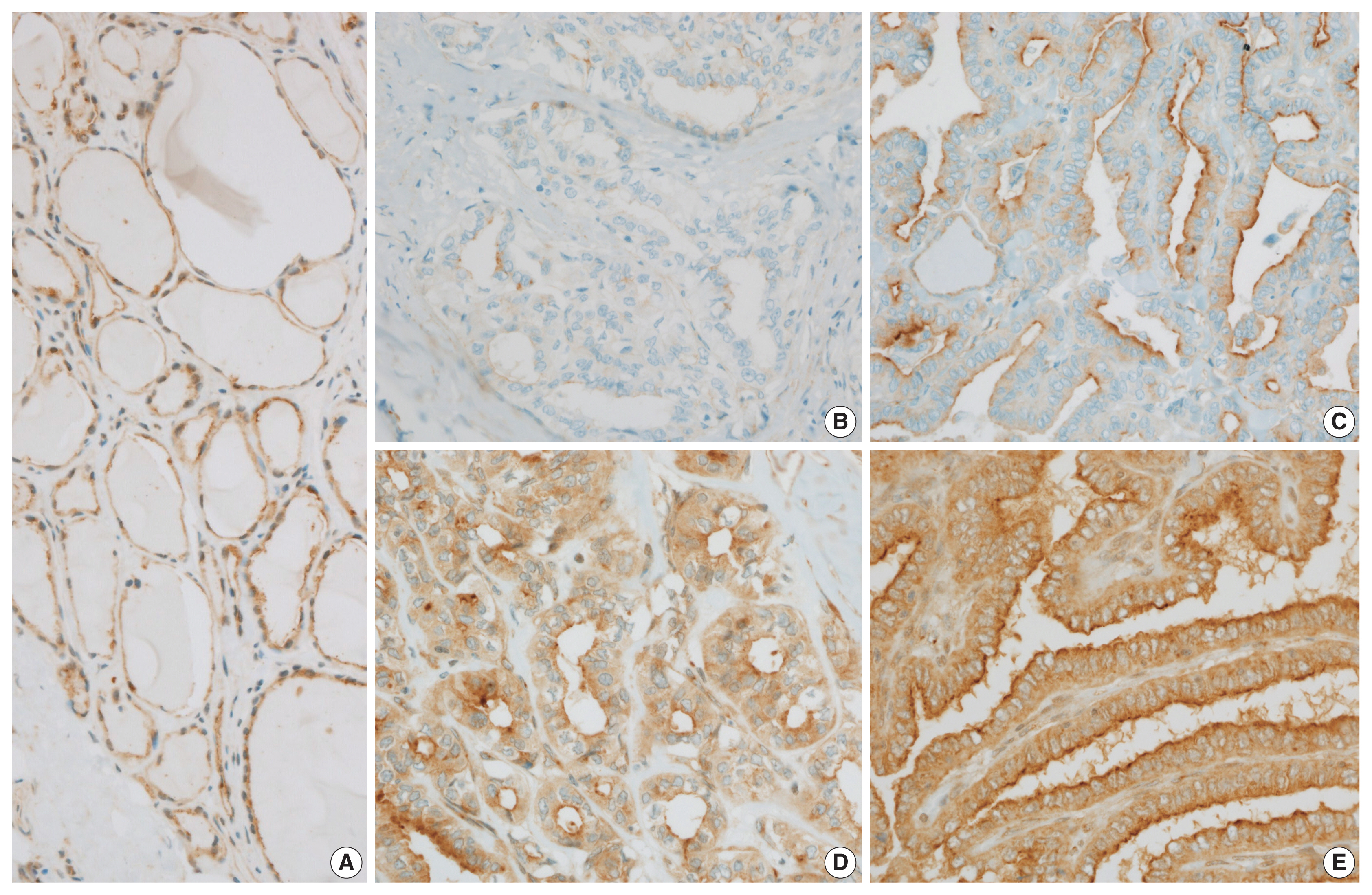
To investigate protein expression, immunohistochemical staining was performed on 4 μm sections from the TMA blocks. The sections were deparaffinized, rehydrated, and incubated in 3% hydrogen peroxide. After heating in 10 mM citrate buffer (pH 6.0), the slides were incubated with a mouse monoclonal primary myoferlin antibody (1:100, 7D2, ab76746, Abcam, Cambridge, UK). 3,3'-Diaminobenzidine staining and counterstaining of hematoxylin were carried out for visualization. The expression of myoferlin in the tumor cells was graded from negative to weak (+1), moderate (+2), and strong (+3) positivity by two experienced pathologists. Normal thyroid follicular cells were used as the internal control. If tumor cells exhibited equivalent intensity of myoferlin expression compared to normal follicular cells, they were graded as moderate positive. Similarly, tumor cells that exhibited stronger or weaker expression were graded as strong and weak positive, respectively.
Statistical analysis
Pearson’s chi square test and Fisher’s exact test were used to evaluate correlations between myoferlin expression level and a range of clinicopathological features including age and sex of patients and the size, extension, lymph node metastasis, and pathologic T category and N category in AJCC staging of the tumors. For all statistical analyses, differences were considered statistically significant at p < .05. IBM SPSS Statistics for Windows, ver. 21.0 (IBM Corp., Armonk, NY, USA) was used for the statistical analysis.
RESULTS
Clinicopathological features
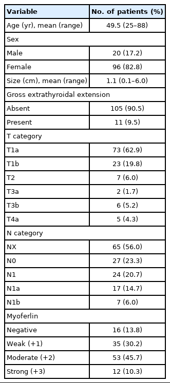
Table 1 summarizes the clinical data and pathological features of the patients enrolled in this study. Of the 116 PTC patients enrolled, the mean age was 49.5 years (range, 25 to 88 years), with 20 male (17.2%) and 96 female (82.8%) patients. The largest diameter tumor sizes ranged from 0.1–6.0 cm (mean, 1.12 cm). Notably, in 11 (9.5%) of these cases, gross tumor extension beyond the thyroid gland was observed. Most patients (96/116, 82.7%) had pT1 carcinomas, whereas seven (6%), eight (6.9%), and five (4.3%) were observed for pT2, pT3, and pT4 tumors, respectively. Lymph node dissection was performed in 51 (44.0%) patients, of whom 24 (20.7%) were found to have lymph node PTC metastasis. There were 17 (14.7%) pN1a tumors and seven (6.0%) pN1b tumors. No distant metastasis was observed in any of the cases.
Myoferlin expression in PTC
Normal thyroid follicular cells exhibited myoferlin expression in the cell cytoplasm and membrane (Fig. 1A). Myoferlin expression in PTC cells was observed in 100 cases (86.2%), showing a even expression pattern involving the cytoplasm and membrane (Fig. 1B–E). Myoferlin expression level was graded as weak (+1) in 35 cases (30.2%), moderate (+2) in 53 cases (45.75%), and strong (+3) in 12 cases (10.3%) (Table 1). To facilitate reproducibility, these grading categories were further simplified into two categories: low (negative or only weakly positive, +1) and high (moderate, +2 to strong, +3) expression levels. Of all patients, 51 (44.0%) exhibited negative or weak myoferlin expression level, whereas 65 (56.0%) showed moderate to strong expression.
Correlation of myoferlin expression with T category and N category of PTC
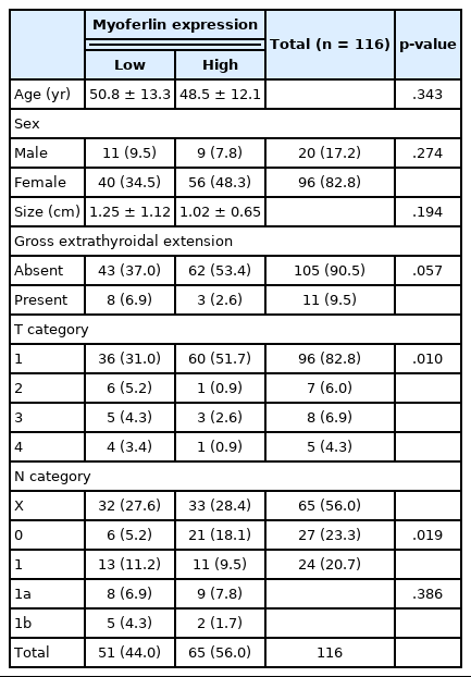
Table 2 summarizes the associations between myoferlin expression level and the clinicopathological features of patients. Patient age and sex, tumor size, and presence of gross extrathyroidal extensions did not demonstrate a significant correlation with myoferlin expression of tumors. In pT1 tumors, a larger number exhibited a high (moderate or strong) level of myoferlin expression (n = 60, 51.7%) compared to low (negative or weak) expression (n = 36, 31.0%). In pT4 tumors, it was the opposite, as the number of tumors with a low level of expression (n = 4, 3.4%) outnumbered those with a high level of expression (n = 1, 0.9%). Overall, the T category and myoferlin expression level of tumors exhibited the following tendency: tumors of the higher T category group showed a correlation with a lower level of myoferlin expression (p = .010).
Among the cases that underwent lymph node dissection, in lymph node metastasis-free (pN0) tumors, six (5.2%) exhibited low myoferlin expression level, whereas high levels were observed in 21 (18.1%). Among lymph node metastatic tumors (pN1), 13 (11.2%) indicated low myoferlin expression levels, whereas 11 (9.5%) cases revealed high levels. Statistically, pN1 tumors with lymph node metastasis were significantly correlated with low myoferlin expression levels (p = .019). Whether the tumor of the pN1 category was pN1a or pN1b showed no significant association with myoferlin expression (p = .386).
DISCUSSION
This study confirmed that myoferlin is expressed in PTC cells. In previous studies, myoferlin overexpression was indicative of poor disease survival in various types of carcinomas including non-small cell lung carcinoma [16], pancreatic adenocarcinoma [17], and clear cell renal cell carcinoma [18]. However, our retrospective study data showed a statistically significant correlation of loss of myoferlin expression with a higher T category of tumor and lymph node PTC metastasis. With both features being risk factors for poor survival and disease recurrence, this correlation counters the majority of recent research observations that have reported an evident correlation between higher myoferlin level and unfavorable prognosis.
In a study from our institution on endometrioid carcinoma, loss of myoferlin expression level correlated with high histologic grade [20]. This result also counters the previously described correlation of high myoferlin expression with poor prognosis in some carcinomas. The continuous cyclic regeneration of normal endometrial tissue was suggested as a possible cause of this correlation. Myoferlin is a multifunctional protein in normal cells, although its role in various cancer cells is unknown. Research findings suggest that myoferlin expression has different tendencies due to its various roles in different types of carcinomas.
No previous myoferlin studies were performed on endocrine cell–derived tumors. Myoferlin is expressed in normal thyroid follicular cells, but its specific roles in normal cells or oncogenic roles have yet to be studied. Therefore, one plausible hypothesis is that the endocrine activity of thyroid follicular cells influences the distinctive pattern of myoferlin expression in the thyroid. The difference in key functions of myoferlin could possibly be the cause of its expression tendency differing in PTC from lung and pancreatic epithelial tumors. Further molecular studies on the role of myoferlin in different types of tumor cells are needed to clarify the specific function of this protein.
Of the 116 patients enrolled, there were only four cases with recurrent disease and no patient death during follow-up. With regard to these findings, owing to the limited sample size in the present study, an analysis of a larger cohort is necessary to confirm correlations with survival rates.
In differentiated papillary carcinoma, therapeutic neck dissection is strongly recommended and routinely performed in clinically palpable or biopsy-proven nodal disease, as it is well established that nodal metastasis is associated with disease recurrence [6]. However, the use of prophylactic neck dissection for clinically node-negative PTC is controversial. If prediction of occult lymph node metastatic status is determinable by pre-surgical cytology or biopsy specimens, a justification could be made for more individualized disease management, which could include determining whether to perform neck dissection or to closely follow-up on the case. The correlation proposed in the present study points to a potential prognostic role of myoferlin in lymph node PTC metastasis.
Conclusively, our study demonstrated correlation of myoferlin expression with pathological features in papillary thyroid carcinoma. Loss of expression of myoferlin was correlated with a higher T category of tumor and the presence of lymph node metastasis in PTC. This result shows the potential prognostic role of myoferlin in PTC as a helpful biomarker in therapeutic management.
Notes
Ethics Statement
This study was approved by the Institutional Review Board of Gyeongsang National University Hospital, and the need for informed consent was waived (GNUH-10-026).
Availability of Data and Material
The datasets generated or analyzed during the study are available from the corresponding author on reasonable request.
Code Availability
Not applicable.
Author Contributions
Conceptualization: DCK, DHS. Data curation: JMN, DCK, JHL, JSL, JWY, HJA, MHK. Investigation: DCK, DHS, JHL, JSL, JWY, MHK. Methodology: JMN, DCK. Project administration: DCK, DHS. Resources: DHS. Supervision: DHS, MHK, JWY. Validation: DCK, HJA, HMK. Visualization: JMN. Writing—original draft: JMN, DCK. Writing—review & editing: JMN, DCK, JWY. Approval of final manuscript: all authors.
Conflicts of Interest
The authors declare that they have no potential conflicts of interest.
Funding Statement
This work was supported by the fund of research promotion program, Gyeongsang National University, 2015.


