Evaluation of the characteristics of multiple human papillomavirus (HPV) infections identified using the BD Onclarity HPV assay and comparison with those of single HPV infection
Article information
Abstract
Background
Human papillomavirus (HPV) infection is a major cause of cervical cancer and associated precursor lesions. Multiple HPV genotype infections have been reported. However, their clinicopathological characteristics still remain elusive.
Methods
For this study, 814 consecutive patients who had undergone colposcopy and HPV genotyping test using BD Onclarity HPV assay were retrospectively selected. Clinicopathological parameters of multiple HPV infections were compared with those of single HPV infection.
Results
Multiple HPV infections were found in 110 out of 814 cases (13.5%). Multiple HPV infections were associated with a significantly higher incidence of high-grade intraepithelial lesions (HSILs) compared with single HPV infection. Other high-risk HPV genotypes, in addition to HPV 16, were found more frequently in the multiple HPV infections group; these included HPV 51, 52, 33/58, 56/59/66, and 35/39/68. No specific coinfection pattern was not identified. Additionally, the number of HPV genotypes in multiple HPV infections was not associated with the progression to HSIL or squamous cell carcinoma.
Conclusions
Multiple HPV infections have distinct clinicopathological characteristics (compared with single HPV infection). As their biological behavior is uncertain, close and frequent follow-up is warranted.
Human papillomavirus (HPV) is the major cause of anogenital cancers and their precursor lesions [1]. Sensitive molecular HPV detection methods revealed that nearly all squamous intraepithelial lesions are associated with HPV [2–4]. According to the phylogenetic classification of papillomaviruses, HPVs are categorized in the genus Alphapapillomavirus [5]. Among these, A9 clade and A7 clade, which include HPV 16, and HPV 18 and 45, respectively, are particularly noteworthy [3]. To date, more than 200 HPV genotypes have been identified and listed in the National Institutes of Health’s HPV database (PaVE: The papillomavirus Episteme) [6]. However, only ~20 HPV genotypes are commonly encountered in anogenital lesions. Currently, 12 HPV genotypes, i.e., HPV 16, 18, 31, 33, 35, 39, 45, 51, 52, 56, 58, and 59, are classified as group 1 carcinogens by the International Agency for Research on Cancer [7]. Although HPV 16 and 18 are the predominant causative agents of cervical cancer and their precursor lesions, other high-risk HPVs are also associated with significant oncogenic risk [8].
HPV genotyping tests have also revealed coinfection with multiple HPV genotypes in variable frequencies [9]. The incidence of multiple HPV infections varies depending on the region, ethnicity, and the detection method [10–13]. Although multiple HPV infections have been detected in cervical lesions, their clinicopathological significance remains elusive. While some studies have demonstrated that multiple HPV infections may be associated with cervical carcinogenesis [14,15], others have reported contradicting results [16].
BD Onclarity HPV assay, an U.S. Food and Drug Administration–approved HPV screening test, is a semi-quantitative real-time polymerase chain reaction–based test that can simultaneously detect multiple HPV genotypes, including HPV 16, 18, 31, 45, 51, 52, 33/35, 56/59/66, and 35/39/68 [17]. It is widely used in many hospitals in Korea [18]. In this study, we have investigated the clinicopathological aspects of multiple HPV infections using the BD Onclarity HPV assay.
MATERIALS AND METHODS
Study population
We retrospectively selected 814 consecutive patients who had undergone colonoscopy as well as HPV genotyping tests in the department of pathology, Kyungpook National University Chilgok Hospital in 2019. These patients underwent HPV genotyping assays owing to abnormal cytology or biopsy results, or during routine health check-ups. The clinicopathological data, including age and biopsy results, of the patients were retrieved from the electronic medical records of the hospital.
Histological evaluation
Specimens obtained during colposcopy were fixed in 10% neutral-buffered formalin and embedded in paraffin blocks. Paraffin blocks were cut into 4 μm sections and stained with hematoxylin and eosin. Two independent pathologists with specialization in gynecological pathology (M.K. and J.Y.P.) reviewed all the samples. All patients were diagnosed and classified in accordance to the recommendations of the LAST project working group [19].
HPV genotyping test
Onclarity testing uses the automated BD Viper Lt platform (BD Diagnostics, Sparks, MD, USA), the full workflow for which has been described in detail in a previous paper [20]. Briefly, 0.5 mL of original resuspended SurePath material was aliquoted into a BD tube containing 1.7 mL of sample medium. The samples were prewarmed for 30 minutes at 120°C. They were then transferred to the fully automated BD Viper Lt platform and tested using Onclarity according to the manufacturer’s instructions.
Statistical analysis
All statistical analyses were performed using IBM SPSS for Windows ver. 23.0 (IBM Corp., Armonk, NY, USA). The association between clinicopathological parameters was analyzed using the chi-square test or Fisher exact test. p-values < .05 were considered significant. Adjusted p-values using Bonferroni correction were used for multiple comparisons.
RESULTS
HPV genotype prevalence in multiple and single HPV infections
Table 1 presents HPV genotype prevalence in multiple and single HPV infections, respectively. Detailed clinicopathological characteristics of the patient cohorts are listed in Supplementary Table S1. Among 814 cases wherein HPV genotyping tests were performed, multiple HPV infection was found in 110 cases (13.5%, 110/814) and single HPV infections were found in 361 cases (44.3%, 361/814). The mean age of the patients was 58.2 years in the multiple HPV infections group and 55.5 years in the single HPV infection group. In the multiple HPV infections group, HPV 35/39/68 was most frequently detected, followed by 56/59/66, 16, and 52. In the single HPV infection group, HPV 16 was most frequently detected, followed by 56/59/66, 35/39/68, and 52. HPV 31 (p = .005), 51 (p < .001), 52 (p < .001), 33/58 (p < .001), 56/59/66 (p < .001), and 35/39/68 (p < .001) were significantly more frequently detected in the multiple HPV infections group. Multiple infections among grouped HPV strains, for example, HPV 33 and 58, can possibly happen. However, such possible cases were counted as single HPV infection, based on the previous study [17]. The result of full HPV genotyping assay in a different cohort using Seegene Anyplex II also suggested that multiple infections within three bulk groups (33/35, 56/59/66, and 35/39/68) were not frequent (Supplementary Table S2).
Association of multiple HPV infection with histology
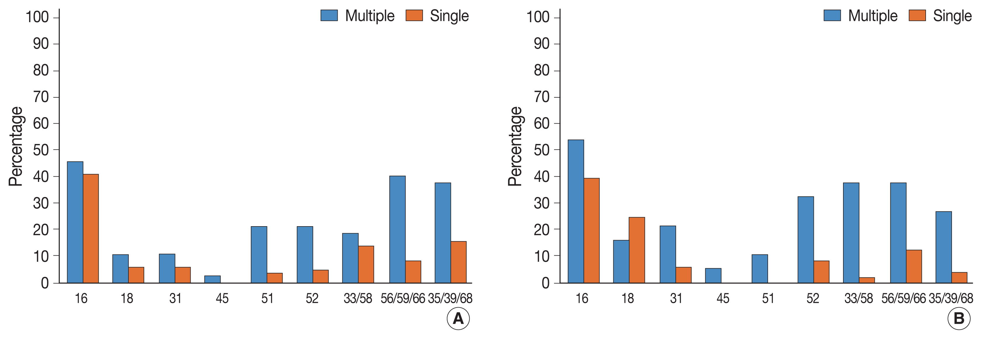
We then investigated the association between HPV infection status and histological diagnosis of cervical lesions. Of note, multiple HPV infections showed more significant association with high-grade squamous intraepithelial lesions (HSILs) compared with single HPV infection (p = .033) (Table 2). Low-grade squamous intraepithelial lesions (LSILs), squamous cell carcinoma (SQCC), and adenocarcinoma (ADC) were also more frequent in the multiple HPV infections group, albeit this was not significant. In the multiple HPV infections group, HPV 16 was the most frequently detected HPV genotype in LSILs, HSILs, SQCC, and ADC (Table 3). In the single HPV infection group, HPV 16 was the most frequently detected HPV genotype in HSILs and SQCC. In addition to HPV 16, other high-risk HPV genotypes were also detected more frequently in the patients with ≥ HSIL lesions (HSIL and SQCC), which suggests clinical significance. Among these, HPV 51, 52, 33/58, 56/59/66, and 35/39/68 were more frequently found in the multiple HPV infections group than in the single HPV infection group (Table 4, Fig. 1). In the multiple HPV group, there was no correlation between the number of infected HPV strains and the progression to cervical cancer and their precursor lesions (Supplementary Table S3). Although HPV 16 was the most frequently detected HPV genotype in multiple HPV infections, the presence of HPV 16 genotype in multiple HPV infections was not associated with the progression to cervical neoplastic lesions (compared with non-HPV 16 type of multiple HPV infections) (Supplementary Table S4). We further divided the study cases into two groups according to the HPV 16 or/and HPV 18 infection status (group 1: HPV 16 or/and HPV18 infection vs. group 2: non–HPV 16 and non–HPV 18 high-risk HPV). In both groups, there were no statistically significant correlation between multiple HPV infection and cervical lesions. However, HSIL tended to occur more frequently in group 2 (Supplementary Tables S5, S6).

Association between ≥ HSIL lesion (HSIL and SQCC) and HPV genotypes (multiple and single HPV infections)
DISCUSSION
In this study, we demonstrated the clinicopathological implications of multiple HPV infections in Korean patients, using the BD Onclarity HPV assay. Multiple HPV infections were more closely related to HSILs than single HPV infection. In addition, we report that high-risk HPV genotypes other than HPV 16 were detected more frequently in the multiple HPV infections group. In our previous study, using Seegene Anyplex II HPV28 detection kit, we showed that multiple HPV infections were related to HSIL and persistent HPV infection [21]. However, Seegene Anyplex II and BD Onclarity assays have different ranges and use different probes for the detection of HPV [22]. The two HPV genotyping assays, thus, might have different performance capacity for the detection of multiple HPV infections. Therefore, we re-investigated the clinicopathological aspects of multiple HPV infections using the BD Onclarity HPV assay.
The clinicopathological significance of multiple HPV infections still remains debatable. Overall, HPV 31, 51, 52, 33/58, 56/59/66, and 35/39/68 genotypes were more frequently detected in multiple HPV infections than in single HPV infection. The incidence of HPV genotypes in multiple HPV infections greatly differs across studies, depending on regional variation, ethnicity, and participant characteristics [12,14,23].
In this study, multiple HPV infection was significantly associated with HSIL, which was consistent with the results from our previous study. Oncogenic risk of multiple HPV infections is still unclear. While some previous studies have shown that multiple HPV infections contribute to cervical carcinogenesis, other studies have shown that multiple HPV infections do not confer additional carcinogenic effect (compared to single HPV infection) [10,16, 24,25]. Thus, a larger cohort study may be instrumental in validating the oncogenic potential of multiple HPV infection.
In multiple HPV infection, HPV 16 was the most prevalent HPV genotype in SIL and SQCC. However, other high-risk HPV genotypes were also more frequently detected in multiple HPV infections than in single HPV infection. In particular, HPV 51, 52, 33/58, 56/59/66, and 35/39/68 showed significant association with ≥HSILs in multiple HPV infections. There was no difference between the HPV 16–positive multiple HPV infections group and the HPV 16–negative multiple HPV infections group. We also divided study cases into HPV 16 or/and HPV18 infection group and non–HPV 16 and non–HPV 18 high-risk group, and further investigated the association between the multiple infection and the histology of cervical lesions in both groups; however, significant associations were not found. In non–HPV 16 and non–HPV 18 high-risk HPV group, HSIL tended to occur more frequently among multiple HPV infections. Thus, coinfection with non–HPV 16 and non–HPV 18 high-risk HPV genotypes might have synergistic effect on cervical carcinogenesis. Subsequent larger cohort study or expanded full genotyping assay should be followed to validate this hypothesis.
This study has a few limitations. First, this study has a relatively small sample size. Subsequent studies with larger cohort would be valuable to further strengthen the major findings of this study. In addition, we were unable to evaluate the effect of multiple HPV genotypes on the duration of HPV infection, as we could not perform the follow-up HPV genotyping test using BD Onclarity HPV assay.
In conclusion, multiple HPV infections have distinct clinicopathological characteristics. As their clinicopathological characteristics are still uncertain, close follow-up is warranted for the patients with multiple HPV infections.
Supplementary Information
The Data Supplement is available with this article at https://doi.org/10.4132/jptm.2022.08.02.
Notes
Ethics Statement
The study was conducted according to the guidelines of the Declaration of Helsinki and approved by the Institutional review board of Kyungpook National University Chilgok Hospital (KNUCH 2019-04-002-002). The requirement for written informed consent from the patients was waived due to the retrospective nature of the study.
Availability of Data and Material
All data generated or analyzed during the study are included in this published article (and its supplementary information files).
Code Availability
Not applicable.
Conflicts of Interest
The authors declare that they have no potential conflicts of interest.
Author Contributions
Conceptualization: JYP. Data curation: JK, MK. Formal analysis: JK, MK. Funding acquisition: JYP. Investigation: JK. Methodology: JK, MK. Supervision: JYP. Writing—original draft: JK. Writing—review & editing: MK, JYP. Approval of final manuscript: all authors.
Funding Statement
This research was supported (in part) by The Korean Society for Cytopathology Grant No. 2019-01
Acknowledgements
The authors are grateful for the support provided by the Molecular Pathology Laboratory, Kyungpook National University Chilgok Hospital.


