Obesity-driven adipokine signaling in breast cancer: mechanistic insights, therapeutic challenges, and opportunities for precision oncology
Article information

Abstract
Breast cancer is the leading cause of illness and death among women worldwide, with more than 2.3 million new cases diagnosed each year. The incidence and fatality rates are steadily increasing, notably in Asian countries. Obesity has been established as a major and growing risk factor for breast cancer progression. The World Health Organization reports that adult obesity rates have doubled since 1990. Obesity promotes tumor growth by inhibiting adipokine production and activating cancer-promoting pathways. In obese people, the microenvironment surrounding breast cancer cells is drastically altered. This is mostly due to the malfunctioning of adipocytes (fat cells) and macrophages. These defective cells' adipokines alter signaling pathways required for cancer cell proliferation, survival, and inflammation. This dysregulation has a significant role in tumor development, metastasis, and resistance to traditional cancer treatments, particularly in obese patients. This review highlights the role of adipokines in breast cancer, with a focus on disease progression, therapeutic challenges, and knowledge gaps. Clarifying how obesity alters tumor biology is key to advancing personalized and effective treatments for obese patients.
INTRODUCTION
Breast cancer is the most frequently diagnosed malignancy in women, with more than 2.3 million new cases reported annually. It continues to be a leading contributor to global cancer-related morbidity and mortality [1]. An increasing incidence and mortality have been reported in Asian countries [2]. Obesity is on the rise, and studies suggest that breast cancer patients with obesity may have a reduced response to treatment. However, this remains debatable and is not currently factored into treatment decisions due to gaps in scientific understanding. Breast cancer risk and outcomes are influenced by multiple factors, including age, family history, genetic predisposition, obesity, alcohol use, and smoking. While obesity is considered a modifiable risk factor, some studies report contradictory findings [3]. Obesity is characterized by excess fat buildup and malfunctioning adipocytes that produce aberrant adipokines into the tumor microenvironment, activating oncogenic pathways such as JAK, signal transducer and activator of transcription 3 (STAT3), nuclear factor кB (NF-κB), and Akt, which promote breast cancer growth [4,5].
The tumor microenvironment plays a key role in cancer progression by supporting cell proliferation, angiogenesis, and survival [6]. In breast cancer, it is composed mainly of adipocytes, macrophages, and tumor cells. Among these, adipocytes are the most abundant and metabolically active, particularly in obese individuals, and significantly influence tumor cell behavior [7].
Breast cancer cells penetrate the mammary fat pad early in the tumor growth process and interact with surrounding adipocytes via secreted chemicals [8]. Obesity has been shown in clinical research to affect breast cancer outcomes, with obese patients frequently presenting with larger, more aggressive tumors and lower survival rates [9,10]. Understanding the link between obesity and breast cancer is critical for determining the molecular mechanisms behind this association. Fig. 1 provides a graphical overview of obesity-associated remodeling of the breast tumor microenvironment. It illustrates the altered secretion from adipocytes and deregulation of oncogenic signaling in tumor cells.
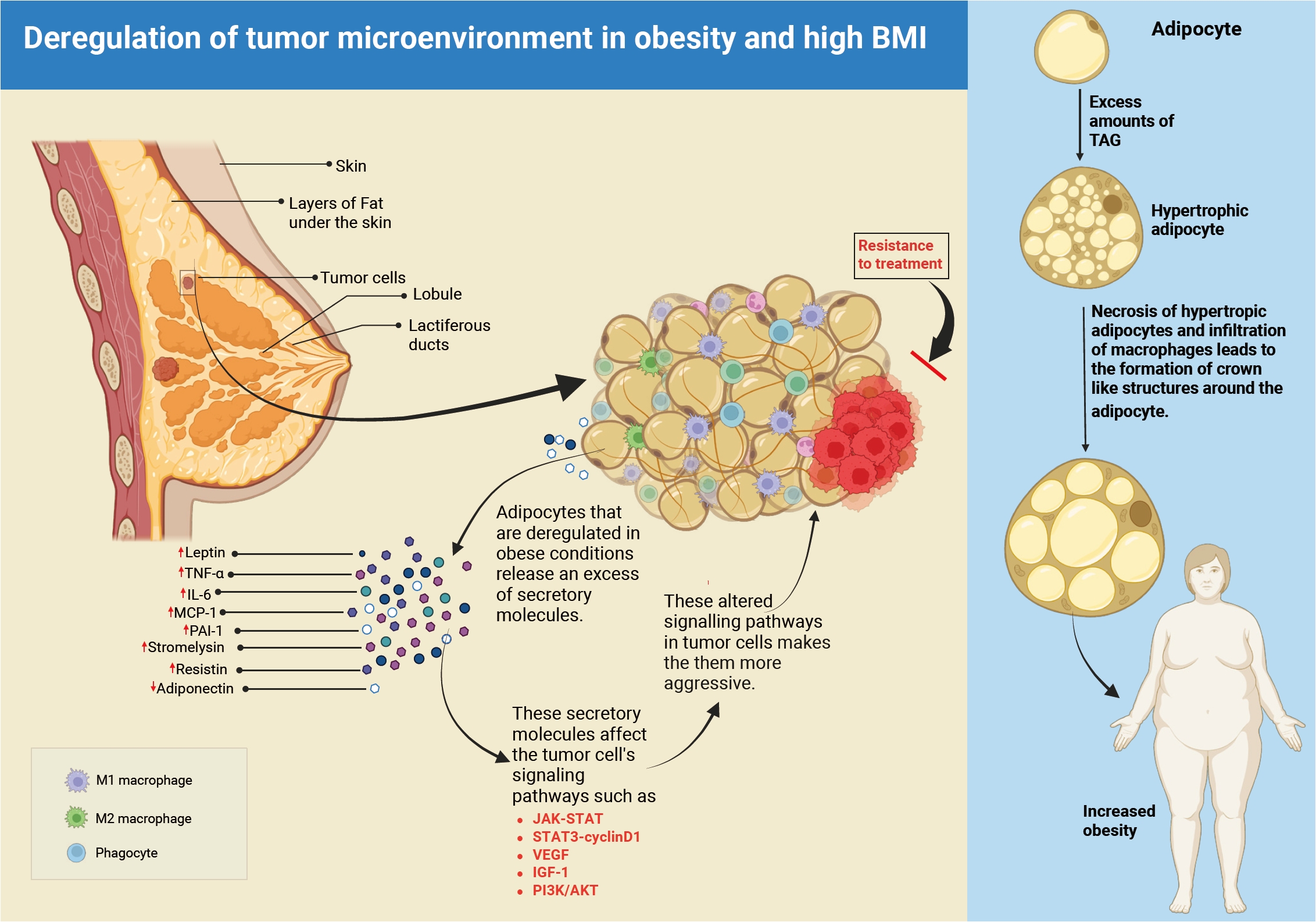
Obesity-associated tumor microenvironment in obese breast cancer patients. The right-hand side of the image presents a sequence of normal adipocytes developing into hypertrophic and necrotic phenotypes during obesity. The left-hand panel shows the molecular landscape of the breast tumor microenvironment. Altered adipocytes produce molecules including leptin, tumor necrosis factor α (TNF-α), interleukin-6 (IL-6), monocyte chemoattractant protein-1 (MCP-1), and resistin that further alter the signaling pathways (e.g., JAK-STAT, vascular endothelial growth factor [VEGF], and phosphoinositide 3-kinase [PI3K]/Akt) in the tumor cells. It encourages aggressiveness of the tumor, promotes cell survival and invasion, metastasis, and resistance to chemotherapy. BMI, body mass index; IGF-1, insulin-like growth factor 1; PAI-1, plasminogen activator inhibitor-1; STAT3, signal transducer and activator of transcription 3; TAG, triacylglycerol.
In this review, we have discussed the various signaling pathways involved and their translational importance, with a special emphasis on the involvement of adipokines released by dysfunctional, hypertrophic adipocytes in the tumor microenvironment. We have also mentioned the current knowledge gaps and suggest future research directions for establishing customized therapy regimens for obese breast cancer patients.
ADIPOKINES AT THE CROSSROADS OF OBESITY AND BREAST CANCER
Adipose tissue secretes adipokines, which act as essential regulators of metabolic and inflammatory pathways [11]. These include hormones (leptin), cytokines (interleukin [IL]-6, tumor necrosis factor [TNF], and IL-1β), and angiogenic agents (vascular endothelial growth factor [VEGF]). These mediators play a role in the development, progression, and treatment resistance of obesity-related malignancies, notably breast cancer [12]. Although adiponectin has anti-inflammatory properties, other adipokines such as resistin, monocyte chemoattractant protein-1 (MCP-1), leptin, IL-6, transforming growth factor-β, IL-10, plasminogen activator inhibitor-1 (PAI-1), and stromelysin increase pro-inflammatory responses that contribute to tumor formation [13].
Adipokines act like hormones, serving as growth factors that regulate insulin resistance and influence lipid and glucose metabolism. In obesity, excessive fat and dysfunctional adipocytes disrupt adipokine production and secretion, leading to altered immune responses and etiology of various diseases [14]. Adipokines are involved in both pro- and anti-inflammatory responses, and their dysregulation in the cancer niche significantly affects tumor growth and metastasis.
In breast cancer, adipokines independently and synergistically activate key signaling pathways that drive tumor growth, proliferation, invasion, metastasis, and survival. Fig. 2 illustrates different oncogenic pathway deregulation via the adipocyte secretory molecules and promoting tumor progression. For example, leptin stimulates cancer cell growth and survival, while TNF-α and IL-6 promote inflammation and tumor progression [15]. Adiponectin's anti-inflammatory properties can inhibit cancer cell proliferation and spread. Maintaining a balance of pro- and anti-inflammatory adipokines in the tumor microenvironment is therefore critical. An imbalance impairs adipose tissue homeostasis, resulting in persistent inflammation. In obesity, adipokine imbalances have an important role in regulating metabolism and signaling, eventually impacting tumor progression and treatment response.
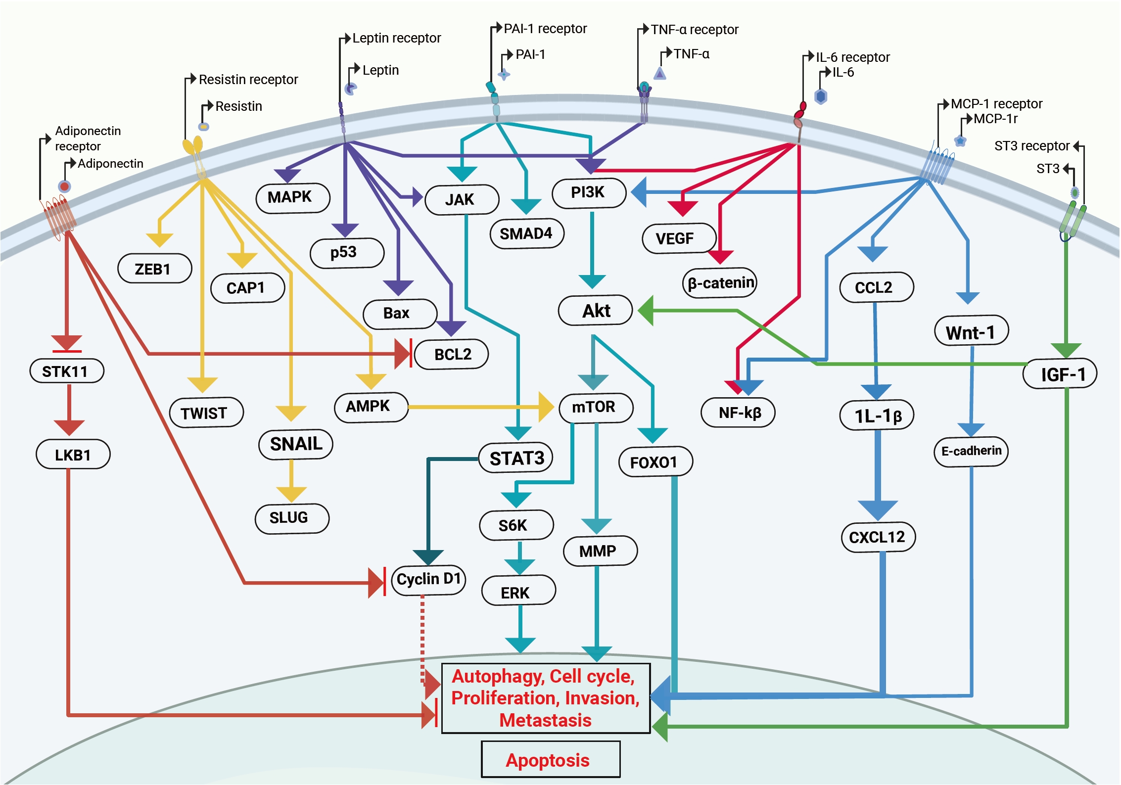
The signaling pathways activated by deregulated adipocytes in obesity-associated breast cancer. JAK/signal transducer and activator of transcription 3 (STAT3), phosphoinositide 3-kinase (PI3K)/Akt/mechanistic target of rapamycin (mTOR), mitogen-activated protein kinase (MAPK), nuclear factor кB (NF-κB), vascular endothelial growth factor (VEGF), and β-catenin signaling pathways activated by the altered secretion of leptin, interleukin-6 (IL-6), tumor necrosis factor α (TNF-α), plasminogen activator inhibitor-1 (PAI-1), resistin, monocyte chemoattractant protein-1 (MCP-1), and stromelysin-3 (ST3) from adipocytes. This activated pathway alters cellular functions like cell proliferation, survival, angiogenesis, invasion, epithelial–mesenchymal transition, and metastasis. AMPK, AMP-activated protein kinase; CAP1, cyclase-associated protein 1; CCL2, C-C motif chemokine ligand 2; CXCL12, C-X-C motif chemokine ligand 12; ERK, extracellular signal-regulated kinase; FOXO1, forkhead box O1; IGF-1, insulin-like growth factor 1; LKB1, liver kinase B1; MMP, matrix metalloproteinase; STK11, serine/threonine kinase 11; ZEB1, zinc finger E-box-binding homeobox 1.
TUMOR NICHE IN OBESITY: A DRIVER OF CANCER PROGRESSION AND DIVERSITY
Normal niches are made up of fibroblastic cells, immunological cells, endothelium and perivascular cells (or their progenitors), extracellular matrix (ECM) components, and signaling molecules like cytokines and growth factors. The tumor niche is a component of the tumor microenvironment (TME), which consists of stromal and non-cancerous cells that interact with tumor cells. Thus, tumor behavior is regulated by both the biology of the tumor cells and the features of their microenvironment. Fig. 3 shows the impact of adipokines on breast cancer progression in an obesity-associated tumor niche. Increased expression of leptin, TNF-α, IL-6, MCP-1, resistin, PAI-1, and stromelysin-3, together with reduced adiponectin, promotes key hallmarks of cancer, including enhanced proliferation, invasion, angiogenesis, stemness, epithelial-mesenchymal transition, and therapeutic resistance.
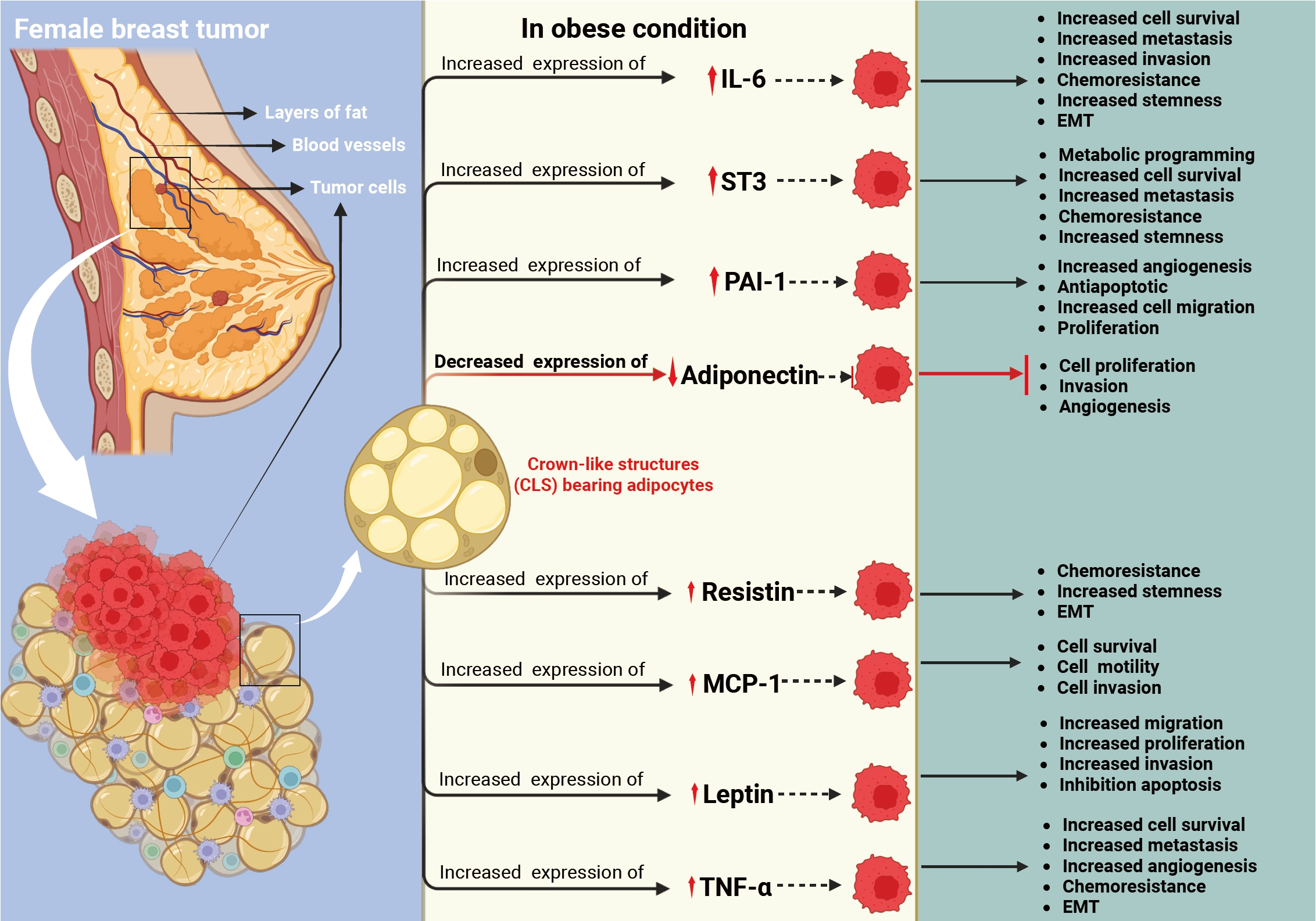
This figure shows deregulated adipokines and their role in the progression of breast cancer. The left panel is highlighted with fat layers, the crown-like structures (CLS) of adipocytes in an obese condition within the breast and the tumor cells. The right panel indicates the upregulation of interleukin-6 (IL-6), stromelysin-3 (ST3), plasminogen activator inhibitor-1 (PAI-1), resistin, monocyte chemoattractant protein-1 (MCP-1), leptin, and tumor necrosis factor α (TNF-α), downregulation of adiponectin, and its association with metastasis, cell survival, chemoresistance, and epithelial-mesenchymal transition (EMT).
Intratumoral heterogeneity accelerates disease development, lowers therapy efficacy, and affects patient survival. Tumor heterogeneity is caused by genetic and epigenetic alterations inside tumor cells, as well as effects from the TME. In obesity, adipocyte dysfunction and hypertrophy further disturb the TME by affecting the release of over 600 bioactive signaling molecules [16].
These peptides are a diverse group that influences tumor cell behavior by affecting survival pathways, metabolism, and energy balance [17]. Interestingly, heterogeneity exists both within a single tumor (intratumoral) and between different tumors (intertumoral). This may explain why tumors with similar genomic profiles respond differently to treatment. In addition, obesity-related changes in the tumor environment can alter the epigenetic landscape and gene expression of tumor cells. As adipocytes grow and become hypertrophic, they stiffen, interrupting membrane signaling and leading to decreased cell function and metabolic dysregulation.
Adipokine secretion is directly related to adipocyte size; as adiposity increases, pro-inflammatory adipokines are overproduced and anti-inflammatory adipokines are underproduced [18,19]. To understand the biology of breast cancer and treatment outcomes, it is vital to understand the alterations in the breast cancer niche caused by unregulated adipocytes, which contribute to cancer growth and metastasis (Fig. 2).
LEPTIN AND BREAST CANCER: FROM SATIETY HORMONE TO ONCOGENIC SIGNAL—A PARADIGM SHIFT WITH OBESITY
Leptin is a 16-kD peptide hormone created mostly by adipocytes, and the amount of its production is positively related to obesity and higher in obese people [20,21]. Leptin binds to a specific leptin receptor (LEPR) on the cell surface. Wang et al. [22] found that 70.5% of tumor tissues show high levels of LEPR. It was much greater than the benign breast tissues (56.3%) and normal para-tumoral tissues (44%). Higher levels of leptin in the tumor environment may influence signaling in tumor cells. Although adipose tissue is strongly linked to breast cancer, the role of leptin in the disease is still debated. Both in-vivo and in-vitro studies suggest that leptin can affect key cellular functions such as motility, migration, and tumor progression [23,24]. The findings suggest that in obesity, leptin may promote tumor invasion and angiogenesis, leading to metastasis and reduced treatment efficacy. This could explain the higher cancer mortality risk in overweight patients.
One of the most difficult aspects of breast cancer treatment is distant metastasis, which is caused by tumor cells undergoing epithelial-mesenchymal transition (EMT). He et al. [25] discovered that leptin enhances invasion and migration in triple-negative breast cancer (TNBC) by activating PLOD2 (a protein involved in the hydroxylation of lysyl residues in collagen-like peptides), which stimulates the phosphoinositide 3-kinase (PI3K)/Akt and JAK/STAT3 pathways. Another study utilizing TNBC and estrogen receptor (ER)+ cells demonstrated that leptin promotes production and activation of matrix metalloproteinase (MMP)-2 and MMP-9, as well as FAK activation via Src, resulting in higher invasion and phenotypic alterations in TNBC cells [26]. Strong et al. [27], on the other hand, proposed that leptin is more effective on ER+ cells. They discovered that genes related to cell cycle, apoptosis, and migration (CDKN2A, GSTP1, SFRP1, PLAU, THBS1, and CSF) were changed in ER+ cells via the ERK1/ERK2 signal and cross-talk between LEPR and ER [27-29]. In obese women, a high dose of leptin generates estradiol and estrogen via increasing aromatase activity [30]. Leptin promotes tumor recurrence and poor prognosis in TNBC by enhancing the self-renewal capacity of cancer cells [31]. In other cancers, leptin has been shown to activate VEGF through NF-κB/hypoxia-inducible factor-1α signaling via both canonical and non-canonical pathways. Beyond its role in EMT, leptin also supports cancer cell proliferation and survival by activating multiple signaling pathways, including insulin-like growth factor 1 (IGF-1)/activator protein 1 (AP-1), NOTCH/survivin, PI3K/AKT/sterol regulatory element-binding protein 2/acetyl-CoA acetyl transferase 2(ACAT2)/STAT3/cyclin D1, extracellular signal-regulated kinase 1/2, AP-1, mitogen-activated protein kinase (MAPK), and PI3K [32,33]. Despite significant research demonstrating that changing leptin affects tumor pathways such as PI3K/Akt, JAK/STAT3, and MAPK in breast cancer, clinical translation remains elusive. This disparity could be attributed to (1) the varied behavior of ER+, TNBC, and human epidermal growth factor receptor 2 (HER2)+ malignancies in respect to leptin, (2) a lack of knowledge of normal leptin ranges in clinical practice, (3) a lack of effective therapeutic agents, and (4) an insufficient understanding of the tumor microenvironment.
TNF-α AND BREAST CANCER: A CHANGING ROLE IN TME REMODELING DURING OBESITY
TNF-α, the first inflammatory cytokine identified as an adipokine, acts in both autocrine and paracrine ways. It influences processes such as apoptosis, MMP production, and angiogenesis, which can promote tumor cell survival and metastasis [34,35]. Obesity has been linked to elevated levels of TNF-α in adipose tissue [36,37]. TNF-α promotes estrogen production by increasing aromatase expression in adipose tissue [38]. It may contribute to tumor formation in ER+ breast cancer by stimulating ER signaling even more. High TNF-α levels have been linked to metastases and advanced clinical stage in both HER2+ and ER+ breast cancers [39,40]. Wolczyk et al. [41] found that TNF-α increases cell migration in ER+ and TNBC cells by upregulating MMP9, CD26, and fibroblast activation protein-α. Gangliosides (GD3) play a role in cell development, differentiation, and proliferation. TNF-α has been shown to regulate GD3 synthase (GD3S) in breast cancer [42]. TNF-α plays a key role in tumor cell invasion and EMT. It activates major signaling pathways such as MAPK, PI3K/Akt, and ZEB1/ZEB2, and is influenced by the migration-inhibitory factor. TNFα also increases the production of MMPs and α2β1 integrin, which promote tumor invasion, migration, and vascular inflammation [43-45]. Besides promoting tumor migration and invasion, TNF-α also suppresses the anti-tumor immune response. A recent review reported that clinical studies have shown that TNF-α inhibition can enhance the effectiveness of immune checkpoint inhibitors in cancer therapy [46]. Many studies indicate that TNF-α has a significant influence on tumor growth and medication resistance. However, major challenges remain. (1) TNFα is produced not only by adipose tissue but also by various other cell types. (2) It has a dual nature with no reliable marker to predict its function or mode of action. (3) There is limited understanding of its role in the interaction between breast cancer and adipose cells within the TME in clinical settings.
OBESITY-DRIVEN IL-6 PATHWAYS IN BREAST CANCER: LINKING TUMOR BIOLOGY AND THERAPEUTIC FAILURE
Human IL-6 is a 26-kDa glycoprotein that mainly acts as a pro-inflammatory molecule, though some studies also show it can have anti-inflammatory effects [47]. IL-6 is secreted by many cells, with mature adipose tissue being a major source. Studies have shown that obese individuals have higher plasma levels of IL-6 [48]. IL-6 has a multifunctional effect on tumor growth, cell proliferation, stemness, metastasis, and tumor immunology [47,49]. He et al. [25] demonstrated that adipocyte-derived IL-6 enhances TNBC metastasis by boosting PLOD2 expression via PI3K/Akt and JAK/STAT3 activation. IL-6 also plays a key role in estrogen production; therefore, it may have a large influence in ER+ breast cancer. IL-6 promotes the activities of aromatase, estradiol 17beta-hydroxysteroid dehydrogenase, and estrone sulfatase [50]. A study reported that trastuzumab resistance in HER2-overexpressing cells is linked to increased IL-6 expression [51]. IL-6 signaling is complex and occurs through three modes: classical signaling, trans-signaling, and cluster (trans-presentation) signaling [52]. Sindhu et al. [53] found that obesity elevates IL-6R and IL-6 expression in subcutaneous adipose tissue. This suggests that, in obese individuals, the TME may alter IL-6 signaling through these pathways, potentially contributing to more aggressive tumor progression and greater tumor heterogeneity via cluster signaling. Deregulation of the IL-6/JAK/STAT3 signaling pathway has emerged as a major cause of therapy resistance in HER2+ breast cancer and TNBC. Tocilizumab and Sarilumab are IL-6Ra inhibitors in various stages of clinical trials (NCT03135171, NCT04333706) to treat breast cancer. It is worth noting that clinical trials with IL-6Ra inhibitors have begun in breast cancer, although the potential for use in obesity-associated breast cancer requires further investigation. Further investigation is needed to create a procedure for identifying the subgroup of obese patients who respond best to IL-6–targeted therapy.
DECODING THE DUAL ROLE OF PAI-1 IN OBESITY-ASSOCIATED BREAST CANCER
PAI-1 is a 45-kDa single-chain glycoprotein secreted by adipose tissue. It promotes breast tumor growth by enhancing cell proliferation, angiogenesis, and migration, while reducing apoptosis [54,55]. Elevated levels of free fatty acids, triglycerides, and TNF-α increase circulating PAI-1 [56,57]. PAI-1 inhibits the serine proteases urokinase-type plasminogen activator (uPA) and tissue-type plasminogen activator (tPA), thereby blocking plasminogen-to-plasmin conversion and subsequent MMP activation [58]. Additionally, the PAI-1–uPA heterodimer can exert non-proteolytic, pro-tumor effects by increasing neutrophil accumulation in the tumor niche [59]. Although the exact mechanism is unclear, blocking this heterodimer could be a potential therapeutic strategy for obesity-related breast cancer. However, studies have demonstrated that altered PAI-1 is involved in the radioresistance via DNA repair, STAT3 mediated metastasis, migration [60-64] Studies (in-vivo and in-vitro) show that obesity-related resistance in TNBC cells is mediated by PAI-1. Further clinical trials are needed to confirm these findings. PAI-1 plays a key role in the interaction between adipocytes and cancer cells. It is involved in cytoskeleton remodeling and SMAD4 signaling dysregulation, leading to invasion and EMT in breast cancer. PAI-1 also activates PI3K-Akt signaling through integrin αVβ3. Overall, in breast cancer, PAI-1 promotes proliferation, migration, invasion, metastasis, and angiogenesis, reduces apoptosis, and is linked with poor prognosis [65-67]. Clinical data have revealed that obesity increases the level of PAI-1, which is associated with a poor prognosis in breast cancer [54]. In other investigations, higher PAI-1 expression has been linked to decreased relapse-free survival [68-70]. In summary, PAI-1–uPA–tPA is a very complicated axis-related signaling that requires particular attention to its origin and downstream signaling. After understanding its role and manner of regulation in different subgroups, it may be effectively exploited in clinical settings. PAI-1–uPA's dual proteolytic and non-proteolytic activities are poorly known, which limits its therapeutic applicability. Given the significant role the tumor niche plays in regulating tumor heterogeneity and prognosis, it is essential to investigate the involvement of PAI-1–uPA in the axis between adipocytes and tumor cells in obese conditions.
STROMELYSIN-3 (MMP11): A KEY MEDIATOR OF METASTASIS IN THE ALTERED TME OF OBESITY-DRIVEN BREAST CANCER
Stromelysin-3 (ST3), or MMP11, has been linked to poor treatment outcomes and an invasive kind of malignancy [71]. ST3 has been linked to tumor growth since its inception [72]. Clinical studies have revealed that patients with poor overall survival and relapse-free survival had increased stromal expression of MMP11 [73,74]. ST3/MMP11 expression has also been linked to local tumor aggressiveness [75,76]. In obese situations, MMPs play a crucial role in adipose and cancer cell interaction, notably in the early stages of cancer cell invasion into connective tissue [77-79]. Another investigation found that ST3-mediated paracrine signaling enhances tumor migration to connective tissues [80]. Supporting these findings, investigations have revealed that in obese individuals, ST3 levels are higher, as is invasive angiogenesis [81,82], as well as local aggressive tumors [75,76]. Increased MMP11 expression has also been linked to lapatinib resistance in breast cancer cells [83]. Roscilli et al. [84] propose MMP11 as a viable marker for cancer immunotherapy techniques for breast cancer. In vitro studies have shown that ST3 regulates pathways such as SMAD2 and IGF-1/Akt/forkhead box O1, which affect cell proliferation, survival, and invasion [85,86]. Antiapoptotic impact of p42/p44 MAPK and Akt in HER2-positive breast cancer [84,87,88]. Several studies have shown that ST3 inhibitors can target cancer cells, but therapeutic success is still elusive, owing to a lack of knowledge of ST3's role in ectopic and paracrine regulation, as well as its impact on the TME (Fig. 3).
ADIPONECTIN: AN ANTI-PROLIFERATIVE GELATIN-BINDING HORMONE—LOST IN OBESITY
The ADIPOQ gene encodes the 30-kDa, 244-aa protein known as adiponectin, which is mostly released by adipocytes [89]. It acts as an anti-proliferative agent since its secretion is inversely correlated with body weight [90,91]. Adiponectin expression is also inversely correlated with other pro-tumorigenic adipokines, such as TNF-α and IL-6 [92,93]. Reduced levels of circulating adiponectin have been linked to an increased risk of breast cancer, according to a meta-analysis of 119 studies [94]. Reduced adiponectin levels in obesity have been reported to be positively correlated with physiologically aggressive phenotypes [95-97]. Similarly, it has been discovered that carriers of the BRCA mutation who have lower levels of adiponectin are more likely to develop breast cancer [98]. In terms of function, adiponectin inhibits Bcl2 and Bag1 and activates p53, caspase 8, and Bax to reduce tumor neovascularization and promote death. Therefore, obese people with lower adiponectin levels are more prone to tumor initiation and progression. In breast cancer, serum adiponectin levels may also function as a predictive biomarker for survival and disease aggressiveness, possibly directing the escalation or intensification of treatment [99]. It has been shown that adiponectin causes cell cycle arrest and inhibits the growth of TNBC through caspase 1, 3, 8, and 9 [100-104]. Similarly, additional in vitro research has demonstrated that unregulated Wnt and Akt pathways [105], PI3K/Akt signaling [106], and AMP-activated protein kinase (AMPK) [107], as well as serine/threonine kinase 11 (STK11) change the production and secretion of adipokines in TNBC. By enhancing PTP1B and inhibiting leptin-induced JAK2/STAT3 oncogenic signaling, adiponectin prevents breast cancer invasion by activating protein phosphatase 2A via the STK11/liver kinase B1 (LKB1)–AMPK–unc-51-like kinase 1 axis [107-110]. Although there is enough evidence to support the detrimental function of adiponectin in tumor progression, it is still rarely used in clinical settings because of a few questions, such as (1) what are the baseline levels of adiponectin in patients with lean and obese breast cancer?, (2) influence on several molecular groupings, (3) how it relates to various therapies, and (4) the role it plays in altering the tumor niche.
MCP-1 IN THE TME DRIVING MACROPHAGE RECRUITMENT- LINKING OBESITY, AND BREAST CANCER
A 99-amino acid precursor MCP-1 protein is encoded by the MCP-1 gene (SCYA2). The mature 76-amino acid MCP-1 protein is formed by cleaving its N-terminal region; additional glycosylation, whether present or absent, divides it into two isoforms, designated I and P [111]. It has been shown that obese people have higher levels of circulating MCP-1 [112,113]. Adipose-derived MCP-1 has been linked to mammary carcinogenesis, according to in-vivo research [114]. Altered MCP-1 level is linked to tumor aggressive growth and metastasis altering the biological phenotype of the disease [115], MCP-1 also plays a role in the infiltration of macrophages into the TME, which is linked to metastasis and poor clinical outcomes [116-118]. Clinical studies have shown that in human breast cancer, macrophage infiltration is linked to a worse prognosis and a higher risk of metastatic disease [119]. In TNBC, high levels of MCP-1 in the TME are linked to increased metastasis, invasion, and a worse clinical outcome [120,121]. MCP-1–induced macrophage and crown-like structure development in obese people increases angiogenesis through C-C motif chemokine ligand 2/IL-1β/C-X-C motif chemokine ligand 12 and VEGF/VEGF receptor signaling pathways [122]. Additional research has shown that MCP-1 enhances breast cancer cell survival and motility by inhibiting E-cadherin via the TWIST, Wnt1, Smad3, and MAPK signaling cascades [123,124]. MCP-1 levels have been demonstrated to impact ERK/glycogen synthase kinase-3β/Snail signaling in MCF7 cells [125]. Tamoxifen resistance has also been documented in breast cancer patients with raised MCP-1 levels via the PI3K/Akt/mechanistic target of rapamycin (mTOR) signaling pathway [126]. MCP-1 neutralizing antibodies, such as C1142 and MAB279, are receiving interest as possible treatments; however, they are still in the preclinical stage [127]. However, additional study is required to fully understand MCP-1's involvement in cancer progression and to find the most effective therapy targets and techniques.
RESISTIN AND THE BREAST CANCER NICHE: A MODULATOR IN OBESITY-ASSOCIATED BREAST CANCER
Resistin is a pro-inflammatory cytokine that was initially identified for its function in insulin resistance, but recent research has linked it to cancer development and therapeutic resistance. Resistin, which is produced predominantly by adipocytes and adipose-derived macrophages, has been shown to be higher in obese breast cancer patients [128]. Resistin causes breast cancer by increasing pro-inflammatory cytokines through the NF-κB pathway and initiating tumors through CAP1 [129]. Breast cancer cells use the Toll-like receptor 4 (TLR4)/NF-κB/STAT3 signaling pathway to produce EMT, increase cancer stemness, and facilitate metastasis [130,131].
Resistin also promotes cellular invasion by increasing ezrin/radixin/moesin protein phosphorylation and upregulating EMT markers (SNAIL, SLUG, ZEB1, TWIST1, fibronectin, and vimentin), while decreasing epithelial indicators such as E-cadherin and claudin 1 [132-134]. Resistin levels have been related to increased tumor size, grade, lymph node invasion, and other undesirable clinical characteristics [135-138], indicating its potential as an independent prognostic biomarker and a candidate for targeted therapy.
Preclinical findings indicate resistin's significance in breast cancer; however, clinical translation utilization remains limited. (1) There are no standardized methods for assessing resistin in patients, (2) its involvement in various molecular subtypes of breast cancer, and (3) its prognostic usefulness for therapy response have yet to be clearly demonstrated.
CONCLUSION
Obesity is a well-known factor associated with breast cancer, and research has focused on understanding how obesity-related factors influence cancer development and progression. Excess fat tissue (accumulation of adipocytes) leads to prolonged low-grade inflammation, which affects the tumor microenvironment. Increased adipose tissue alters the secretion of molecules such as TNF-α, leptin, IL-6, PAI-1, ST3, adiponectin, MCP-1, and resistin. Together, these are involved in the modification of the tumor niche, providing a condition that promotes the initiation, progression, resistance, and aggressiveness of breast cancer.
Adipokines exert their combined and independent effects on the major intracellular signaling networks involved in breast cancer progression, particularly in the obesity context. They are generally secreted in a group or as a set of adipokines and they function in a complex and interconnected signaling network. In Fig. 2, it is shown that signaling pathways triggered by leptin, TNF-α, IL-6, MCP-1, resistin, and PAI-1 affect the cell cycle, invasion, and metastasis via JAK-STAT, mTOR-like pathway. The niche biology does not depend only on one or two adipokines; it also depends on the ratio of pro- and anti-tumor adipokines present in the niche. Hence, the studies based on a single or a few selected adipokines may not give a clear picture of tumor niche alteration. Much deeper investigation on the pro- and anti-tumor adipokines’ orchestration in the tumor niche is needed for translational development.
In our review, we have discussed in detail the obesity-associated adipokines and tumor niche remodeling. We have explained in detail about each pro- and anti-tumorigenic adipokine and its regulatory roles. However, our understanding of tumor niche and its translational application is partial due to limitations in conventional 2D cell culture models for co-culture, which can be addressed in future by using the 3D co-culture models.
These obesity-related variables serve as niche modifiers, forming a microenvironment that promotes breast cancer development and progression. By affecting inflammation, cell signaling, and the ECM, these chemicals promote tumor genesis, survival, and metastasis. The current evidence demonstrates the important connection between obesity and tumor development, emphasizing the importance of adipokines released by adipocytes. In Fig. 4, it has been shown how adipocyte secretory molecules change in normal and obese physiological conditions in a breast cancer niche. This understanding has yet to be properly applied in clinical practice. Few clinical trials, observational studies, and systematic reviews have been performed to explore the changes in adipokines among breast cancer patients/survivors with or without various interventions (Table 1) [139-146]. The interventions mainly involve the use of metformin and/or weight loss mechanisms using exercise, which have also demonstrated changes in adipokine profiles. However, to date, no standardized clinical protocol has been developed to segregate the obesity-associated aggressive breast cancer group and separate therapeutic or clinical management protocols for that group. It is vital to critically evaluate existing data, identify gaps, and create targeted research to fill those gaps and enable clinical application. Major gaps include the lack of established baseline values for these markers, inadequate evaluation of their robustness across different patient populations, unclear behavior across various molecular subtypes of breast cancer, and a lack of clinical data supporting therapeutic interventions targeting these markers. To close the translational gap, it is necessary to design and perform well-structured, multicenter clinical studies that can systematically analyze these markers and establish their clinical importance.
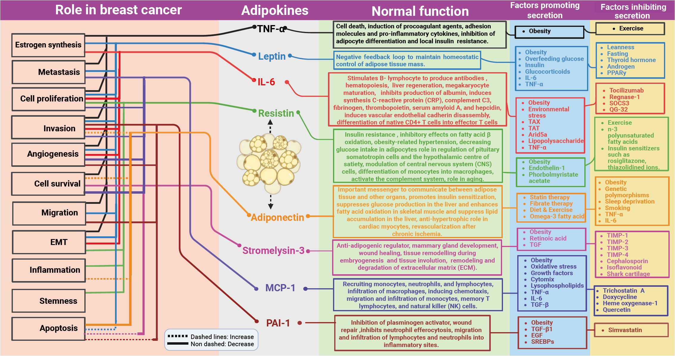
The integrated figure showing the function of adipokines during different physiological conditions—normal physiological functions of adipocytes and obesity-associated adipokines in breast cancer. Factors that can regulate the secretion of adipokines are also indicated in the figure. EGF, epidermal growth factor; EMT, epithelial-mesenchymal transition; IL-6, interleukin-6; MCP-1, monocyte chemoattractant protein-1; PAI-1, plasminogen activator inhibitor-1; PPARγ, peroxisome proliferator-activated receptor γ; SREBP, sterol regulatory element-binding protein; TGF-β, transforming growth factor β; TIMP, tissue inhibitor of metalloproteinases; TNF-α, tumor necrosis factor α.
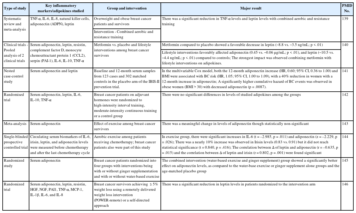
Clinical trials, observational studies, and systematic reviews investigating changes in adipokines among breast cancer patients/survivors with or without various interventions
Notes
Ethics Statement
Not applicable.
Availability of Data and Material
Data sharing not applicable to this article as no datasets were generated or analyzed during the study.
Code Availability
Not applicable.
Author Contributions
Conceptualization: Soonu VK. Data curation: Soonu VK. Formal analysis: Soonu VK, Sreejith K, Deepak Roshan VG. Funding acquisition: Soonu VK. Investigation: Soonu VK, Praveen Kumar Shenoy, Deepak Roshan VG, Vipin Gopinath, Sreejith K. Project administration: Sreejith K. Resources: Sreejith K. Supervision: Sreejith K. Visualization: Soonu VK. Writing—original draft: Soonu VK. Writing—review & editing: Praveen Kumar Shenoy, Vanithapriyaa Karthickeyan, Deepak Roshan VG, Vipin Gopinath, Sreejith K. Approval of final manuscript: all authors.
Conflicts of Interest
The authors declare that they have no potential conflicts of interest.
Funding Statement
No funding to declare.
Acknowledgments
First author Soonu VK was a recipient of a WOS-A grant from DST, Govt of India. For digitizing the images, we acknowledge Biodesk India. Dr Vanithapriyaa Karthickeyan, founder and director, LEYA ART NTELZNX PRIVATE LIMITED, for language editing.