
 E-submission
Articles
- Page Path
- HOME > J Pathol Transl Med > Forthcoming articles > Article
-
Review Article
Obesity-driven adipokine signaling in breast cancer: mechanistic insights, therapeutic challenges, and opportunities for precision oncology -
Vadakke Kunnumma Soonu1
 , Praveen Kumar Shenoy2, Valiyaparampil Gopi Deepak Roshan3, Vipin Gopinath3, Krishnan Sreejith1
 -
DOI: https://doi.org/10.4132/jptm.2026.06.30
Published online: September 9, 2026
1Department of Biotechnology and Microbiology, Dr. Janaki Ammal Campus, Kannur University, Thalassery, India
2Division of Medical Oncology, Department of Clinical Hematology and Medical Oncology, Malabar Cancer Centre, Thalassery, India
3Division of Genetics and Cytogenetics, Department of Clinical Laboratory Service and Translational Research, Malabar Cancer Centre, Thalassery, India
- Corresponding Author: Sreejith Krishnan, PhD Department of Biotechnology and Microbiology, Dr. Janaki Ammal Campus, Kannur University, Thalassery, Kerala - 670661, India Tel: +91-9446870675, Fax: +91-4972711460, E-mail: sreejithkrishnan@hotmail.com
© The Korean Society of Pathologists/The Korean Society for Cytopathology
This is an Open Access article distributed under the terms of the Creative Commons Attribution Non-Commercial License (https://creativecommons.org/licenses/by-nc/4.0) which permits unrestricted non-commercial use, distribution, and reproduction in any medium, provided the original work is properly cited.
- 39 Views
- 1 Download


- Abstract
- INTRODUCTION
- ADIPOKINES AT THE CROSSROADS OF OBESITY AND BREAST CANCER
- TUMOR NICHE IN OBESITY: A DRIVER OF CANCER PROGRESSION AND DIVERSITY
- LEPTIN AND BREAST CANCER: FROM SATIETY HORMONE TO ONCOGENIC SIGNAL—A PARADIGM SHIFT WITH OBESITY
- TNF-α AND BREAST CANCER: A CHANGING ROLE IN TME REMODELING DURING OBESITY
- OBESITY-DRIVEN IL-6 PATHWAYS IN BREAST CANCER: LINKING TUMOR BIOLOGY AND THERAPEUTIC FAILURE
- DECODING THE DUAL ROLE OF PAI-1 IN OBESITY-ASSOCIATED BREAST CANCER
- STROMELYSIN-3 (MMP11): A KEY MEDIATOR OF METASTASIS IN THE ALTERED TME OF OBESITY-DRIVEN BREAST CANCER
- ADIPONECTIN: AN ANTI-PROLIFERATIVE GELATIN-BINDING HORMONE—LOST IN OBESITY
- MCP-1 IN THE TME DRIVING MACROPHAGE RECRUITMENT- LINKING OBESITY, AND BREAST CANCER
- RESISTIN AND THE BREAST CANCER NICHE: A MODULATOR IN OBESITY-ASSOCIATED BREAST CANCER
- CONCLUSION
- NOTES
- REFERENCES
Abstract
- Breast cancer is the leading cause of illness and death among women worldwide, with more than 2.3 million new cases diagnosed each year. The incidence and fatality rates are steadily increasing, notably in Asian countries. Obesity has been established as a major and growing risk factor for breast cancer progression. The World Health Organization reports that adult obesity rates have doubled since 1990. Obesity promotes tumor growth by inhibiting adipokine production and activating cancer-promoting pathways. In obese people, the microenvironment surrounding breast cancer cells is drastically altered. This is mostly due to the malfunctioning of adipocytes (fat cells) and macrophages. These defective cells' adipokines alter signaling pathways required for cancer cell proliferation, survival, and inflammation. This dysregulation has a significant role in tumor development, metastasis, and resistance to traditional cancer treatments, particularly in obese patients. This review highlights the role of adipokines in breast cancer, with a focus on disease progression, therapeutic challenges, and knowledge gaps. Clarifying how obesity alters tumor biology is key to advancing personalized and effective treatments for obese patients.
- Breast cancer is the most frequently diagnosed malignancy in women, with more than 2.3 million new cases reported annually. It continues to be a leading contributor to global cancer-related morbidity and mortality [1]. An increasing incidence and mortality have been reported in Asian countries [2]. Obesity is on the rise, and studies suggest that breast cancer patients with obesity may have a reduced response to treatment. However, this remains debatable and is not currently factored into treatment decisions due to gaps in scientific understanding. Breast cancer risk and outcomes are influenced by multiple factors, including age, family history, genetic predisposition, obesity, alcohol use, and smoking. While obesity is considered a modifiable risk factor, some studies report contradictory findings [3]. Obesity is characterized by excess fat buildup and malfunctioning adipocytes that produce aberrant adipokines into the tumor microenvironment, activating oncogenic pathways such as JAK, signal transducer and activator of transcription 3 (STAT3), nuclear factor кB (NF-κB), and Akt, which promote breast cancer growth [4,5].
- The tumor microenvironment plays a key role in cancer progression by supporting cell proliferation, angiogenesis, and survival [6]. In breast cancer, it is composed mainly of adipocytes, macrophages, and tumor cells. Among these, adipocytes are the most abundant and metabolically active, particularly in obese individuals, and significantly influence tumor cell behavior [7].
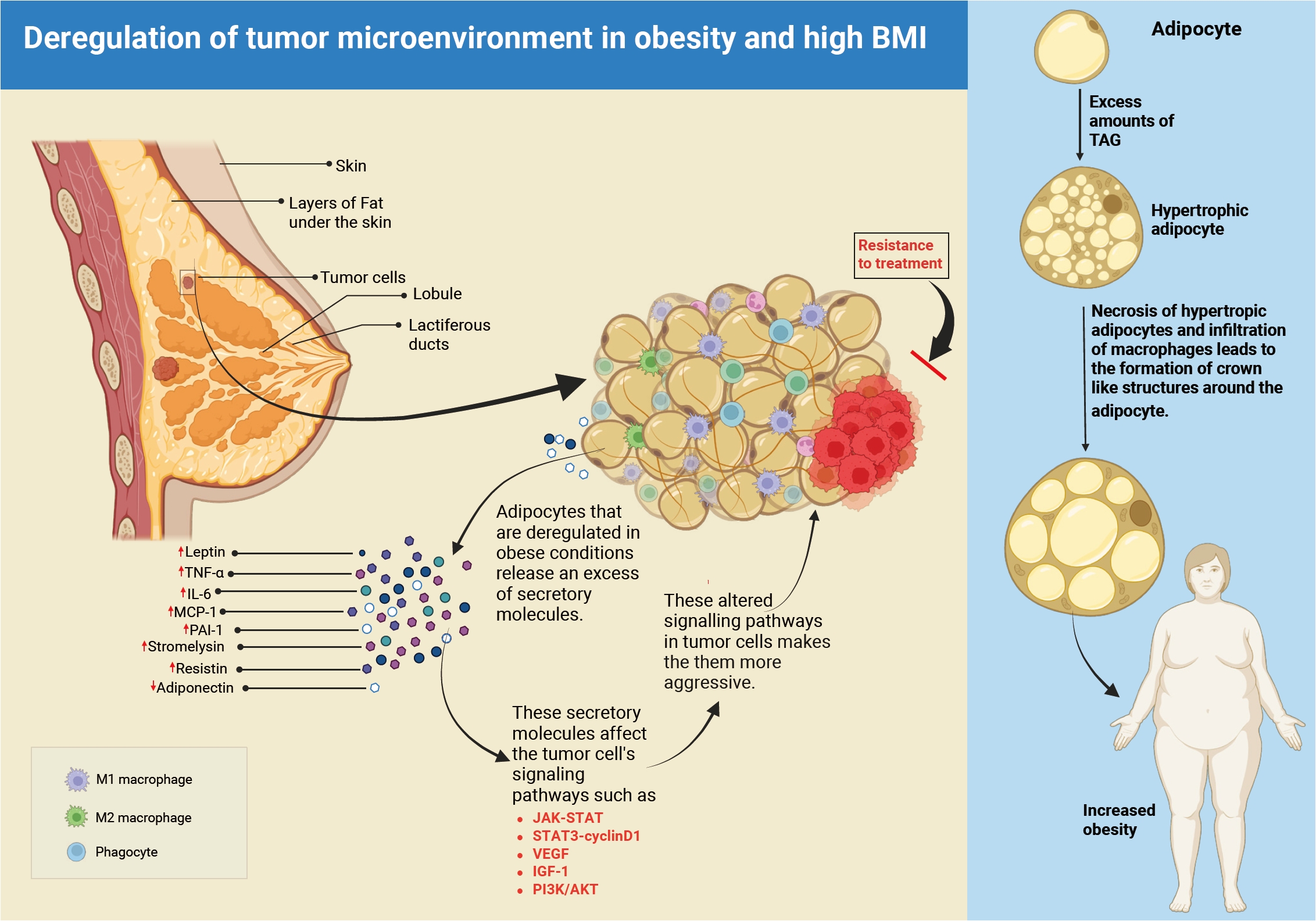
- Breast cancer cells penetrate the mammary fat pad early in the tumor growth process and interact with surrounding adipocytes via secreted chemicals [8]. Obesity has been shown in clinical research to affect breast cancer outcomes, with obese patients frequently presenting with larger, more aggressive tumors and lower survival rates [9,10]. Understanding the link between obesity and breast cancer is critical for determining the molecular mechanisms behind this association. Fig. 1 provides a graphical overview of obesity-associated remodeling of the breast tumor microenvironment. It illustrates the altered secretion from adipocytes and deregulation of oncogenic signaling in tumor cells.
- In this review, we have discussed the various signaling pathways involved and their translational importance, with a special emphasis on the involvement of adipokines released by dysfunctional, hypertrophic adipocytes in the tumor microenvironment. We have also mentioned the current knowledge gaps and suggest future research directions for establishing customized therapy regimens for obese breast cancer patients.
INTRODUCTION
- Adipose tissue secretes adipokines, which act as essential regulators of metabolic and inflammatory pathways [11]. These include hormones (leptin), cytokines (interleukin [IL]-6, tumor necrosis factor [TNF], and IL-1β), and angiogenic agents (vascular endothelial growth factor [VEGF]). These mediators play a role in the development, progression, and treatment resistance of obesity-related malignancies, notably breast cancer [12]. Although adiponectin has anti-inflammatory properties, other adipokines such as resistin, monocyte chemoattractant protein-1 (MCP-1), leptin, IL-6, transforming growth factor-β, IL-10, plasminogen activator inhibitor-1 (PAI-1), and stromelysin increase pro-inflammatory responses that contribute to tumor formation [13].
- Adipokines act like hormones, serving as growth factors that regulate insulin resistance and influence lipid and glucose metabolism. In obesity, excessive fat and dysfunctional adipocytes disrupt adipokine production and secretion, leading to altered immune responses and etiology of various diseases [14]. Adipokines are involved in both pro- and anti-inflammatory responses, and their dysregulation in the cancer niche significantly affects tumor growth and metastasis.
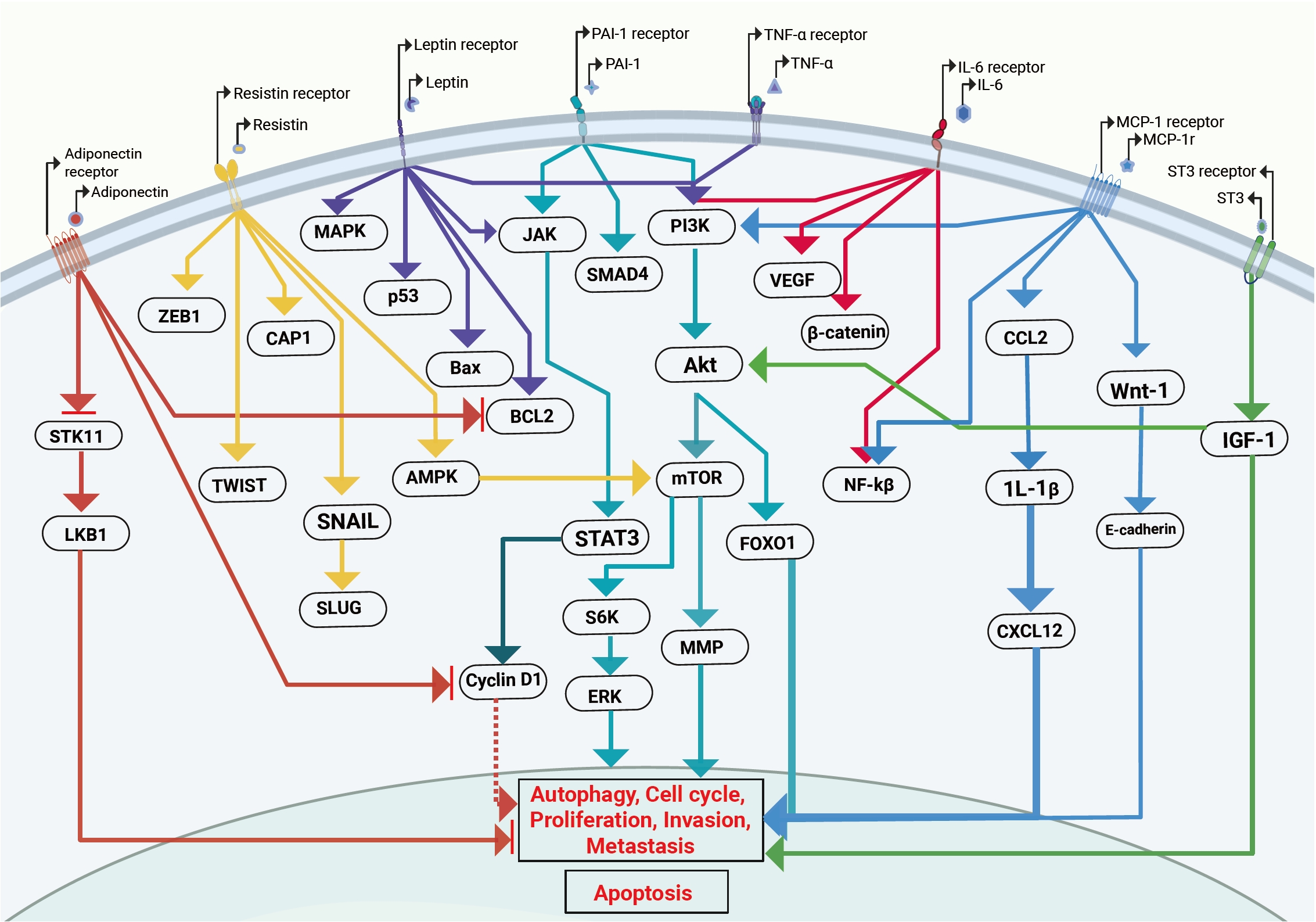
- In breast cancer, adipokines independently and synergistically activate key signaling pathways that drive tumor growth, proliferation, invasion, metastasis, and survival. Fig. 2 illustrates different oncogenic pathway deregulation via the adipocyte secretory molecules and promoting tumor progression. For example, leptin stimulates cancer cell growth and survival, while TNF-α and IL-6 promote inflammation and tumor progression [15]. Adiponectin's anti-inflammatory properties can inhibit cancer cell proliferation and spread. Maintaining a balance of pro- and anti-inflammatory adipokines in the tumor microenvironment is therefore critical. An imbalance impairs adipose tissue homeostasis, resulting in persistent inflammation. In obesity, adipokine imbalances have an important role in regulating metabolism and signaling, eventually impacting tumor progression and treatment response.
ADIPOKINES AT THE CROSSROADS OF OBESITY AND BREAST CANCER
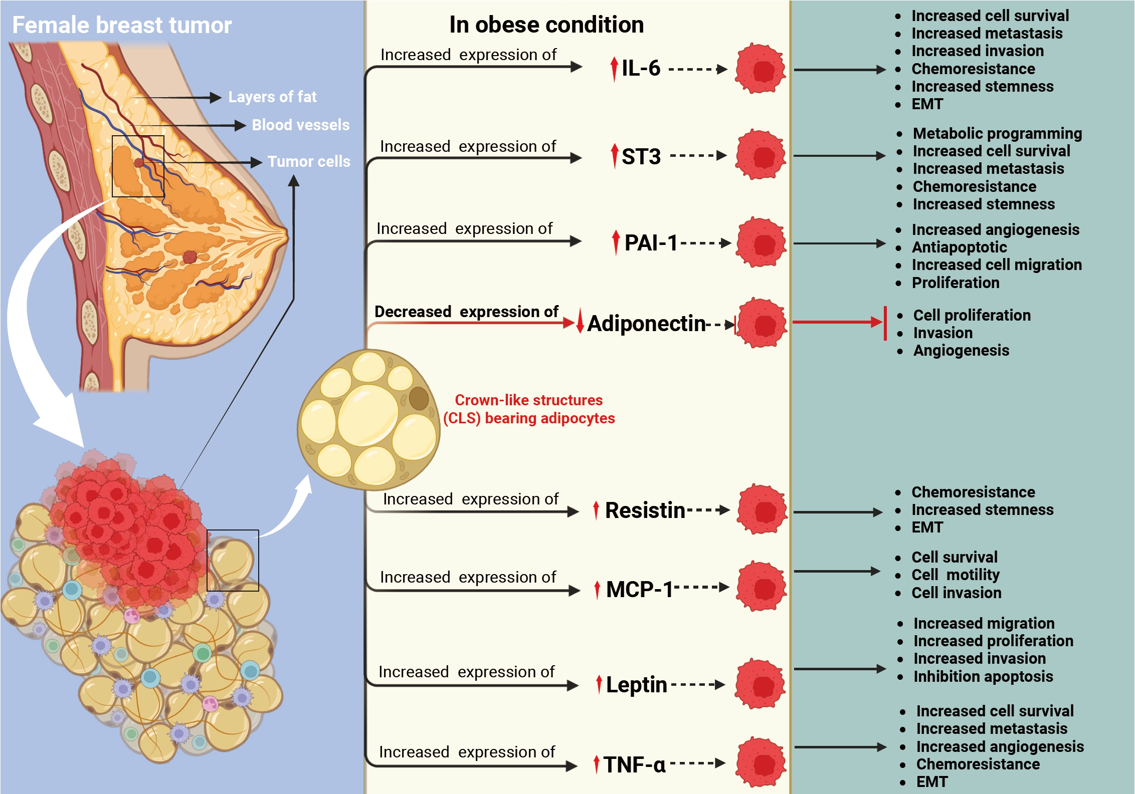
- Normal niches are made up of fibroblastic cells, immunological cells, endothelium and perivascular cells (or their progenitors), extracellular matrix (ECM) components, and signaling molecules like cytokines and growth factors. The tumor niche is a component of the tumor microenvironment (TME), which consists of stromal and non-cancerous cells that interact with tumor cells. Thus, tumor behavior is regulated by both the biology of the tumor cells and the features of their microenvironment. Fig. 3 shows the impact of adipokines on breast cancer progression in an obesity-associated tumor niche. Increased expression of leptin, TNF-α, IL-6, MCP-1, resistin, PAI-1, and stromelysin-3, together with reduced adiponectin, promotes key hallmarks of cancer, including enhanced proliferation, invasion, angiogenesis, stemness, epithelial-mesenchymal transition, and therapeutic resistance.
- Intratumoral heterogeneity accelerates disease development, lowers therapy efficacy, and affects patient survival. Tumor heterogeneity is caused by genetic and epigenetic alterations inside tumor cells, as well as effects from the TME. In obesity, adipocyte dysfunction and hypertrophy further disturb the TME by affecting the release of over 600 bioactive signaling molecules [16].
- These peptides are a diverse group that influences tumor cell behavior by affecting survival pathways, metabolism, and energy balance [17]. Interestingly, heterogeneity exists both within a single tumor (intratumoral) and between different tumors (intertumoral). This may explain why tumors with similar genomic profiles respond differently to treatment. In addition, obesity-related changes in the tumor environment can alter the epigenetic landscape and gene expression of tumor cells. As adipocytes grow and become hypertrophic, they stiffen, interrupting membrane signaling and leading to decreased cell function and metabolic dysregulation.
- Adipokine secretion is directly related to adipocyte size; as adiposity increases, pro-inflammatory adipokines are overproduced and anti-inflammatory adipokines are underproduced [18,19]. To understand the biology of breast cancer and treatment outcomes, it is vital to understand the alterations in the breast cancer niche caused by unregulated adipocytes, which contribute to cancer growth and metastasis (Fig. 2).
TUMOR NICHE IN OBESITY: A DRIVER OF CANCER PROGRESSION AND DIVERSITY
- Leptin is a 16-kD peptide hormone created mostly by adipocytes, and the amount of its production is positively related to obesity and higher in obese people [20,21]. Leptin binds to a specific leptin receptor (LEPR) on the cell surface. Wang et al. [22] found that 70.5% of tumor tissues show high levels of LEPR. It was much greater than the benign breast tissues (56.3%) and normal para-tumoral tissues (44%). Higher levels of leptin in the tumor environment may influence signaling in tumor cells. Although adipose tissue is strongly linked to breast cancer, the role of leptin in the disease is still debated. Both in-vivo and in-vitro studies suggest that leptin can affect key cellular functions such as motility, migration, and tumor progression [23,24]. The findings suggest that in obesity, leptin may promote tumor invasion and angiogenesis, leading to metastasis and reduced treatment efficacy. This could explain the higher cancer mortality risk in overweight patients.
- One of the most difficult aspects of breast cancer treatment is distant metastasis, which is caused by tumor cells undergoing epithelial-mesenchymal transition (EMT). He et al. [25] discovered that leptin enhances invasion and migration in triple-negative breast cancer (TNBC) by activating PLOD2 (a protein involved in the hydroxylation of lysyl residues in collagen-like peptides), which stimulates the phosphoinositide 3-kinase (PI3K)/Akt and JAK/STAT3 pathways. Another study utilizing TNBC and estrogen receptor (ER)+ cells demonstrated that leptin promotes production and activation of matrix metalloproteinase (MMP)-2 and MMP-9, as well as FAK activation via Src, resulting in higher invasion and phenotypic alterations in TNBC cells [26]. Strong et al. [27], on the other hand, proposed that leptin is more effective on ER+ cells. They discovered that genes related to cell cycle, apoptosis, and migration (CDKN2A, GSTP1, SFRP1, PLAU, THBS1, and CSF) were changed in ER+ cells via the ERK1/ERK2 signal and cross-talk between LEPR and ER [27-29]. In obese women, a high dose of leptin generates estradiol and estrogen via increasing aromatase activity [30]. Leptin promotes tumor recurrence and poor prognosis in TNBC by enhancing the self-renewal capacity of cancer cells [31]. In other cancers, leptin has been shown to activate VEGF through NF-κB/hypoxia-inducible factor-1α signaling via both canonical and non-canonical pathways. Beyond its role in EMT, leptin also supports cancer cell proliferation and survival by activating multiple signaling pathways, including insulin-like growth factor 1 (IGF-1)/activator protein 1 (AP-1), NOTCH/survivin, PI3K/AKT/sterol regulatory element-binding protein 2/acetyl-CoA acetyl transferase 2(ACAT2)/STAT3/cyclin D1, extracellular signal-regulated kinase 1/2, AP-1, mitogen-activated protein kinase (MAPK), and PI3K [32,33]. Despite significant research demonstrating that changing leptin affects tumor pathways such as PI3K/Akt, JAK/STAT3, and MAPK in breast cancer, clinical translation remains elusive. This disparity could be attributed to (1) the varied behavior of ER+, TNBC, and human epidermal growth factor receptor 2 (HER2)+ malignancies in respect to leptin, (2) a lack of knowledge of normal leptin ranges in clinical practice, (3) a lack of effective therapeutic agents, and (4) an insufficient understanding of the tumor microenvironment.
LEPTIN AND BREAST CANCER: FROM SATIETY HORMONE TO ONCOGENIC SIGNAL—A PARADIGM SHIFT WITH OBESITY
- TNF-α, the first inflammatory cytokine identified as an adipokine, acts in both autocrine and paracrine ways. It influences processes such as apoptosis, MMP production, and angiogenesis, which can promote tumor cell survival and metastasis [34,35]. Obesity has been linked to elevated levels of TNF-α in adipose tissue [36,37]. TNF-α promotes estrogen production by increasing aromatase expression in adipose tissue [38]. It may contribute to tumor formation in ER+ breast cancer by stimulating ER signaling even more. High TNF-α levels have been linked to metastases and advanced clinical stage in both HER2+ and ER+ breast cancers [39,40]. Wolczyk et al. [41] found that TNF-α increases cell migration in ER+ and TNBC cells by upregulating MMP9, CD26, and fibroblast activation protein-α. Gangliosides (GD3) play a role in cell development, differentiation, and proliferation. TNF-α has been shown to regulate GD3 synthase (GD3S) in breast cancer [42]. TNF-α plays a key role in tumor cell invasion and EMT. It activates major signaling pathways such as MAPK, PI3K/Akt, and ZEB1/ZEB2, and is influenced by the migration-inhibitory factor. TNFα also increases the production of MMPs and α2β1 integrin, which promote tumor invasion, migration, and vascular inflammation [43-45]. Besides promoting tumor migration and invasion, TNF-α also suppresses the anti-tumor immune response. A recent review reported that clinical studies have shown that TNF-α inhibition can enhance the effectiveness of immune checkpoint inhibitors in cancer therapy [46]. Many studies indicate that TNF-α has a significant influence on tumor growth and medication resistance. However, major challenges remain. (1) TNFα is produced not only by adipose tissue but also by various other cell types. (2) It has a dual nature with no reliable marker to predict its function or mode of action. (3) There is limited understanding of its role in the interaction between breast cancer and adipose cells within the TME in clinical settings.
TNF-α AND BREAST CANCER: A CHANGING ROLE IN TME REMODELING DURING OBESITY
- Human IL-6 is a 26-kDa glycoprotein that mainly acts as a pro-inflammatory molecule, though some studies also show it can have anti-inflammatory effects [47]. IL-6 is secreted by many cells, with mature adipose tissue being a major source. Studies have shown that obese individuals have higher plasma levels of IL-6 [48]. IL-6 has a multifunctional effect on tumor growth, cell proliferation, stemness, metastasis, and tumor immunology [47,49]. He et al. [25] demonstrated that adipocyte-derived IL-6 enhances TNBC metastasis by boosting PLOD2 expression via PI3K/Akt and JAK/STAT3 activation. IL-6 also plays a key role in estrogen production; therefore, it may have a large influence in ER+ breast cancer. IL-6 promotes the activities of aromatase, estradiol 17beta-hydroxysteroid dehydrogenase, and estrone sulfatase [50]. A study reported that trastuzumab resistance in HER2-overexpressing cells is linked to increased IL-6 expression [51]. IL-6 signaling is complex and occurs through three modes: classical signaling, trans-signaling, and cluster (trans-presentation) signaling [52]. Sindhu et al. [53] found that obesity elevates IL-6R and IL-6 expression in subcutaneous adipose tissue. This suggests that, in obese individuals, the TME may alter IL-6 signaling through these pathways, potentially contributing to more aggressive tumor progression and greater tumor heterogeneity via cluster signaling. Deregulation of the IL-6/JAK/STAT3 signaling pathway has emerged as a major cause of therapy resistance in HER2+ breast cancer and TNBC. Tocilizumab and Sarilumab are IL-6Ra inhibitors in various stages of clinical trials (NCT03135171, NCT04333706) to treat breast cancer. It is worth noting that clinical trials with IL-6Ra inhibitors have begun in breast cancer, although the potential for use in obesity-associated breast cancer requires further investigation. Further investigation is needed to create a procedure for identifying the subgroup of obese patients who respond best to IL-6–targeted therapy.
OBESITY-DRIVEN IL-6 PATHWAYS IN BREAST CANCER: LINKING TUMOR BIOLOGY AND THERAPEUTIC FAILURE
- PAI-1 is a 45-kDa single-chain glycoprotein secreted by adipose tissue. It promotes breast tumor growth by enhancing cell proliferation, angiogenesis, and migration, while reducing apoptosis [54,55]. Elevated levels of free fatty acids, triglycerides, and TNF-α increase circulating PAI-1 [56,57]. PAI-1 inhibits the serine proteases urokinase-type plasminogen activator (uPA) and tissue-type plasminogen activator (tPA), thereby blocking plasminogen-to-plasmin conversion and subsequent MMP activation [58]. Additionally, the PAI-1–uPA heterodimer can exert non-proteolytic, pro-tumor effects by increasing neutrophil accumulation in the tumor niche [59]. Although the exact mechanism is unclear, blocking this heterodimer could be a potential therapeutic strategy for obesity-related breast cancer. However, studies have demonstrated that altered PAI-1 is involved in the radioresistance via DNA repair, STAT3 mediated metastasis, migration [60-64] Studies (in-vivo and in-vitro) show that obesity-related resistance in TNBC cells is mediated by PAI-1. Further clinical trials are needed to confirm these findings. PAI-1 plays a key role in the interaction between adipocytes and cancer cells. It is involved in cytoskeleton remodeling and SMAD4 signaling dysregulation, leading to invasion and EMT in breast cancer. PAI-1 also activates PI3K-Akt signaling through integrin αVβ3. Overall, in breast cancer, PAI-1 promotes proliferation, migration, invasion, metastasis, and angiogenesis, reduces apoptosis, and is linked with poor prognosis [65-67]. Clinical data have revealed that obesity increases the level of PAI-1, which is associated with a poor prognosis in breast cancer [54]. In other investigations, higher PAI-1 expression has been linked to decreased relapse-free survival [68-70]. In summary, PAI-1–uPA–tPA is a very complicated axis-related signaling that requires particular attention to its origin and downstream signaling. After understanding its role and manner of regulation in different subgroups, it may be effectively exploited in clinical settings. PAI-1–uPA's dual proteolytic and non-proteolytic activities are poorly known, which limits its therapeutic applicability. Given the significant role the tumor niche plays in regulating tumor heterogeneity and prognosis, it is essential to investigate the involvement of PAI-1–uPA in the axis between adipocytes and tumor cells in obese conditions.
DECODING THE DUAL ROLE OF PAI-1 IN OBESITY-ASSOCIATED BREAST CANCER
- Stromelysin-3 (ST3), or MMP11, has been linked to poor treatment outcomes and an invasive kind of malignancy [71]. ST3 has been linked to tumor growth since its inception [72]. Clinical studies have revealed that patients with poor overall survival and relapse-free survival had increased stromal expression of MMP11 [73,74]. ST3/MMP11 expression has also been linked to local tumor aggressiveness [75,76]. In obese situations, MMPs play a crucial role in adipose and cancer cell interaction, notably in the early stages of cancer cell invasion into connective tissue [77-79]. Another investigation found that ST3-mediated paracrine signaling enhances tumor migration to connective tissues [80]. Supporting these findings, investigations have revealed that in obese individuals, ST3 levels are higher, as is invasive angiogenesis [81,82], as well as local aggressive tumors [75,76]. Increased MMP11 expression has also been linked to lapatinib resistance in breast cancer cells [83]. Roscilli et al. [84] propose MMP11 as a viable marker for cancer immunotherapy techniques for breast cancer. In vitro studies have shown that ST3 regulates pathways such as SMAD2 and IGF-1/Akt/forkhead box O1, which affect cell proliferation, survival, and invasion [85,86]. Antiapoptotic impact of p42/p44 MAPK and Akt in HER2-positive breast cancer [84,87,88]. Several studies have shown that ST3 inhibitors can target cancer cells, but therapeutic success is still elusive, owing to a lack of knowledge of ST3's role in ectopic and paracrine regulation, as well as its impact on the TME (Fig. 3).
STROMELYSIN-3 (MMP11): A KEY MEDIATOR OF METASTASIS IN THE ALTERED TME OF OBESITY-DRIVEN BREAST CANCER
- The ADIPOQ gene encodes the 30-kDa, 244-aa protein known as adiponectin, which is mostly released by adipocytes [89]. It acts as an anti-proliferative agent since its secretion is inversely correlated with body weight [90,91]. Adiponectin expression is also inversely correlated with other pro-tumorigenic adipokines, such as TNF-α and IL-6 [92,93]. Reduced levels of circulating adiponectin have been linked to an increased risk of breast cancer, according to a meta-analysis of 119 studies [94]. Reduced adiponectin levels in obesity have been reported to be positively correlated with physiologically aggressive phenotypes [95-97]. Similarly, it has been discovered that carriers of the BRCA mutation who have lower levels of adiponectin are more likely to develop breast cancer [98]. In terms of function, adiponectin inhibits Bcl2 and Bag1 and activates p53, caspase 8, and Bax to reduce tumor neovascularization and promote death. Therefore, obese people with lower adiponectin levels are more prone to tumor initiation and progression. In breast cancer, serum adiponectin levels may also function as a predictive biomarker for survival and disease aggressiveness, possibly directing the escalation or intensification of treatment [99]. It has been shown that adiponectin causes cell cycle arrest and inhibits the growth of TNBC through caspase 1, 3, 8, and 9 [100-104]. Similarly, additional in vitro research has demonstrated that unregulated Wnt and Akt pathways [105], PI3K/Akt signaling [106], and AMP-activated protein kinase (AMPK) [107], as well as serine/threonine kinase 11 (STK11) change the production and secretion of adipokines in TNBC. By enhancing PTP1B and inhibiting leptin-induced JAK2/STAT3 oncogenic signaling, adiponectin prevents breast cancer invasion by activating protein phosphatase 2A via the STK11/liver kinase B1 (LKB1)–AMPK–unc-51-like kinase 1 axis [107-110]. Although there is enough evidence to support the detrimental function of adiponectin in tumor progression, it is still rarely used in clinical settings because of a few questions, such as (1) what are the baseline levels of adiponectin in patients with lean and obese breast cancer?, (2) influence on several molecular groupings, (3) how it relates to various therapies, and (4) the role it plays in altering the tumor niche.
ADIPONECTIN: AN ANTI-PROLIFERATIVE GELATIN-BINDING HORMONE—LOST IN OBESITY
- A 99-amino acid precursor MCP-1 protein is encoded by the MCP-1 gene (SCYA2). The mature 76-amino acid MCP-1 protein is formed by cleaving its N-terminal region; additional glycosylation, whether present or absent, divides it into two isoforms, designated I and P [111]. It has been shown that obese people have higher levels of circulating MCP-1 [112,113]. Adipose-derived MCP-1 has been linked to mammary carcinogenesis, according to in-vivo research [114]. Altered MCP-1 level is linked to tumor aggressive growth and metastasis altering the biological phenotype of the disease [115], MCP-1 also plays a role in the infiltration of macrophages into the TME, which is linked to metastasis and poor clinical outcomes [116-118]. Clinical studies have shown that in human breast cancer, macrophage infiltration is linked to a worse prognosis and a higher risk of metastatic disease [119]. In TNBC, high levels of MCP-1 in the TME are linked to increased metastasis, invasion, and a worse clinical outcome [120,121]. MCP-1–induced macrophage and crown-like structure development in obese people increases angiogenesis through C-C motif chemokine ligand 2/IL-1β/C-X-C motif chemokine ligand 12 and VEGF/VEGF receptor signaling pathways [122]. Additional research has shown that MCP-1 enhances breast cancer cell survival and motility by inhibiting E-cadherin via the TWIST, Wnt1, Smad3, and MAPK signaling cascades [123,124]. MCP-1 levels have been demonstrated to impact ERK/glycogen synthase kinase-3β/Snail signaling in MCF7 cells [125]. Tamoxifen resistance has also been documented in breast cancer patients with raised MCP-1 levels via the PI3K/Akt/mechanistic target of rapamycin (mTOR) signaling pathway [126]. MCP-1 neutralizing antibodies, such as C1142 and MAB279, are receiving interest as possible treatments; however, they are still in the preclinical stage [127]. However, additional study is required to fully understand MCP-1's involvement in cancer progression and to find the most effective therapy targets and techniques.
MCP-1 IN THE TME DRIVING MACROPHAGE RECRUITMENT- LINKING OBESITY, AND BREAST CANCER
- Resistin is a pro-inflammatory cytokine that was initially identified for its function in insulin resistance, but recent research has linked it to cancer development and therapeutic resistance. Resistin, which is produced predominantly by adipocytes and adipose-derived macrophages, has been shown to be higher in obese breast cancer patients [128]. Resistin causes breast cancer by increasing pro-inflammatory cytokines through the NF-κB pathway and initiating tumors through CAP1 [129]. Breast cancer cells use the Toll-like receptor 4 (TLR4)/NF-κB/STAT3 signaling pathway to produce EMT, increase cancer stemness, and facilitate metastasis [130,131].
- Resistin also promotes cellular invasion by increasing ezrin/radixin/moesin protein phosphorylation and upregulating EMT markers (SNAIL, SLUG, ZEB1, TWIST1, fibronectin, and vimentin), while decreasing epithelial indicators such as E-cadherin and claudin 1 [132-134]. Resistin levels have been related to increased tumor size, grade, lymph node invasion, and other undesirable clinical characteristics [135-138], indicating its potential as an independent prognostic biomarker and a candidate for targeted therapy.
- Preclinical findings indicate resistin's significance in breast cancer; however, clinical translation utilization remains limited. (1) There are no standardized methods for assessing resistin in patients, (2) its involvement in various molecular subtypes of breast cancer, and (3) its prognostic usefulness for therapy response have yet to be clearly demonstrated.
RESISTIN AND THE BREAST CANCER NICHE: A MODULATOR IN OBESITY-ASSOCIATED BREAST CANCER
- Obesity is a well-known factor associated with breast cancer, and research has focused on understanding how obesity-related factors influence cancer development and progression. Excess fat tissue (accumulation of adipocytes) leads to prolonged low-grade inflammation, which affects the tumor microenvironment. Increased adipose tissue alters the secretion of molecules such as TNF-α, leptin, IL-6, PAI-1, ST3, adiponectin, MCP-1, and resistin. Together, these are involved in the modification of the tumor niche, providing a condition that promotes the initiation, progression, resistance, and aggressiveness of breast cancer.
- Adipokines exert their combined and independent effects on the major intracellular signaling networks involved in breast cancer progression, particularly in the obesity context. They are generally secreted in a group or as a set of adipokines and they function in a complex and interconnected signaling network. In Fig. 2, it is shown that signaling pathways triggered by leptin, TNF-α, IL-6, MCP-1, resistin, and PAI-1 affect the cell cycle, invasion, and metastasis via JAK-STAT, mTOR-like pathway. The niche biology does not depend only on one or two adipokines; it also depends on the ratio of pro- and anti-tumor adipokines present in the niche. Hence, the studies based on a single or a few selected adipokines may not give a clear picture of tumor niche alteration. Much deeper investigation on the pro- and anti-tumor adipokines’ orchestration in the tumor niche is needed for translational development.
- In our review, we have discussed in detail the obesity-associated adipokines and tumor niche remodeling. We have explained in detail about each pro- and anti-tumorigenic adipokine and its regulatory roles. However, our understanding of tumor niche and its translational application is partial due to limitations in conventional 2D cell culture models for co-culture, which can be addressed in future by using the 3D co-culture models.
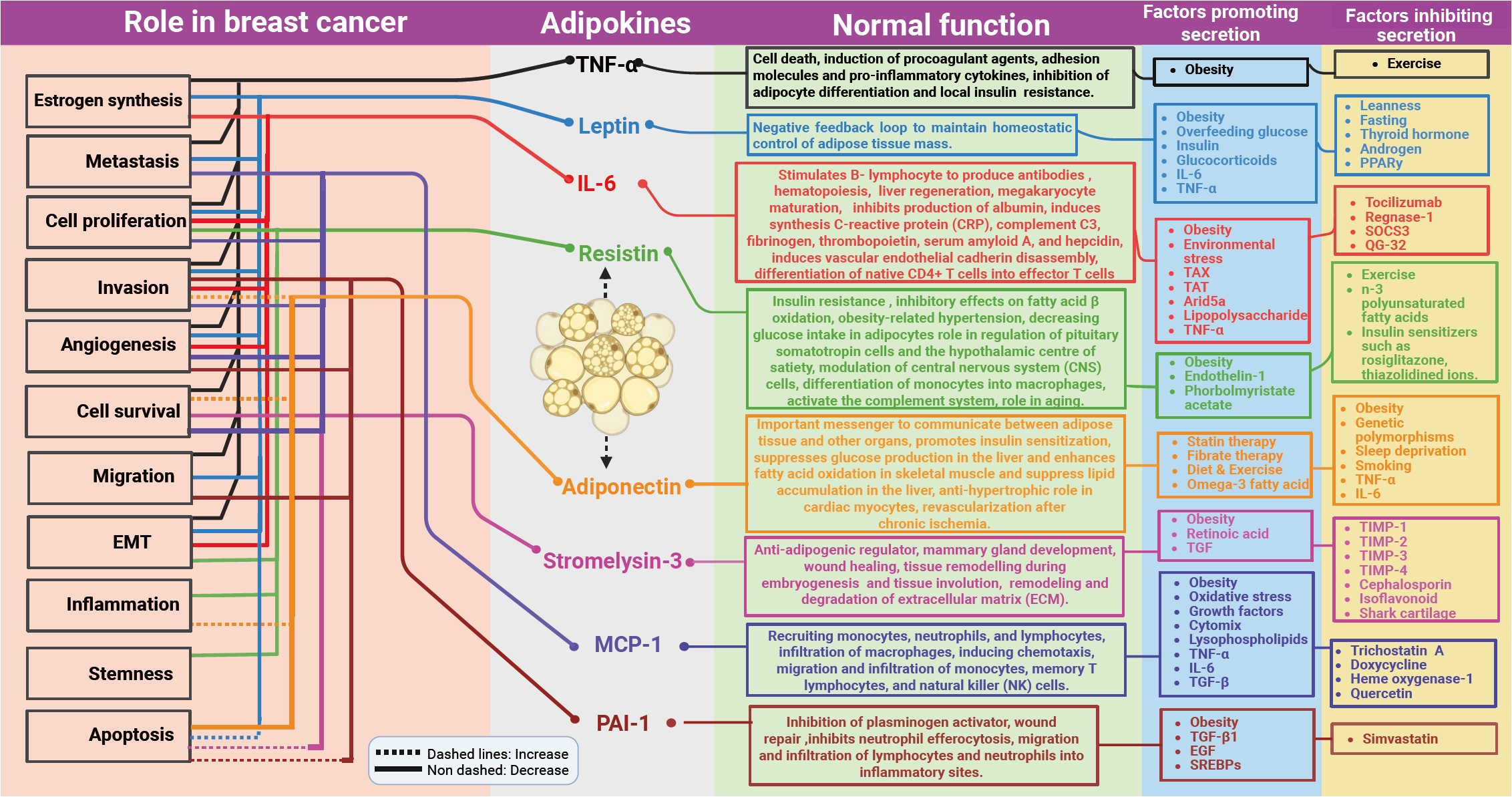
- These obesity-related variables serve as niche modifiers, forming a microenvironment that promotes breast cancer development and progression. By affecting inflammation, cell signaling, and the ECM, these chemicals promote tumor genesis, survival, and metastasis. The current evidence demonstrates the important connection between obesity and tumor development, emphasizing the importance of adipokines released by adipocytes. In Fig. 4, it has been shown how adipocyte secretory molecules change in normal and obese physiological conditions in a breast cancer niche. This understanding has yet to be properly applied in clinical practice. Few clinical trials, observational studies, and systematic reviews have been performed to explore the changes in adipokines among breast cancer patients/survivors with or without various interventions (Table 1) [139-146]. The interventions mainly involve the use of metformin and/or weight loss mechanisms using exercise, which have also demonstrated changes in adipokine profiles. However, to date, no standardized clinical protocol has been developed to segregate the obesity-associated aggressive breast cancer group and separate therapeutic or clinical management protocols for that group. It is vital to critically evaluate existing data, identify gaps, and create targeted research to fill those gaps and enable clinical application. Major gaps include the lack of established baseline values for these markers, inadequate evaluation of their robustness across different patient populations, unclear behavior across various molecular subtypes of breast cancer, and a lack of clinical data supporting therapeutic interventions targeting these markers. To close the translational gap, it is necessary to design and perform well-structured, multicenter clinical studies that can systematically analyze these markers and establish their clinical importance.
CONCLUSION
Ethics Statement
Not applicable.
Availability of Data and Material
Data sharing not applicable to this article as no datasets were generated or analyzed during the study.
Code Availability
Not applicable.
Author Contributions
Conceptualization: Soonu VK. Data curation: Soonu VK. Formal analysis: Soonu VK, Sreejith K, Deepak Roshan VG. Funding acquisition: Soonu VK. Investigation: Soonu VK, Praveen Kumar Shenoy, Deepak Roshan VG, Vipin Gopinath, Sreejith K. Project administration: Sreejith K. Resources: Sreejith K. Supervision: Sreejith K. Visualization: Soonu VK. Writing—original draft: Soonu VK. Writing—review & editing: Praveen Kumar Shenoy, Vanithapriyaa Karthickeyan, Deepak Roshan VG, Vipin Gopinath, Sreejith K. Approval of final manuscript: all authors.
Conflicts of Interest
The authors declare that they have no potential conflicts of interest.
Funding Statement
No funding to declare.
Acknowledgments
First author Soonu VK was a recipient of a WOS-A grant from DST, Govt of India. For digitizing the images, we acknowledge Biodesk India. Dr Vanithapriyaa Karthickeyan, founder and director, LEYA ART NTELZNX PRIVATE LIMITED, for language editing.
| Type of study | Key inflammatory markers/adipokines studied | Group and intervention | Major result | Reference |
|---|---|---|---|---|
| Systematic review and meta-analysis | TNF-α, IL-6, IL-8, natural killer cells, adiponectin (ADPN), leptin | Overweight and obese breast cancer patients and survivors | There was a significant reduction in TNF-α levels and leptin levels with combined aerobic and resistance training | [139] |
| Intervention - Combined aerobic and resistance training | ||||
| Clinical trials - Pooled analysis of 2 clinical trials | Serum adiponectin, leptin, resistin, complement factor D, monocyte chemoattractant protein 1 (CCL2), serpin (PAI-1), IL-6, IL-10, TNF-α | Metformin vs. placebo and lifestyle interventions among breast cancer survivors | Metformin compared to placebo showed a favorable decrease in leptin (−8.8 vs. −3.5 ng/mL; p < .01) | [140] |
| Lifestyle interventions favorably affected adiponectin (0.45 vs. −0.06 µg/mL; p < .01), and leptin (−10.5 vs. −4.4 ng/mL; p < .01) compared to controls; The strongest impact was observed combining metformin with lifestyle interventions on adipokines | ||||
| Nested case-control study | Serum adiponectin and leptin | Baseline and 12-month serum samples from 123 cases and 302 matched controls in the placebo arm of the IBIS-II prevention trial | In the multivariable Cox model, both the 12-month adiponectin increase (HR, 0.60; 95% CI, 0.36 to 1.00) and BMI were associated with BC risk (HR, 1.05; 95% CI, 1.00 to 1.09), with a 40% reduction in women with a 12-month increase in adiponectin; A significantly higher cumulative hazard of BC events was observed in obese women (BMI > 30) with decreased adiponectin (p = .0087) | [141] |
| Randomized trial | Serum adiponectin, leptin, IL-6, IL-10, TNF-α | Breast cancer patients on adjuvant hormones were randomized to high-intensity interval training, moderate-intensity continuous training or a control group | There were no significant differences in levels of studied adipokines among the groups | [142] |
| Meta-analysis | Serum adiponectin | Effect of exercise among breast cancer survivors | There was a meaningful change in levels of adiponectin though statistically non-significant | [143] |
| Single-blinded prospective controlled trial | Circulating serum biomarkers of IL-6, irisin, leptin, and adiponectin levels were measured before chemotherapy and after the last chemotherapy cycle | Aerobic exercise among patients receiving chemotherapy; breast cancer patients also were part of this study | In exercise group, there were significant increases in IL-6 (t = –2.985, p = .011) and adiponectin (z = –2.229, p = .026); There was a nearly 10% increase in irisin levels (0.83 vs. 0.91) but it did not reach statistical significance (t = 0.840, p = .416); The correlation between Δ of leptin and adiponectin (r = –0.635, p = .015) and the correlation between Δ of leptin and irisin (r = 0.802, p = .001) were found significant | [144] |
| Randomized study | Serum adiponectin | Breast cancer patients randomized into four groups with interventions being with or without ginger supplementation and with or without water-based exercise | The combined intervention (water-based exercise and ginger supplement) group showed a significantly better effect on adiponectin levels, as compared to the water-base exercise or ginger supplement alone groups and the age-matched placebo group | [145] |
| Randomized trial | Serum adiponectin, leptin, resistin, HGF, NGF, PAI-1, TNF-α, MCP-1, IL-1β, IL-6, and IL-8 | Breast cancer survivors achieving ≥5% weight loss using a remotely delivered weight loss intervention (POWER-remote) or a self-directed approach | There was a significant reduction in leptin levels in patients randomized to the intervention arm | [146] |
TNF-α, tumor necrosis factor α; IL, interleukin; CCL2, C-C motif chemokine ligand 2, PAI-1, plasminogen activator inhibitor-1; HR, hazard ratio; CI, confidence interval; BMI, body mass index; BC, breast cancer; HGF, hepatocyte growth factor; NGF, nerve growth factor; MCP-1, monocyte chemoattractant protein-1.
- 1. Bray F, Laversanne M, Sung H, et al. Global cancer statistics 2022: GLOBOCAN estimates of incidence and mortality worldwide for 36 cancers in 185 countries. CA Cancer J Clin 2024; 74: 229-63. ArticlePubMedPMC
- 2. Huang J, Ngai CH, Deng Y, et al. Cancer incidence and mortality in Asian countries: a trend analysis. Cancer Control 2022; 29: 10732748221095955.ArticlePubMedPMCPDF
- 3. Cohen SY, Stoll CR, Anandarajah A, Doering M, Colditz GA. Modifiable risk factors in women at high risk of breast cancer: a systematic review. Breast Cancer Res 2023; 25: 45.ArticlePubMedPMCPDF
- 4. Horwitz A, Birk R. Adipose tissue hyperplasia and hypertrophy in common and syndromic obesity: the case of BBS obesity. Nutrients 2023; 15: 3445.ArticlePubMedPMC
- 5. Arjunan A, Song J. Pharmacological and physiological roles of adipokines and myokines in metabolic-related dementia. Biomed Pharmacother 2023; 163: 114847.ArticlePubMed
- 6. Wang Q, Shao X, Zhang Y, et al. Role of tumor microenvironment in cancer progression and therapeutic strategy. Cancer Med 2023; 12: 11149-65. ArticlePubMedPMCPDF
- 7. Bernard JJ, Wellberg EA. The tumor promotional role of adipocytes in the breast cancer microenvironment and macroenvironment. Am J Pathol 2021; 191: 1342-52. ArticlePubMedPMC
- 8. Blucher C, Stadler SC. Obesity and breast cancer: current insights on the role of fatty acids and lipid metabolism in promoting breast cancer growth and progression. Front Endocrinol (Lausanne) 2017; 8: 293.ArticlePubMedPMC
- 9. Protani M, Coory M, Martin JH. Effect of obesity on survival of women with breast cancer: systematic review and meta-analysis. Breast Cancer Res Treat 2010; 123: 627-35. ArticlePubMedPDF
- 10. Acevedo F, Walbaum B, Muniz S, et al. Obesity is associated with early recurrence on breast cancer patients that achieved pathological complete response to neoadjuvant chemotherapy. Sci Rep 2022; 12: 21145.ArticlePubMedPMCPDF
- 11. Taylor EB. The complex role of adipokines in obesity, inflammation, and autoimmunity. Clin Sci (Lond) 2021; 135: 731-52. ArticlePubMedPMCPDF
- 12. Pereira S, Alvarez-Leite J. Adipokines: biological functions and metabolically healthy obese profile. J Receptor Ligand Channel Res 2014; 7: 15-25. Article
- 13. Clemente-Suarez VJ, Redondo-Florez L, Beltran-Velasco AI, et al. The role of adipokines in health and disease. Biomedicines 2023; 11: 1290.ArticlePubMedPMC
- 14. Christodoulatos GS, Spyrou N, Kadillari J, Psallida S, Dalamaga M. The role of adipokines in breast cancer: current evidence and perspectives. Curr Obes Rep 2019; 8: 413-33. ArticlePubMedPDF
- 15. Newman G, Gonzalez-Perez RR. Leptin-cytokine crosstalk in breast cancer. Mol Cell Endocrinol 2014; 382: 570-82. ArticlePubMed
- 16. Lehr S, Hartwig S, Sell H. Adipokines: a treasure trove for the discovery of biomarkers for metabolic disorders. Proteomics Clin Appl 2012; 6: 91-101. ArticlePubMedPDF
- 17. Zhou X, Zhang J, Lv W, et al. The pleiotropic roles of adipocyte secretome in remodeling breast cancer. J Exp Clin Cancer Res 2022; 41: 203.ArticlePubMedPMCPDF
- 18. Skurk T, Alberti-Huber C, Herder C, Hauner H. Relationship between adipocyte size and adipokine expression and secretion. J Clin Endocrinol Metab 2007; 92: 1023-33. ArticlePubMed
- 19. Stenkula KG, Erlanson-Albertsson C. Adipose cell size: importance in health and disease. Am J Physiol Regul Integr Comp Physiol 2018; 315: R284-95. ArticlePubMed
- 20. Vilarino-Garcia T, Polonio-Gonzalez ML, Perez-Perez A, et al. Role of leptin in obesity, cardiovascular disease, and type 2 diabetes. Int J Mol Sci 2024; 25: 2338.ArticlePubMedPMC
- 21. Irwin ML, McTiernan A, Bernstein L, et al. Relationship of obesity and physical activity with C-peptide, leptin, and insulin-like growth factors in breast cancer survivors. Cancer Epidemiol Biomarkers Prev 2005; 14: 2881-8. ArticlePubMedPMCPDF
- 22. Wang Y, Du L, Jing J, Zhao X, Wang X, Hou S. Leptin and leptin receptor expression as biomarkers for breast cancer: a retrospective study. BMC Cancer 2023; 23: 260.ArticlePubMedPMCPDF
- 23. Niu J, Jiang L, Guo W, Shao L, Liu Y, Wang L. The association between leptin level and breast cancer: a meta-analysis. PLoS One 2013; 8: e67349.ArticlePubMedPMC
- 24. Dubois V, Delort L, Billard H, Vasson MP, Caldefie-Chezet F. Breast cancer and obesity: in vitro interferences between adipokines and proangiogenic features and/or antitumor therapies? PLoS One 2013; 8: e58541. ArticlePubMedPMC
- 25. He JY, Wei XH, Li SJ, et al. Adipocyte-derived IL-6 and leptin promote breast cancer metastasis via upregulation of lysyl hydroxylase-2 expression. Cell Commun Signal 2018; 16: 100.ArticlePubMedPMCPDF
- 26. Juarez-Cruz JC, Zuniga-Eulogio MD, Olea-Flores M, et al. Leptin induces cell migration and invasion in a FAK-Src-dependent manner in breast cancer cells. Endocr Connect 2019; 8: 1539-52. ArticlePubMedPMC
- 27. Strong AL, Ohlstein JF, Biagas BA, et al. Leptin produced by obese adipose stromal/stem cells enhances proliferation and metastasis of estrogen receptor positive breast cancers. Breast Cancer Res 2015; 17: 112.ArticlePubMedPMCPDF
- 28. Catalano S, Mauro L, Marsico S, et al. Leptin induces, via ERK1/ERK2 signal, functional activation of estrogen receptor alpha in MCF-7 cells. J Biol Chem 2004; 279: 19908-15. ArticlePubMed
- 29. Fusco R, Galgani M, Procaccini C, et al. Cellular and molecular crosstalk between leptin receptor and estrogen receptor-alpha in breast cancer: molecular basis for a novel therapeutic setting. Endocr Relat Cancer 2010; 17: 373-82. ArticlePubMed
- 30. Geisler J, Haynes B, Ekse D, Dowsett M, Lonning PE. Total body aromatization in postmenopausal breast cancer patients is strongly correlated to plasma leptin levels. J Steroid Biochem Mol Biol 2007; 104: 27-34. ArticlePubMed
- 31. Miyoshi Y, Funahashi T, Tanaka S, et al. High expression of leptin receptor mRNA in breast cancer tissue predicts poor prognosis for patients with high, but not low, serum leptin levels. Int J Cancer 2006; 118: 1414-9. ArticlePubMed
- 32. Min DY, Jung E, Kim J, Lee YH, Shin SY. Leptin stimulates IGF-1 transcription by activating AP-1 in human breast cancer cells. BMB Rep 2019; 52: 385-90. ArticlePubMedPMC
- 33. Knight BB, Oprea-Ilies GM, Nagalingam A, et al. Survivin upregulation, dependent on leptin-EGFR-Notch1 axis, is essential for leptin-induced migration of breast carcinoma cells. Endocr Relat Cancer 2011; 18: 413-28. ArticlePubMedPMC
- 34. Coppack SW. Pro-inflammatory cytokines and adipose tissue. Proc Nutr Soc 2001; 60: 349-56. ArticlePubMed
- 35. Balkwill F. Tumour necrosis factor and cancer. Nat Rev Cancer 2009; 9: 361-71. ArticlePubMedPDF
- 36. Cawthorn WP, Sethi JK. TNF-alpha and adipocyte biology. FEBS Lett 2008; 582: 117-31. ArticlePubMedPMC
- 37. Hotamisligil GS, Arner P, Caro JF, Atkinson RL, Spiegelman BM. Increased adipose tissue expression of tumor necrosis factor-alpha in human obesity and insulin resistance. J Clin Invest 1995; 95: 2409-15. ArticlePubMedPMC
- 38. Macdiarmid F, Wang D, Duncan LJ, Purohit A, Ghilchick MW, Reed MJ. Stimulation of aromatase activity in breast fibroblasts by tumor necrosis factor alpha. Mol Cell Endocrinol 1994; 106: 17-21. ArticlePubMed
- 39. Ma Y, Ren Y, Dai ZJ, Wu CJ, Ji YH, Xu J. IL-6, IL-8 and TNF-alpha levels correlate with disease stage in breast cancer patients. Adv Clin Exp Med 2017; 26: 421-6. ArticlePubMed
- 40. Antoon JW, Lai R, Struckhoff AP, et al. Altered death receptor signaling promotes epithelial-to-mesenchymal transition and acquired chemoresistance. Sci Rep 2012; 2: 539.ArticlePubMedPMCPDF
- 41. Wolczyk D, Zaremba-Czogalla M, Hryniewicz-Jankowska A, et al. TNF-alpha promotes breast cancer cell migration and enhances the concentration of membrane-associated proteases in lipid rafts. Cell Oncol (Dordr) 2016; 39: 353-63. ArticlePubMedPMC
- 42. Dewald JH, Cavdarli S, Steenackers A, et al. TNF differentially regulates ganglioside biosynthesis and expression in breast cancer cell lines. PLoS One 2018; 13: e0196369. ArticlePubMedPMC
- 43. Pavitra E, Kancharla J, Gupta VK, et al. The role of NF-kappaB in breast cancer initiation, growth, metastasis, and resistance to chemotherapy. Biomed Pharmacother 2023; 163: 114822.ArticlePubMed
- 44. Chua HL, Bhat-Nakshatri P, Clare SE, Morimiya A, Badve S, Nakshatri H. NF-kappaB represses E-cadherin expression and enhances epithelial to mesenchymal transition of mammary epithelial cells: potential involvement of ZEB-1 and ZEB-2. Oncogene 2007; 26: 711-24. ArticlePubMedPDF
- 45. Hagemann T, Wilson J, Kulbe H, et al. Macrophages induce invasiveness of epithelial cancer cells via NF-kappa B and JNK. J Immunol 2005; 175: 1197-205. ArticlePubMed
- 46. Chen AY, Wolchok JD, Bass AR. TNF in the era of immune checkpoint inhibitors: friend or foe? Nat Rev Rheumatol 2021; 17: 213-23. ArticlePubMedPMCPDF
- 47. Chen J, Wei Y, Yang W, et al. IL-6: the link between inflammation, immunity and breast cancer. Front Oncol 2022; 12: 903800.ArticlePubMedPMC
- 48. Vozarova B, Weyer C, Hanson K, Tataranni PA, Bogardus C, Pratley RE. Circulating interleukin-6 in relation to adiposity, insulin action, and insulin secretion. Obes Res 2001; 9: 414-7. ArticlePubMed
- 49. Knupfer H, Preiss R. Significance of interleukin-6 (IL-6) in breast cancer (review). Breast Cancer Res Treat 2007; 102: 129-35. ArticlePubMedPDF
- 50. Purohit A, Newman SP, Reed MJ. The role of cytokines in regulating estrogen synthesis: implications for the etiology of breast cancer. Breast Cancer Res 2002; 4: 65-9. ArticlePubMedPMCPDF
- 51. Korkaya H, Kim GI, Davis A, et al. Activation of an IL6 inflammatory loop mediates trastuzumab resistance in HER2+ breast cancer by expanding the cancer stem cell population. Mol Cell 2012; 47: 570-84. ArticlePubMedPMC
- 52. Manore SG, Doheny DL, Wong GL, Lo HW. IL-6/JAK/STAT3 signaling in breast cancer metastasis: biology and treatment. Front Oncol 2022; 12: 866014.ArticlePubMedPMC
- 53. Sindhu S, Thomas R, Shihab P, Sriraman D, Behbehani K, Ahmad R. Obesity is a positive modulator of IL-6R and IL-6 expression in the subcutaneous adipose tissue: significance for metabolic inflammation. PLoS One 2015; 10: e0133494. ArticlePubMedPMC
- 54. Carter JC, Church FC. Obesity and breast cancer: the roles of peroxisome proliferator-activated receptor-gamma and plasminogen activator inhibitor-1. PPAR Res 2009; 2009: 345320.ArticlePubMedPMC
- 55. Kubala MH, DeClerck YA. The plasminogen activator inhibitor-1 paradox in cancer: a mechanistic understanding. Cancer Metastasis Rev 2019; 38: 483-92. ArticlePubMedPMCPDF
- 56. Masquio DC, de Piano A, Campos RM, et al. Saturated fatty acid intake can influence increase in plasminogen activator inhibitor-1 in obese adolescents. Horm Metab Res 2014; 46: 245-51. ArticlePubMed
- 57. Kishore P, Li W, Tonelli J, et al. Adipocyte-derived factors potentiate nutrient-induced production of plasminogen activator inhibitor-1 by macrophages. Sci Transl Med 2010; 2: 20ra15.ArticlePubMed
- 58. Ghosh AK, Vaughan DE. PAI-1 in tissue fibrosis. J Cell Physiol 2012; 227: 493-507. ArticlePubMedPMC
- 59. Uhl B, Mittmann LA, Dominik J, et al. uPA-PAI-1 heteromerization promotes breast cancer progression by attracting tumorigenic neutrophils. EMBO Mol Med 2021; 13: e13110. ArticlePubMedPMCPDF
- 60. Su YH, Wu YZ, Ann DK, Chen JL, Kuo CY. Obesity promotes radioresistance through SERPINE1-mediated aggressiveness and DNA repair of triple-negative breast cancer. Cell Death Dis 2023; 14: 53.ArticlePubMedPMCPDF
- 61. Li SJ, Wei XH, Zhan XM, et al. Adipocyte-derived leptin promotes PAI-1-mediated breast cancer metastasis in a STAT3/miR-34a dependent manner. Cancers (Basel) 2020; 12: 3864.ArticlePubMedPMC
- 62. Humphries BA, Buschhaus JM, Chen YC, et al. Plasminogen activator inhibitor 1 (PAI1) promotes actin cytoskeleton reorganization and glycolytic metabolism in triple-negative breast cancer. Mol Cancer Res 2019; 17: 1142-54. ArticlePubMedPMCPDF
- 63. Byon CH, Hardy RW, Ren C, et al. Free fatty acids enhance breast cancer cell migration through plasminogen activator inhibitor-1 and SMAD4. Lab Invest 2009; 89: 1221-8. ArticlePubMedPMCPDF
- 64. Wolff C, Malinowsky K, Berg D, et al. Signalling networks associated with urokinase-type plasminogen activator (uPA) and its inhibitor PAI-1 in breast cancer tissues: new insights from protein microarray analysis. J Pathol 2011; 223: 54-63. ArticlePubMed
- 65. Bajou K, Noel A, Gerard RD, et al. Absence of host plasminogen activator inhibitor 1 prevents cancer invasion and vascularization. Nat Med 1998; 4: 923-8. ArticlePubMedPDF
- 66. Carter JC, Church FC. Mature breast adipocytes promote breast cancer cell motility. Exp Mol Pathol 2012; 92: 312-7. ArticlePubMed
- 67. Bajou K, Maillard C, Jost M, et al. Host-derived plasminogen activator inhibitor-1 (PAI-1) concentration is critical for in vivo tumoral angiogenesis and growth. Oncogene 2004; 23: 6986-90. ArticlePubMedPDF
- 68. Janicke F, Schmitt M, Graeff H. Clinical relevance of the urokinase-type and tissue-type plasminogen activators and of their type 1 inhibitor in breast cancer. Semin Thromb Hemost 1991; 17: 303-12. ArticlePubMed
- 69. Ruszkowska-Ciastek B, Kwiatkowska K, Bielawska S, Robakowska M, Bielawski K, Rhone P. Evaluation of the prognostic value of fibrinolytic elements in invasive breast carcinoma patients. Neoplasma 2020; 67: 1146-56. ArticlePubMed
- 70. Palmirotta R, Ferroni P, Savonarola A, et al. Prognostic value of pre-surgical plasma PAI-1 (plasminogen activator inhibitor-1) levels in breast cancer. Thromb Res 2009; 124: 403-8. ArticlePubMed
- 71. Andarawewa KL, Motrescu ER, Chenard MP, et al. Stromelysin-3 is a potent negative regulator of adipogenesis participating to cancer cell-adipocyte interaction/crosstalk at the tumor invasive front. Cancer Res 2005; 65: 10862-71. ArticlePubMedPDF
- 72. Noel AC, Lefebvre O, Maquoi E, et al. Stromelysin-3 expression promotes tumor take in nude mice. J Clin Invest 1996; 97: 1924-30. ArticlePubMedPMC
- 73. Eiro N, Cid S, Fernandez B, et al. MMP11 expression in intratumoral inflammatory cells in breast cancer. Histopathology 2019; 75: 916-30. ArticlePubMedPDF
- 74. Ahmad A, Hanby A, Dublin E, et al. Stromelysin 3: an independent prognostic factor for relapse-free survival in node-positive breast cancer and demonstration of novel breast carcinoma cell expression. Am J Pathol 1998; 152: 721-8. PubMedPMC
- 75. Wolf C, Chenard MP, Durand de Grossouvre P, Bellocq JP, Chambon P, Basset P. Breast-cancer-associated stromelysin-3 gene is expressed in basal cell carcinoma and during cutaneous wound healing. J Invest Dermatol 1992; 99: 870-2. ArticlePubMed
- 76. Muller D, Wolf C, Abecassis J, et al. Increased stromelysin 3 gene expression is associated with increased local invasiveness in head and neck squamous cell carcinomas. Cancer Res 1993; 53: 165-9. PubMed
- 77. Basset P, Bellocq JP, Wolf C, et al. A novel metalloproteinase gene specifically expressed in stromal cells of breast carcinomas. Nature 1990; 348: 699-704. ArticlePubMedPDF
- 78. Wang CS, Tetu B. Stromelysin-3 expression by mammary tumor-associated fibroblasts under in vitro breast cancer cell induction. Int J Cancer 2002; 99: 792-9. ArticlePubMed
- 79. Boulay A, Masson R, Chenard MP, et al. High cancer cell death in syngeneic tumors developed in host mice deficient for the stromelysin-3 matrix metalloproteinase. Cancer Res 2001; 61: 2189-93. PubMed
- 80. Masson R, Lefebvre O, Noel A, et al. In vivo evidence that the stromelysin-3 metalloproteinase contributes in a paracrine manner to epithelial cell malignancy. J Cell Biol 1998; 140: 1535-41. ArticlePubMedPMCPDF
- 81. Nakai K, Tanaka H, Yamanaka K, et al. Effects of C-reactive protein on the expression of matrix metalloproteinases and their inhibitors via Fcgamma receptors on 3T3-L1 adipocytes. Int J Med Sci 2017; 14: 484-93. ArticlePubMedPMC
- 82. Arcidiacono B, Chiefari E, Laria AE, et al. Expression of matrix metalloproteinase-11 is increased under conditions of insulin resistance. World J Diabetes 2017; 8: 422-8. ArticlePubMedPMC
- 83. Wu X, Ren Y, Yao R, Zhou L, Fan R. Circular RNA circ-MMP11 contributes to lapatinib resistance of breast cancer cells by regulating the miR-153-3p/ANLN axis. Front Oncol 2021; 11: 639961.ArticlePubMedPMC
- 84. Roscilli G, Cappelletti M, De Vitis C, et al. Circulating MMP11 and specific antibody immune response in breast and prostate cancer patients. J Transl Med 2014; 12: 54.ArticlePubMedPMCPDF
- 85. Zhuang Y, Li X, Zhan P, Pi G, Wen G. MMP11 promotes the proliferation and progression of breast cancer through stabilizing Smad2 protein. Oncol Rep 2021; 45: 16.ArticlePubMedPMC
- 86. Tan B, Jaulin A, Bund C, et al. Matrix metalloproteinase-11 promotes early mouse mammary gland tumor growth through metabolic reprogramming and increased IGF1/AKT/FoxO1 signaling pathway, enhanced ER stress and alteration in mitochondrial UPR. Cancers (Basel) 2020; 12: 2357.ArticlePubMedPMC
- 87. Han J, Choi YL, Kim H, et al. MMP11 and CD2 as novel prognostic factors in hormone receptor-negative, HER2-positive breast cancer. Breast Cancer Res Treat 2017; 164: 41-56. ArticlePubMedPMCPDF
- 88. Fromigue O, Louis K, Wu E, et al. Active stromelysin-3 (MMP-11) increases MCF-7 survival in three-dimensional Matrigel culture via activation of p42/p44 MAP-kinase. Int J Cancer 2003; 106: 355-63. ArticlePubMed
- 89. Nguyen TM. Adiponectin: role in physiology and pathophysiology. Int J Prev Med 2020; 11: 136.ArticlePubMedPMC
- 90. Arita Y, Kihara S, Ouchi N, et al. Paradoxical decrease of an adipose-specific protein, adiponectin, in obesity. 1999. Biochem Biophys Res Commun 2012; 425: 560-4. ArticlePubMed
- 91. Diez JJ, Iglesias P. The role of the novel adipocyte-derived hormone adiponectin in human disease. Eur J Endocrinol 2003; 148: 293-300. ArticlePubMed
- 92. Fasshauer M, Kralisch S, Klier M, et al. Adiponectin gene expression and secretion is inhibited by interleukin-6 in 3T3-L1 adipocytes. Biochem Biophys Res Commun 2003; 301: 1045-50. ArticlePubMed
- 93. Bruun JM, Lihn AS, Verdich C, et al. Regulation of adiponectin by adipose tissue-derived cytokines: in vivo and in vitro investigations in humans. Am J Physiol Endocrinol Metab 2003; 285: E527-33. ArticlePubMed
- 94. Gui Y, Pan Q, Chen X, Xu S, Luo X, Chen L. The association between obesity related adipokines and risk of breast cancer: a meta-analysis. Oncotarget 2017; 8: 75389-99. ArticlePubMedPMC
- 95. Korner A, Pazaitou-Panayiotou K, Kelesidis T, et al. Total and high-molecular-weight adiponectin in breast cancer: in vitro and in vivo studies. J Clin Endocrinol Metab 2007; 92: 1041-8. ArticlePubMed
- 96. Yu Z, Tang S, Ma H, Duan H, Zeng Y. Association of serum adiponectin with breast cancer: a meta-analysis of 27 case-control studies. Medicine (Baltimore) 2019; 98: e14359. ArticlePubMedPMC
- 97. Miyoshi Y, Funahashi T, Kihara S, et al. Association of serum adiponectin levels with breast cancer risk. Clin Cancer Res 2003; 9: 5699-704. PubMed
- 98. Sambiasi D, De Summa S, Digennaro M, Pilato B, Paradiso A, Tommasi S. Adipokines in hereditary breast cancer patients and healthy relatives. Oncotarget 2017; 8: 101255-61. ArticlePubMedPMC
- 99. Guven HE, Dogan L, Gulcelik MA, Gulcelik NE. Adiponectin: a predictor for breast cancer survival? Eur J Breast Health 2019; 15: 13-7. ArticlePubMedPMC
- 100. Wang Y, Lam JB, Lam KS, et al. Adiponectin modulates the glycogen synthase kinase-3beta/beta-catenin signaling pathway and attenuates mammary tumorigenesis of MDA-MB-231 cells in nude mice. Cancer Res 2006; 66: 11462-70. ArticlePubMed
- 101. Nakayama S, Miyoshi Y, Ishihara H, Noguchi S. Growth-inhibitory effect of adiponectin via adiponectin receptor 1 on human breast cancer cells through inhibition of S-phase entry without inducing apoptosis. Breast Cancer Res Treat 2008; 112: 405-10. ArticlePubMedPDF
- 102. Dieudonne MN, Bussiere M, Dos Santos E, Leneveu MC, Giudicelli Y, Pecquery R. Adiponectin mediates antiproliferative and apoptotic responses in human MCF7 breast cancer cells. Biochem Biophys Res Commun 2006; 345: 271-9. ArticlePubMed
- 103. Brakenhielm E, Veitonmaki N, Cao R, et al. Adiponectin-induced antiangiogenesis and antitumor activity involve caspase-mediated endothelial cell apoptosis. Proc Natl Acad Sci U S A 2004; 101: 2476-81. ArticlePubMedPMC
- 104. Jarde T, Caldefie-Chezet F, Goncalves-Mendes N, et al. Involvement of adiponectin and leptin in breast cancer: clinical and in vitro studies. Endocr Relat Cancer 2009; 16: 1197-210. ArticlePubMed
- 105. Liu J, Lam JB, Chow KH, et al. Adiponectin stimulates Wnt inhibitory factor-1 expression through epigenetic regulations involving the transcription factor specificity protein 1. Carcinogenesis 2008; 29: 2195-202. ArticlePubMed
- 106. Lam JB, Chow KH, Xu A, et al. Adiponectin haploinsufficiency promotes mammary tumor development in MMTV-PyVT mice by modulation of phosphatase and tensin homolog activities. PLoS One 2009; 4: e4968. ArticlePubMedPMC
- 107. Chung SJ, Nagaraju GP, Nagalingam A, et al. ADIPOQ/adiponectin induces cytotoxic autophagy in breast cancer cells through STK11/LKB1-mediated activation of the AMPK-ULK1 axis. Autophagy 2017; 13: 1386-403. ArticlePubMedPMC
- 108. Kim KY, Baek A, Hwang JE, et al. Adiponectin-activated AMPK stimulates dephosphorylation of AKT through protein phosphatase 2A activation. Cancer Res 2009; 69: 4018-26. ArticlePubMedPDF
- 109. Taliaferro-Smith L, Nagalingam A, Knight BB, Oberlick E, Saxena NK, Sharma D. Integral role of PTP1B in adiponectin-mediated inhibition of oncogenic actions of leptin in breast carcinogenesis. Neoplasia 2013; 15: 23-38. ArticlePubMedPMC
- 110. Lund IK, Hansen JA, Andersen HS, Moller NP, Billestrup N. Mechanism of protein tyrosine phosphatase 1B-mediated inhibition of leptin signalling. J Mol Endocrinol 2005; 34: 339-51. ArticlePubMedPDF
- 111. Wang L, Lan J, Tang J, Luo N. MCP-1 targeting: shutting off an engine for tumor development. Oncol Lett 2022; 23: 26.ArticlePubMedPMC
- 112. Gerhardt CC, Romero IA, Cancello R, Camoin L, Strosberg AD. Chemokines control fat accumulation and leptin secretion by cultured human adipocytes. Mol Cell Endocrinol 2001; 175: 81-92. ArticlePubMed
- 113. Kim CS, Park HS, Kawada T, et al. Circulating levels of MCP-1 and IL-8 are elevated in human obese subjects and associated with obesity-related parameters. Int J Obes (Lond) 2006; 30: 1347-55. ArticlePubMedPDF
- 114. Sundaram S, Yan L. Adipose monocyte chemotactic protein-1 deficiency reduces high-fat diet-enhanced mammary tumorigenesis in MMTV-PyMT mice. J Nutr Biochem 2020; 77: 108313.ArticlePubMed
- 115. Lebrecht A, Grimm C, Lantzsch T, et al. Monocyte chemoattractant protein-1 serum levels in patients with breast cancer. Tumour Biol 2004; 25: 14-7. ArticlePubMed
- 116. Fujimoto H, Sangai T, Ishii G, et al. Stromal MCP-1 in mammary tumors induces tumor-associated macrophage infiltration and contributes to tumor progression. Int J Cancer 2009; 125: 1276-84. ArticlePubMed
- 117. Ueno T, Toi M, Saji H, et al. Significance of macrophage chemoattractant protein-1 in macrophage recruitment, angiogenesis, and survival in human breast cancer. Clin Cancer Res 2000; 6: 3282-9. PubMed
- 118. Yang H, Zhang Q, Xu M, et al. CCL2-CCR2 axis recruits tumor associated macrophages to induce immune evasion through PD-1 signaling in esophageal carcinogenesis. Mol Cancer 2020; 19: 41.ArticlePubMedPMCPDF
- 119. Qian BZ, Li J, Zhang H, et al. CCL2 recruits inflammatory monocytes to facilitate breast-tumour metastasis. Nature 2011; 475: 222-5. ArticlePubMedPMCPDF
- 120. Dutta P, Sarkissyan M, Paico K, Wu Y, Vadgama JV. MCP-1 is overexpressed in triple-negative breast cancers and drives cancer invasiveness and metastasis. Breast Cancer Res Treat 2018; 170: 477-86. ArticlePubMedPMCPDF
- 121. Cranford TL, Velazquez KT, Enos RT, et al. Loss of monocyte chemoattractant protein-1 expression delays mammary tumorigenesis and reduces localized inflammation in the C3(1)/SV40Tag triple negative breast cancer model. Cancer Biol Ther 2017; 18: 85-93. ArticlePubMedPMC
- 122. Arendt LM, McCready J, Keller PJ, et al. Obesity promotes breast cancer by CCL2-mediated macrophage recruitment and angiogenesis. Cancer Res 2013; 73: 6080-93. ArticlePubMedPMCPDF
- 123. Fang WB, Jokar I, Zou A, Lambert D, Dendukuri P, Cheng N. CCL2/CCR2 chemokine signaling coordinates survival and motility of breast cancer cells through Smad3 protein- and p42/44 mitogen-activated protein kinase (MAPK)-dependent mechanisms. J Biol Chem 2012; 287: 36593-608. ArticlePubMedPMC
- 124. Linde N, Casanova-Acebes M, Sosa MS, et al. Macrophages orchestrate breast cancer early dissemination and metastasis. Nat Commun 2018; 9: 21.ArticlePubMedPMCPDF
- 125. Li S, Lu J, Chen Y, et al. MCP-1-induced ERK/GSK-3beta/Snail signaling facilitates the epithelial-mesenchymal transition and promotes the migration of MCF-7 human breast carcinoma cells. Cell Mol Immunol 2017; 14: 621-30. ArticlePubMedPDF
- 126. Li D, Ji H, Niu X, et al. Tumor-associated macrophages secrete CC-chemokine ligand 2 and induce tamoxifen resistance by activating PI3K/Akt/mTOR in breast cancer. Cancer Sci 2020; 111: 47-58. ArticlePubMedPMCPDF
- 127. Loberg RD, Ying C, Craig M, et al. Targeting CCL2 with systemic delivery of neutralizing antibodies induces prostate cancer tumor regression in vivo. Cancer Res 2007; 67: 9417-24. ArticlePubMedPDF
- 128. Gong WJ, Zheng W, Xiao L, et al. Circulating resistin levels and obesity-related cancer risk: a meta-analysis. Oncotarget 2016; 7: 57694-704. ArticlePubMedPMC
- 129. Munoz-Palomeque A, Guerrero-Ramirez MA, Rubio-Chavez LA, et al. Association of RETN and CAP1 SNPs, expression and serum resistin levels with breast cancer in Mexican women. Genet Test Mol Biomarkers 2018; 22: 209-17. ArticlePubMed
- 130. Liu Z, Shi A, Song D, et al. Resistin confers resistance to doxorubicin-induced apoptosis in human breast cancer cells through autophagy induction. Am J Cancer Res 2017; 7: 574-83. PubMedPMC
- 131. Wang CH, Wang PJ, Hsieh YC, et al. Resistin facilitates breast cancer progression via TLR4-mediated induction of mesenchymal phenotypes and stemness properties. Oncogene 2018; 37: 589-600. ArticlePubMedPDF
- 132. Lee JO, Kim N, Lee HJ, et al. Resistin, a fat-derived secretory factor, promotes metastasis of MDA-MB-231 human breast cancer cells through ERM activation. Sci Rep 2016; 6: 18923.ArticlePubMedPMCPDF
- 133. Avtanski D, Garcia A, Caraballo B, et al. Resistin induces breast cancer cells epithelial to mesenchymal transition (EMT) and stemness through both adenylyl cyclase-associated protein 1 (CAP1)-dependent and CAP1-independent mechanisms. Cytokine 2019; 120: 155-64. ArticlePubMed
- 134. Avtanski D, Garcia A, Caraballo B, et al. In vitro effects of resistin on epithelial to mesenchymal transition (EMT) in MCF-7 and MDA-MB-231 breast cancer cells: qRT-PCR and Westen blot analyses data. Data Brief 2019; 25: 104118.ArticlePubMedPMC
- 135. Dalamaga M, Sotiropoulos G, Karmaniolas K, Pelekanos N, Papadavid E, Lekka A. Serum resistin: a biomarker of breast cancer in postmenopausal women? Association with clinicopathological characteristics, tumor markers, inflammatory and metabolic parameters. Clin Biochem 2013; 46: 584-90. ArticlePubMed
- 136. Rosendahl AH, Bergqvist M, Lettiero B, Kimbung S, Borgquist S. Adipocytes and obesity-related conditions jointly promote breast cancer cell growth and motility: associations with CAP1 for prognosis. Front Endocrinol (Lausanne) 2018; 9: 689.ArticlePubMedPMC
- 137. Lee YC, Chen YJ, Wu CC, Lo S, Hou MF, Yuan SS. Resistin expression in breast cancer tissue as a marker of prognosis and hormone therapy stratification. Gynecol Oncol 2012; 125: 742-50. ArticlePubMed
- 138. Assiri AM, Kamel HF, Hassanien MF. Resistin, visfatin, adiponectin, and leptin: risk of breast cancer in pre- and postmenopausal saudi females and their possible diagnostic and predictive implications as novel biomarkers. Dis Markers 2015; 2015: 253519.ArticlePubMedPMCPDF
- 139. Al-Mhanna SB, Batrakoulis A, Norhayati MN, et al. Combined aerobic and resistance training improves body composition, alters cardiometabolic risk, and ameliorates cancer-related indicators in breast cancer patients and survivors with overweight/obesity: a systematic review and meta-analysis of randomized controlled trials. J Sports Sci Med 2024; 23: 366-95. ArticlePubMedPMC
- 140. Johansson H, Bellerba F, Macis D, et al. Effect of metformin and lifestyle intervention on adipokines and hormones in breast cancer survivors: a pooled analysis from two randomized controlled trials. Breast Cancer Res Treat 2024; 205: 49-59. ArticlePubMedPMCPDF
- 141. Macis D, Bellerba F, Aristarco V, et al. A mediation analysis of obesity and adiponectin association with postmenopausal breast cancer risk: a nested cohort study in the International Breast Cancer Intervention Study II (IBIS-II) Prevention Trial. Nutrients 2024; 16: 2098.ArticlePubMedPMC
- 142. Isanejad A, Nazari S, Gharib B, Motlagh AG. Comparison of the effects of high-intensity interval and moderate-intensity continuous training on inflammatory markers, cardiorespiratory fitness, and quality of life in breast cancer patients. J Sport Health Sci 2023; 12: 674-89. ArticlePubMedPMC
- 143. Kang DW, Lee J, Suh SH, Ligibel J, Courneya KS, Jeon JY. Effects of exercise on insulin, IGF axis, adipocytokines, and inflammatory markers in breast cancer survivors: a systematic review and meta-analysis. Cancer Epidemiol Biomarkers Prev 2017; 26: 355-65. ArticlePubMedPDF
- 144. Tugral A, Aribas Z, Kaya Ucar G, et al. The effect of supervised aerobic exercise on adipokine and myokine biomarkers in patients with cancer during systemic chemotherapy: a single-blinded prospective controlled trial. Support Care Cancer 2025; 33: 741.ArticlePubMed
- 145. Karimi N, Roshan VD. Change in adiponectin and oxidative stress after modifiable lifestyle interventions in breast cancer cases. Asian Pac J Cancer Prev 2013; 14: 2845-50. ArticlePubMed
- 146. Santa-Maria CA, Coughlin JW, Sharma D, et al. The effects of a remote-based weight loss program on adipocytokines, metabolic markers, and telomere length in breast cancer survivors: the POWER-Remote Trial. Clin Cancer Res 2020; 26: 3024-34. ArticlePubMedPMCPDF
REFERENCES
Figure & Data
References
Citations

 PubReader
 ePub Link
-
 Cite this Article
- Cite this Article
-
- Close
- Download Citation
- Close
 XML Download
- Figure
-
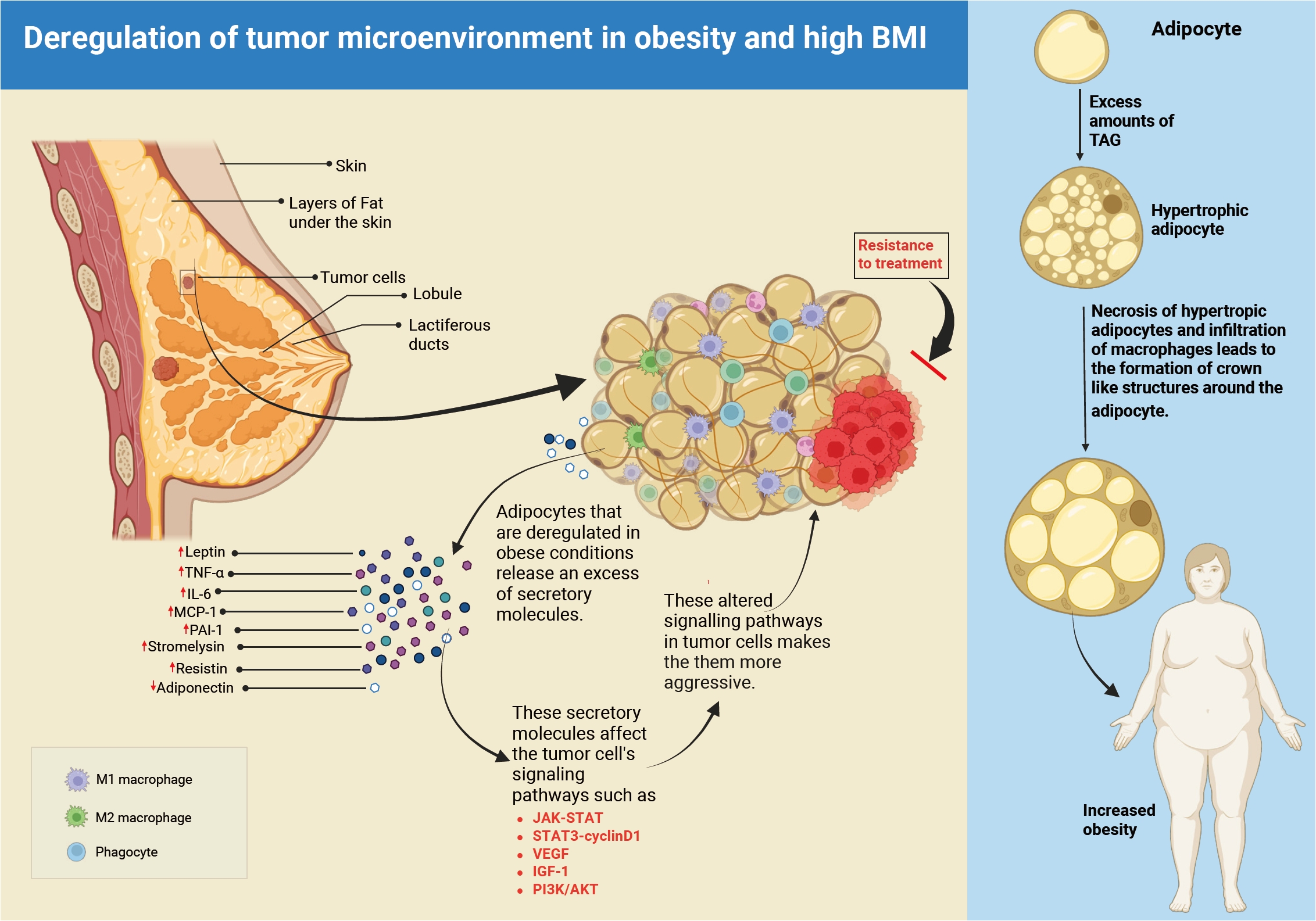
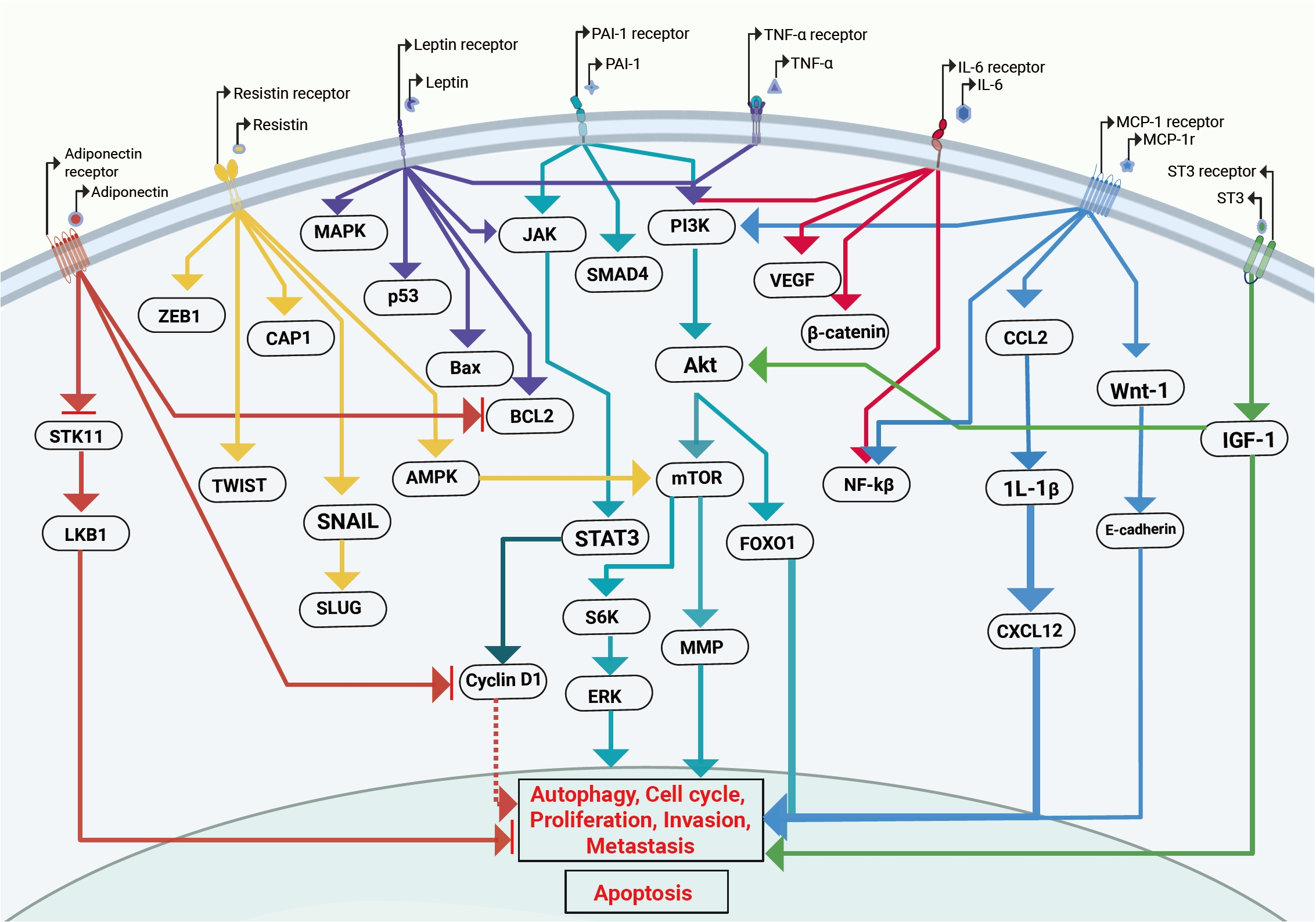
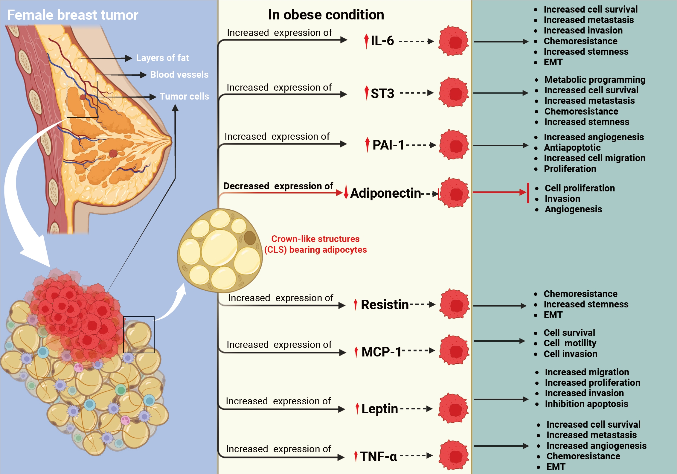
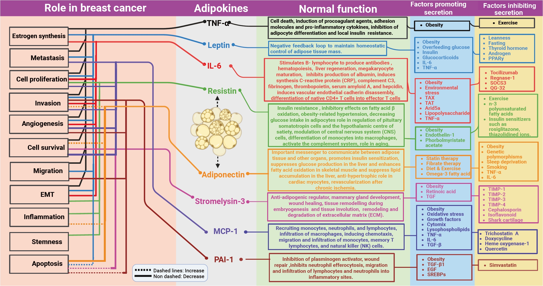
Fig. 1.
Fig. 2.
Fig. 3.
Fig. 4.
| Type of study | Key inflammatory markers/adipokines studied | Group and intervention | Major result | Reference |
|---|---|---|---|---|
| Systematic review and meta-analysis | TNF-α, IL-6, IL-8, natural killer cells, adiponectin (ADPN), leptin | Overweight and obese breast cancer patients and survivors | There was a significant reduction in TNF-α levels and leptin levels with combined aerobic and resistance training | [139] |
| Intervention - Combined aerobic and resistance training | ||||
| Clinical trials - Pooled analysis of 2 clinical trials | Serum adiponectin, leptin, resistin, complement factor D, monocyte chemoattractant protein 1 (CCL2), serpin (PAI-1), IL-6, IL-10, TNF-α | Metformin vs. placebo and lifestyle interventions among breast cancer survivors | Metformin compared to placebo showed a favorable decrease in leptin (−8.8 vs. −3.5 ng/mL; p < .01) | [140] |
| Lifestyle interventions favorably affected adiponectin (0.45 vs. −0.06 µg/mL; p < .01), and leptin (−10.5 vs. −4.4 ng/mL; p < .01) compared to controls; The strongest impact was observed combining metformin with lifestyle interventions on adipokines | ||||
| Nested case-control study | Serum adiponectin and leptin | Baseline and 12-month serum samples from 123 cases and 302 matched controls in the placebo arm of the IBIS-II prevention trial | In the multivariable Cox model, both the 12-month adiponectin increase (HR, 0.60; 95% CI, 0.36 to 1.00) and BMI were associated with BC risk (HR, 1.05; 95% CI, 1.00 to 1.09), with a 40% reduction in women with a 12-month increase in adiponectin; A significantly higher cumulative hazard of BC events was observed in obese women (BMI > 30) with decreased adiponectin (p = .0087) | [141] |
| Randomized trial | Serum adiponectin, leptin, IL-6, IL-10, TNF-α | Breast cancer patients on adjuvant hormones were randomized to high-intensity interval training, moderate-intensity continuous training or a control group | There were no significant differences in levels of studied adipokines among the groups | [142] |
| Meta-analysis | Serum adiponectin | Effect of exercise among breast cancer survivors | There was a meaningful change in levels of adiponectin though statistically non-significant | [143] |
| Single-blinded prospective controlled trial | Circulating serum biomarkers of IL-6, irisin, leptin, and adiponectin levels were measured before chemotherapy and after the last chemotherapy cycle | Aerobic exercise among patients receiving chemotherapy; breast cancer patients also were part of this study | In exercise group, there were significant increases in IL-6 (t = –2.985, p = .011) and adiponectin (z = –2.229, p = .026); There was a nearly 10% increase in irisin levels (0.83 vs. 0.91) but it did not reach statistical significance (t = 0.840, p = .416); The correlation between Δ of leptin and adiponectin (r = –0.635, p = .015) and the correlation between Δ of leptin and irisin (r = 0.802, p = .001) were found significant | [144] |
| Randomized study | Serum adiponectin | Breast cancer patients randomized into four groups with interventions being with or without ginger supplementation and with or without water-based exercise | The combined intervention (water-based exercise and ginger supplement) group showed a significantly better effect on adiponectin levels, as compared to the water-base exercise or ginger supplement alone groups and the age-matched placebo group | [145] |
| Randomized trial | Serum adiponectin, leptin, resistin, HGF, NGF, PAI-1, TNF-α, MCP-1, IL-1β, IL-6, and IL-8 | Breast cancer survivors achieving ≥5% weight loss using a remotely delivered weight loss intervention (POWER-remote) or a self-directed approach | There was a significant reduction in leptin levels in patients randomized to the intervention arm | [146] |
TNF-α, tumor necrosis factor α; IL, interleukin; CCL2, C-C motif chemokine ligand 2, PAI-1, plasminogen activator inhibitor-1; HR, hazard ratio; CI, confidence interval; BMI, body mass index; BC, breast cancer; HGF, hepatocyte growth factor; NGF, nerve growth factor; MCP-1, monocyte chemoattractant protein-1.


