Subclassification of the undifferentiated component in early gastric cancer: the poorly differentiated tubular adenocarcinoma component is associated with lymph node metastasis and local aggressiveness
Article information

Abstract
Background
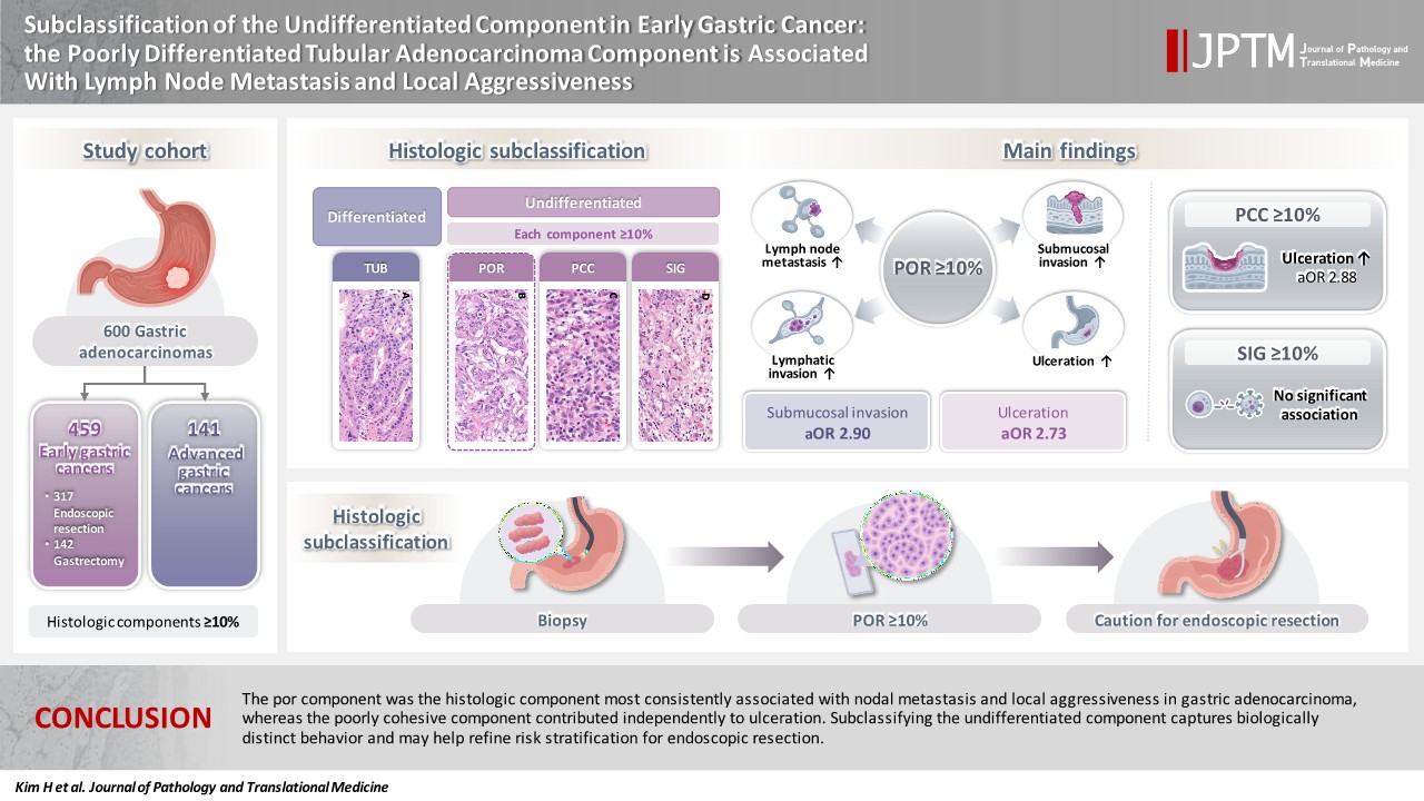
Gastric adenocarcinoma is histologically heterogeneous, and the undifferentiated category encompasses tubular adenocarcinoma, poorly differentiated (por), poorly cohesive carcinoma (pcc), and signet-ring cell carcinoma (sig). These components are often treated as a single entity, and which one drives the aggressive behavior of mixed-type tumors remains unclear. We subclassified the undifferentiated component and examined the association of each histologic component with adverse pathologic features.
Methods
We retrospectively analyzed 600 gastric adenocarcinomas (459 early, 141 advanced). Each histologic component (tub [tubular adenocarcinoma, well to moderately differentiated], por, pcc, sig) was assessed separately in the mucosal and invasive portions and dichotomized at 10%. Associations with lymph node metastasis (LNM), submucosal invasion, lymphatic invasion, and ulceration were evaluated by univariate testing and multivariable logistic regression.
Results
In early gastric cancer, por ≥10% was associated with LNM (p = .006) and lymphatic invasion (p < .001), and was an independent risk factor for submucosal invasion (adjusted odds ratio [OR], 2.90; 95% confidence interval [CI], 1.80 to 4.68; p < .001) and ulceration (adjusted OR, 2.73; 95% CI, 1.40 to 5.34; p = .003). By contrast, pcc ≥10% was independently associated only with ulceration (adjusted OR, 2.88; 95% CI, 1.25 to 6.65; p = .013), and sig showed no association with any adverse feature (all p > .05). Findings in the mucosal component paralleled those in the whole tumor. No histologic component was associated with survival.
Conclusions
The por component was the histologic component most consistently associated with nodal metastasis and local aggressiveness in gastric adenocarcinoma, whereas the poorly cohesive component contributed independently to ulceration. Subclassifying the undifferentiated component captures biologically distinct behavior and may help refine risk stratification for endoscopic resection.
INTRODUCTION
Gastric cancer is one of the most common malignancies worldwide and remains a leading cause of cancer-related death [1]. Because it is a histologically heterogeneous disease, several classification systems have been developed to capture its morphologic diversity. The most widely used are the World Health Organization (WHO) classification (papillary, tubular, mucinous, and poorly cohesive, including signet-ring cell carcinoma) [2] and the Lauren classification (intestinal, diffuse, mixed, and indeterminate) [3], both of which are adopted in Korea [4]. In Japan, a division into differentiated and undifferentiated types is additionally used, and this differentiated–undifferentiated distinction underlies current criteria for endoscopic resection [5].
With an increasing proportion of gastric cancers now detected as early gastric cancer (EGC), endoscopic submucosal dissection (ESD) has become a standard treatment, and eligibility is determined by expanded criteria based on tumor differentiation, size, and the presence of ulceration [6]. Differentiated-type tumors are eligible for ESD under broader size and ulceration criteria, whereas undifferentiated-type tumors are subject to more restrictive criteria. Under this framework, the undifferentiated-type tumor is regarded as a single entity. However, it encompasses morphologically distinct components: tubular adenocarcinoma, poorly differentiated (por), which forms solid or poorly structured glands, poorly cohesive carcinoma, not otherwise specified (pcc), and signet-ring cell carcinoma (sig), which are characterized by dyscohesive or signet-ring cells. Whether these components carry the same risk of nodal metastasis and aggressive local behavior has not been established.
This distinction becomes particularly relevant in mixed-type adenocarcinomas, which contain both differentiated (well to moderately differentiated tubular or papillary) and undifferentiated components. Mixed-type adenocarcinomas account for a substantial proportion of gastric cancers and have consistently been reported to carry a higher risk of lymph node metastasis (LNM) than purely differentiated or undifferentiated types in EGC [7-9]. Aside from nodal metastasis, some studies have shown that mixed-type is a risk factor for non-curative endoscopic resection and for requiring additional surgeries [10,11]. More recent work has suggested that the proportion of undifferentiated components further influences tumor behavior [12]. Nonetheless, although the body of literature is gradually growing, no established guideline specifies how mixed-type EGC should be treated. One reason for this uncertainty is that it remains unclear which histologic component of the mixed-type tumor is related to adverse clinicopathologic outcomes. Recent studies have subdivided the undifferentiated component into poorly differentiated carcinoma and signet-ring cell carcinoma and shown that the former is associated with aggressive tumor behavior, including LNM [13,14]. However, these studies did not further distinguish por from pcc, leaving open the question of which component drives the increased risk attributed to mixed histology.
We hypothesized that the por, pcc, and sig components confer different risks, and that subclassifying the undifferentiated component would clarify the behavior of mixed-type adenocarcinomas. In this study, we examined the associations of each histologic component with LNM, submucosal invasion, lymphatic invasion, and ulceration, and with post-resection survival. We further assessed whether each histologic component in the mucosal portion of the tumor is associated with these outcomes since pretreatment biopsy targets the mucosa.
MATERIALS AND METHODS
Study cohort and clinical setting
In this retrospective study, we included 600 patients who underwent radical gastrectomy with lymph node dissection or endoscopic resection for gastric adenocarcinoma at Korea University Guro Hospital from January 2010 to April 2012. Among 600 cases, 317 cases were EGC treated with endoscopic resection, 142 were EGC treated with gastrectomy, and 141 were advanced gastric cancer (AGC).
Clinicopathologic information including age at diagnosis, sex, depth of invasion, location, size, ulceration, lymphatic invasion, venous invasion, and perineural invasion was retrieved from pathology reports and electronic medical records.
Histologic evaluation
For each case, three representative hematoxylin and eosin slides were scanned with a Pannoramic 250 FLASH III Digital Scanner (3DHISTECH, Budapest, Hungary) and reviewed on SlideViewer (3DHISTECH). Digital slides were reviewed by two pathologists. The percentage of histologic components was assessed separately in mucosal and invasive components since the composition of the whole tumor and mucosal component could be different. The histologic composition of the whole tumor was calculated as the sum of the mucosal and invasive components, weighted by the relative proportion of each. Following the WHO classification, we classified histologic components into tubular adenocarcinoma, well to moderately differentiated (tub), por, pcc, and sig (Fig. 1A–D). Each histologic component was assessed and quantified independently, and the presence of each component was defined by a 10% threshold. Because the components were evaluated independently, a single tumor could be positive for more than one component, and the categories were not mutually exclusive. For mixed adenocarcinoma, mixed-por was defined as tub ≥10% and por ≥10%, mixed-pcc as tub ≥10% and pcc ≥10%, and mixed-sig as tub ≥10% and sig ≥10%. By this definition, a mixed-type tumor containing two different undifferentiated components was counted as positive in two categories (e.g., a tumor with both sig ≥10% and por ≥10% was classified as both mixed-sig and mixed-por).

Representative images of the histologic components. (A) Well to moderately differentiated tubular adenocarcinoma (tub). (B) Poorly differentiated tubular adenocarcinoma (por). (C) Poorly cohesive carcinoma, not otherwise specified (pcc). (D) Signet-ring cell carcinoma (sig). (E) Por forms lumens, although glandular structures are not readily recognizable (black arrow); pcc shows single-layered short cords (red arrow) and dyscohesive single cells (blue arrow), whereas cords composed of more than one cell layer were classified as por (yellow arrow). (F) Por forms slit-like, barely recognizable lumens (black arrowheads), whereas pcc grows as single cells and irregular small clusters (yellow arrowheads). (G) Pcc consists of single cells and haphazardly dispersed small clusters (red arrowhead), whereas por shows confluent growth of multilayered cords and trabeculae (blue arrowhead).
Notably, por and pcc are often difficult to distinguish and are subject to interobserver variability, which may be one reason why relatively few previous studies have attempted to subclassify the undifferentiated type. We distinguished the pcc component from the por component according to the definitions in the WHO classification. Tumors growing as single cells or small clusters without forming glands or tubules, including single-layered cords, were classified as pcc, whereas por was defined as forming barely recognizable tubules or glands, or a solid structure. The WHO classification also notes that por may show an acinar structure. Discordant cases were discussed by the two pathologists until a consensus was reached. Representative examples of por and pcc are presented in Fig. 1E–G.
Statistical analysis
Categorical variables were compared using the chi-square test with Yates’ correction, or Fisher’s exact test when any expected cell count was fewer than five. Independent associations were assessed by multivariable logistic regression, with results expressed as odds ratios and 95% confidence intervals. Disease-free survival was estimated by the Kaplan-Meier method and compared using the log-rank test. All tests were two-sided, and a p-value <.05 was considered significant. Analyses and visualization were performed in Python 3.12 (Python Software Foundation, Beaverton, OR, USA) using the pandas, statsmodels, lifelines, and SciPy packages.
RESULTS
Clinicopathologic characteristics
A total of 600 patients were analyzed (Table 1). The study group consisted of 414 (69.0%) male and 186 (31.0%) female patients, with a median age at diagnosis of 62 years (interquartile range [IQR], 53 to 70). There were 459 EGCs (317 treated by ESD and 142 by radical gastrectomy) and 141 AGCs.
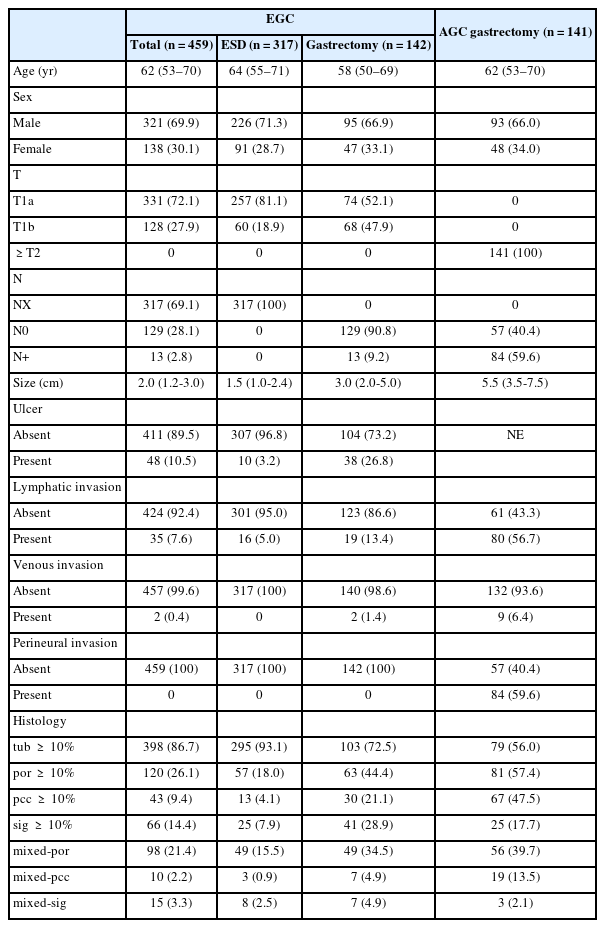
Clinicopathologic characteristics of the study cohort
Among the 459 EGC patients, 321 (69.9%) were male and 138 (30.1%) were female with a median age of 62 years (IQR, 53 to 70 years). The majority of the tumors were mucosal (T1a) tumors (n = 331, 72.1%), with a median size of 2.0 cm (IQR, 1.2 to 3.0). Ulceration was present in 48 (10.5%), lymphatic invasion in 35 (7.6%), and venous invasion in two (0.4%); no perineural invasion was observed. Among the gastrectomy cases (n = 142), 13 (9.2%) had LNM.
Among the 141 AGC patients, 93 (66.0%) were male and 48 (34.0%) were female, with a median age of 62 years (IQR, 53 to 70 years). LNM was present in 84 (59.6%) cases. There were 80 (56.7%) cases of lymphatic invasion, nine (6.4%) of venous invasion and 84 (59.6%) of perineural invasion.
The histologic component assessment showed tub, por, pcc, and sig components (≥10%) in 86.7%, 26.1%, 9.4%, and 14.4% of EGCs and in 56.0%, 57.4%, 47.5%, and 17.7% of AGCs, respectively. Mixed-por, mixed-pcc, and mixed-sig were present in 21.4%, 2.2%, and 3.3% of EGCs and 39.7%, 13.5%, and 2.1% of AGCs, respectively. The distribution of the components within each histologic group is presented in Supplementary Table S1.
Por ≥10% is associated with LNM in both the whole tumor and the mucosa in EGC
In the EGC gastrectomy group (n = 142), 13 (9.2%) cases had LNM (Table 2). All 13 were submucosal invasive tumors with a tub ≥10%. LNM was associated with por ≥10%, mixed-por, submucosal invasion, and ulceration, whereas pcc and sig showed no significant association.
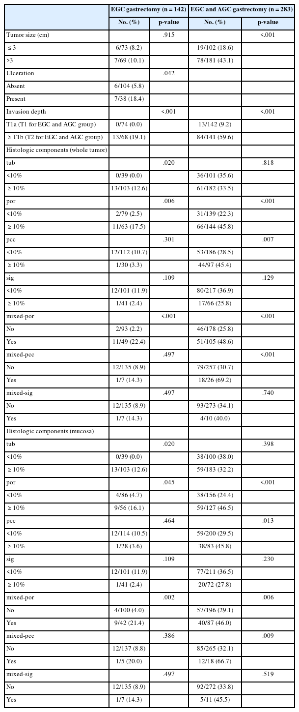
Lymph node metastasis by clinicopathologic and histologic factors
In the gastrectomy group comprising both EGC and AGC (n = 283), LNM was present in 97 (34.3%) cases (Table 2). LNM was associated with por ≥10%, pcc ≥10%, mixed-por, mixed-pcc, tumor size >3 cm, and invasion depth.
We also analyzed the association of mucosal histologic components and LNM, which paralleled the whole-tumor analysis (Table 2). In EGC, all 13 cases with LNM had tub ≥10%. Por ≥10% and mixed-por were associated with LNM. In the EGC and AGC cohort, por ≥10%, pcc ≥10%, mixed-por, and mixed-pcc were associated with LNM.
Multivariable logistic regression was performed using variables significant on univariate analysis (Table 3). The EGC gastrectomy group had too few LNM events (n = 13) to support multivariable analysis. In the EGC and AGC gastrectomy group, which had 97 LNM cases, we included por, pcc, tumor size, and invasion depth as covariates. Mixed-por and mixed-pcc were excluded despite univariate significance because each is a subset of por ≥10% and pcc ≥10%, respectively. In this model, pcc ≥10% and tumor size lost significance whereas por ≥10% and invasion depth remained independently associated with LNM. In a parallel model using mucosal components (por ≥10%, pcc ≥10%, tumor size, and invasion depth), por ≥10% and invasion depth were independently associated with LNM.
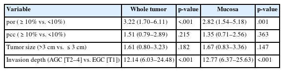
Multivariable logistic regression for lymph node metastasis in EGC and AGC gastrectomy group
Por ≥10% is associated with submucosal invasion, lymphatic invasion, and ulceration
In EGC, we examined the associations between histologic components and other pathologic features affecting ESD eligibility (Table 4). Submucosal invasion was associated with por ≥10%, mixed-por, mixed-pcc, tumor size >3 cm, ulceration, and lymphatic invasion. Lymphatic invasion was associated with por ≥10%, mixed-por, tumor size >3 cm, ulceration, and submucosal invasion. Ulceration was associated with por ≥10%, pcc ≥10%, mixed-por, tumor size >3 cm, invasion depth, and lymphatic invasion, whereas tub ≥10% showed a negative association.
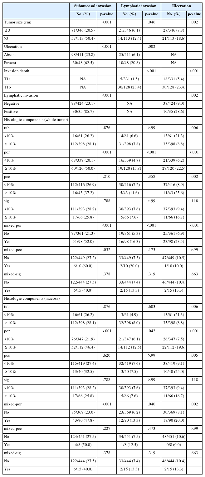
Submucosal invasion, lymphatic invasion, and ulceration by clinicopathologic and histologic factors in EGC
The analysis of mucosal components paralleled the whole-tumor findings, except that mixed-pcc was not associated with submucosal invasion (Table 4). Por ≥10% was associated with submucosal invasion, lymphatic invasion, and ulceration, whereas pcc ≥10% was associated with ulceration. Tub ≥10% showed a negative association with ulceration.
Multivariable logistic regression analysis was performed for each pathologic feature (Table 5). For submucosal invasion, a model including por ≥10%, tumor size >3 cm, and ulceration identified all three as independent risk factors. For lymphatic invasion, a model including por ≥10%, tumor size >3 cm, invasion depth, and ulceration identified invasion depth as an independent risk factor, whereas por ≥10% lost significance. For ulceration, a model including por ≥10%, pcc ≥10%, tumor size, and invasion depth identified por ≥10%, pcc ≥10%, and invasion depth as independent risk factors.

Multivariable logistic regression for submucosal invasion, lymphatic invasion, and ulceration in EGC group
The multivariable results for mucosal components were similar to the whole-tumor analysis except for ulceration. For ulceration, in a model including por ≥10%, pcc ≥10%, tumor size, and invasion depth, pcc ≥10% and invasion depth were independent risk factors, whereas mucosal por ≥10% was not.
Survival
In the EGC cohort, survival events were infrequent (22 disease-free survival and 12 overall survival events). No histologic component or invasion depth was significantly associated with disease-free survival on log-rank testing (Supplementary Fig. S1).
DISCUSSION
In this study, we classified histologic components of gastric adenocarcinoma into tub, por, pcc, and sig, and measured the percentage of each component in the whole tumor and in the mucosal component separately. We found that in EGC, por ≥10% was associated with LNM, submucosal invasion, lymphatic invasion, and ulceration, and these associations were reproducible in both the whole tumor and the mucosal portion. Moreover, por ≥10% was an independent risk factor for submucosal invasion in both the whole tumor and mucosa. In addition, pcc was also an independent risk factor for ulceration, whereas sig showed no significant association with LNM or other locally aggressive features.
While no treatment guideline for mixed adenocarcinoma has been established, several studies have shown that the relative proportion of the differentiated and undifferentiated components influences the behavior of mixed adenocarcinoma. Horiuchi et al. [10] found that the undifferentiated component was a risk factor for additional surgery after endoscopic resection when it predominated in mixed-type adenocarcinoma. Kim et al. [15] found that, even when the undifferentiated component was minor, such tumors showed higher rates of deep submucosal invasion and lymphatic invasion leading to non-curative resection, although their long-term prognosis did not differ from that of purely differentiated tumors. Shimizu et al. [16] reported a significant difference in invasion depth between predominantly differentiated and predominantly undifferentiated mixed adenocarcinomas, but no difference in the LNM rate. Marano et al. [17] showed that the proportion of the sig component is inversely related to aggressive tumor behavior and poor prognosis. Taken together, these findings suggest that although the undifferentiated tumor is known to be associated with adverse clinicopathologic outcomes, each histologic component within this category may confer different risks.
Recent studies have further attempted to separate the undifferentiated component into signet-ring cell carcinoma and poorly differentiated component. These studies have yielded mixed results, but several align with our findings. Chen et al. [13] reported that the presence of a poorly differentiated component increased the LNM rate even when the differentiated component predominated. Hwang et al. [14] and Li et al. [12] further showed that the extent of the poorly differentiated component, but not the signet-ring cell component, was associated with LNM. In contrast, Takizawa et al. [18] found no difference in metastasis between minor sig and poorly differentiated components when the differentiated component predominated. None of these studies, however, separated por and pcc within the poorly differentiated component. By measuring por and pcc separately, we found that they carry distinct risks: por was associated with LNM, submucosal invasion, lymphatic invasion, and ulceration, whereas pcc was associated with ulceration and sig showed no association with any aggressive feature. These findings indicate that the risk attributed to mixed histology is driven specifically by its undifferentiated component, and that subclassifying it—rather than treating it as a single entity—may help reconcile the conflicting results of previous studies and refine risk stratification.
Also, we performed additional analysis with histologic components in the mucosa alone which is the target of pretreatment forceps biopsy. We showed that por ≥10% was likewise associated with LNM, submucosal invasion, lymphatic invasion, and ulceration. Moreover, it was an independent risk factor for submucosal invasion, although its independent association with LNM could not be established because of the scarcity of events. These findings may have implications for the selection of candidates for endoscopic resection. Recognition of a por component in a pretreatment biopsy may therefore serve as a warning that the tumor is more likely to be deeper than it appears, and may warrant further investigation to rule out possible LNM before a treatment plan is made. Endoscopic resection should thus be considered with caution, with careful attention to invasion depth, when a substantial por component is present in the biopsy.
Besides por, pcc also showed an association with LNM in the gastrectomy cohort comprising EGC and AGC. However, this univariate association was not significant anymore after adjustment for invasion depth (Table 3), reflecting its enrichment in advanced tumors rather than an independent effect. Similarly, mixed-pcc was associated with submucosal invasion on univariate analysis but not after adjustment for por, tumor size, and ulceration (adjusted odds ratio, 2.75; 95% confidence interval, 0.68 to 11.20; p = .158) (Supplementary Table S2), reflecting the small size of this group (n = 10) and its enrichment for a coexisting por component. The only outcome for which pcc retained significance was ulceration, where it was an independent risk factor in both the whole-tumor and mucosal-component analyses. Since ulceration is associated with non-curative endoscopic resection, a pcc component may warrant attention in this context; however, unlike the poorly differentiated component, its association was confined to ulceration and did not extend to LNM or depth of invasion. Whether pcc itself, independent of ulceration, is related to non-curative resection remains to be investigated.
At first glance, the univariate association between a tub component ≥10% and a higher rate of LNM appears inconsistent with previous reports linking undifferentiated components to increased metastatic risk. This association, however, is attributable to the depth distribution of the reference group rather than to a specific effect of a tub ≥10%. The reference group (tub <10%) was enriched for mucosa-confined (T1a) tumors (28/39), in which no LNM occurred (Supplementary Table S3). Accordingly, when the analysis was stratified by invasion depth (T1a vs. T1b), the association was no longer significant (p = .107).
This study has several limitations. It was retrospective and conducted at a single institution. Owing to the small number of cases and the inherent scarcity of LNM events within the EGC cohort, we could not perform multivariable regression analysis to determine whether por is an independent risk factor for LNM in EGC alone, and the survival analysis was limited by the small number of events. Despite these limitations, this study is one of the earliest attempts to separate por from pcc within the undifferentiated component, and it demonstrates that these histologic components confer different risks with respect to aggressive tumor behavior. Further investigation in a larger, mixed-type adenocarcinoma cohort is warranted to draw more definitive conclusions.
In conclusion, among the undifferentiated components, a por component ≥10% was associated with adverse features—LNM, submucosal invasion, lymphatic invasion, and ulceration—and was an independent risk factor for submucosal invasion and ulceration. In contrast, pcc and sig components were not associated with adverse features except that pcc independently contributed to ulceration. The association of the tub component with LNM reflected the invasion depth distribution of the reference group rather than a true effect. Notably, the adverse behavior of mixed-type tumors was attributable to their por component: mixed-por tumors carried an increased risk of LNM and submucosal invasion, whereas mixed-pcc did so through a coexisting por component. This indicates that the elevated risk attributed to mixed histology may be driven not by histologic heterogeneity itself, but by the presence of a por component within it and that subclassifying the undifferentiated component may refine risk stratification for endoscopic resection.
Supplementary Information
The Data Supplement is available with this article at https://doi.org/10.4132/jptm.2026.09.15.
Notes
Ethics Statement
This study was approved by the Institutional Review Board of Korea University Guro Hospital (2024GR0387), and the requirement for informed consent was waived because of the retrospective nature of the study and the use of de-identified data.
Availability of Data and Material
The datasets generated or analyzed during the study are not publicly available due to patient privacy concerns but the results of analysis are available from the corresponding author upon reasonable request.
Code Availability
Not applicable.
Author Contributions
Conceptualization: BHK. Data curation: HK. Formal analysis: HK. Methodology: BHK, HK. Supervision: BHK. Writing—original draft: HK. Writing—review & editing: BHK. Approval of final manuscript: all authors.
Conflicts of Interest
The authors declare that they have no potential conflicts of interest.
Funding Statement
No funding to declare.