Squamoid Eccrine Ductal Carcinoma of the Scalp
Article information
Abstract
Squamoid eccrine ductal carcinoma (SEDC) is an exceedingly rare tumor that shows both squamous and adnexal ductal differentiation. We report a case of this unusual tumor occurring on the occiput of a 53-year-old man. A histopathological examination revealed a nodular lesion infiltrating the dermis and subcutaneous tissue with numerous duct-like structure and squamoid differentiation foci. Five months later, the patient presented with a palpable mass at the site of the previous excision and the right side of the neck. Sono-guided fine needle aspiration of the right neck mass was performed and was diagnosed as a metastastasis of a lymph node. A right neck node dissection and re-excision of the occiput was performed. The histopathological findings were similar, but squamoid differentiation was more prominent than that in the previous lesion. Because of the rarity of SEDC, little is known about its biological behavior and optimal treatment.
A wide variety of benign and malignant adnexal tumors occur in the skin. These tumors have hair, sweat, or sebaceous gland origins. Eccrine carcinomas, originating from the eccrine glands, are rare, comprising <0.01% of all cutaneous tumors.1 Eccrine carcinomas usually present as a slow growing infiltrating plaque or nodule on the scalp, extremities, or trunk.2-4 Multiple subtypes of eccrine carcinoma are classified by their distinct histological pattern. Among the multiple subtypes of eccrine carcinomas, squamoid eccrine ductal carcinoma (SEDC) is characterized by prominent squamoid differentiation, resembling squamous cell carcinoma (SCC). SEDC is exceedingly rare, and only a few cases have been reported. About half of all eccrine carcinoma metastasize to other organs1 but given the very few cases of SEDC described to date, only a local recurrent case has been reported and no metastasis of the lymph node was found.5-8 We present a case of local recurrent SEDC of the scalp with metastasis to the lymph nodes.
CASE REPORT
A 53-year-old man presented with a 10-year history of a nodule that had recently started growing and producing an itching sensation on the right occiput. His medical history was unremarkable. A large 2.3×1.5 cm nodular mass was seen, and focal ulceration was noted. A neck computed tomography scan suggested a benign soft tissue mass in the subcutaneous layer, so a local excision was performed. The tumor measured 2.6×1.7×1.5 cm, and there was no evidence of erosion or ulcer on the skin surface. The cut surface showed a relatively well demarcated white solid mass, which was located in the dermis and subcutaneous fat tissue. A focal fibrotic infiltrative border and connection with the epidermis were also noted.
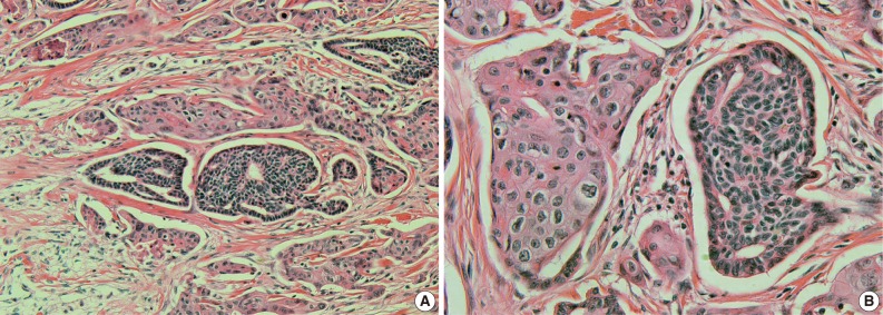
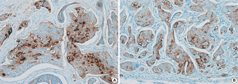
A histopathological examination revealed a nodular lesion composed of duct-like structures lined by cuboidal epithelium (Fig. 1A) in the upper portion of the dermis and numerous nests of squamoid cells with amphophilic cytoplasm and keratinization in the deeper portion (Fig. 1B). The periphery of the lesion revealed more prominent squamoid differentiation and showed plump eosinophilic cytoplasm and keratinization with dense lymphocytic infiltration. Most of the tumor cells were slightly pleomorphic (variation of cellular and nuclear size, high N/C ratio, and hyperchromatic nucleoli), but focally more pleomorphism was seen in the infiltrating lesion. Tumor necrosis foci, lymphatic emboli, and mitotic figures were present. Immunohistochemistry showed positive staining for cytokeratin (CK), CK5/6, CK7, carcinoembryonic antigen (CEA) (Fig. 2A), and epithelial membrane antigen (EMA) (Fig. 2B) but was negative for S-100 protein, gross cystic disease fluid protein-15, and CD15. A diagnosis of SEDC was made. The lateral resection margin was free, but the deep resection margin was very close to the tumor. No further treatments were administered.

A nodular lesion composed of duct-like structures lined by cuboidal epithelium (A) and numerous nests of squamoid cells with amphophilic cytoplasm and keratinization (B).

The tumor cells are immunoreactively positive for carcinoembryonic antigen (A) and epithelial membrane antigen (B).
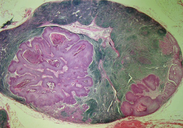
After 5 months, the patient returned with a palpable mass at the previous excision site and on the right side of the neck. Sono-guided fine needle aspiration of the right neck mass revealed a metastatic adenocarcinoma probably from an eccrine ductal adenocarcinoma of the skin. A right neck node dissection and wide local excision of the occiput was performed. The dissected neck lymph nodes were involved with tumor cells and showed ductal structure and squamous differentiation (Fig. 3).
DISCUSSION
Eccrine carcinomas uncommonly originate from eccrine glands. A review at the Mayo Clinic, covering more than 75 years of skin tumors, reported 14 cases of eccrine carcinoma.9 These carcinomas have a slow growth rate and a high potential for recurrence. Diverse variants of eccrine carcinoma exist, including a variant with abundant fibromyxoid stroma, a spindle cell variant with myoepithelial differentiation, a basal cell carcinoma-like variant, and a SEDC variant characterized by squamous metaplasia.1,7 Classification of eccrine neoplasms is problematic because no universally accepted classification scheme exists, and the variable clinical appearance and histological features are often not predictive of clinical behavior.10 Therefore, the diagnostic identification of sweat gland carcinomas is hampered by their rarity and their histological resemblance to various visceral tumors, leading to confusion with metastatic lesions.
SEDC is an exceedingly rare variant of eccrine carcinoma. Wong et al.5 reported the first three cases of SEDC in 1997. Since then, only four additional cases have been reported. SEDC shows a predilection for the head, neck, and extremities and presents most often in the middle-aged and elderly as a hard, usually nonulcerated cutaneous nodule. The histological features of SEDC are duct-like structures resembling the dermal part of the eccrine duct and atypical squamous cell clusters. Follicular or sebaceous differentiation can be seen.11
The histological differential diagnosis for SEDC includes microcystic adnexal carcinoma (MAC) with squamous features. MAC is poorly circumscribed and usually deeply infiltrating. It expresses a constellation of features, including numerous keratinous cysts. Keratinous cysts are often superficially located in the dermis without connection to the epidermis. Deeper components include smaller nests and strands of cells. A dense fibrous stroma surrounds all components. The tumor cells express CEA and EMA.10,12,13 In our case, the absence of a keratinous cyst and a dense fibrous stroma surrounding tumor cells made it less likely that it was a MAC. The differential diagnosis could also be SCC. Immunohistochemical staining with CEA could resolve this problem, as SCCs are typically negative. But if ductal differentiation is not obvious in shave or punch biopsy specimens, SEDC may be mistaken for a SCC.
Recent studies have reported that positive p63 and CK5/6 staining is relatively useful for distinguishing primary adnexal neoplasm with sweat gland differentiation from metastatic adenocarcinoma at various sites.14 p63 is a member of the p53 gene family and may be essential for regulating the differentiation of cells in normal human skin. p63 is normally expressed in basal and spinous cell nuclei of the epidermis, peripheral cells of the eccrine dermal ducts, germinative cells of sebaceous glands, and myoepithelial cells of the terminal portion of the eccrine and apocrine glands.15 CK5/6 is a basic high molecular weight CK. CK5/6 are normally expressed in stratified squamous epithelium, the basal-myoepithelial cell layer of the prostate, the salivary gland, and the breasts, as well as in myoepithelial cells of secretory glands and both layers of the epithelial cells of excretory ducts of eccrine and apocrine glands.16 In our case, the tumor cells were p63 negative but CK5/6 positive.
Limited information is available to manage SEDC because of its rarity. However, a wide surgical excision with a wide negative margin (with or without Mohs's technique) is the treatment of choice. A high rate of local recurrence and subsequent metastasis after conventional surgical excision of eccrine carcinoma is observed. In our case, the SEDC recurred and showed lymph node metastasis 5 months after the first excision.
The significance of squamous differentiation in eccrine carcinoma is unclear.17 Some believe that it shows more aggressive biological behavior, whereas others believe that it is an incidental finding and has no impact on disease biology. A study by Kohda et al.17 reported that both benign and malignant eccrine neoplasms had foci of squamous differentiation.
Our case was interesting for a variety of reasons. First, the relative rarity of SEDC was reported. Second, the tumor recurred and metastasized to a lymph node.
Notes
No potential conflict of interest relevant to this article was reported.
