 E-submission
Search
- Page Path
- HOME > Search
Original Article
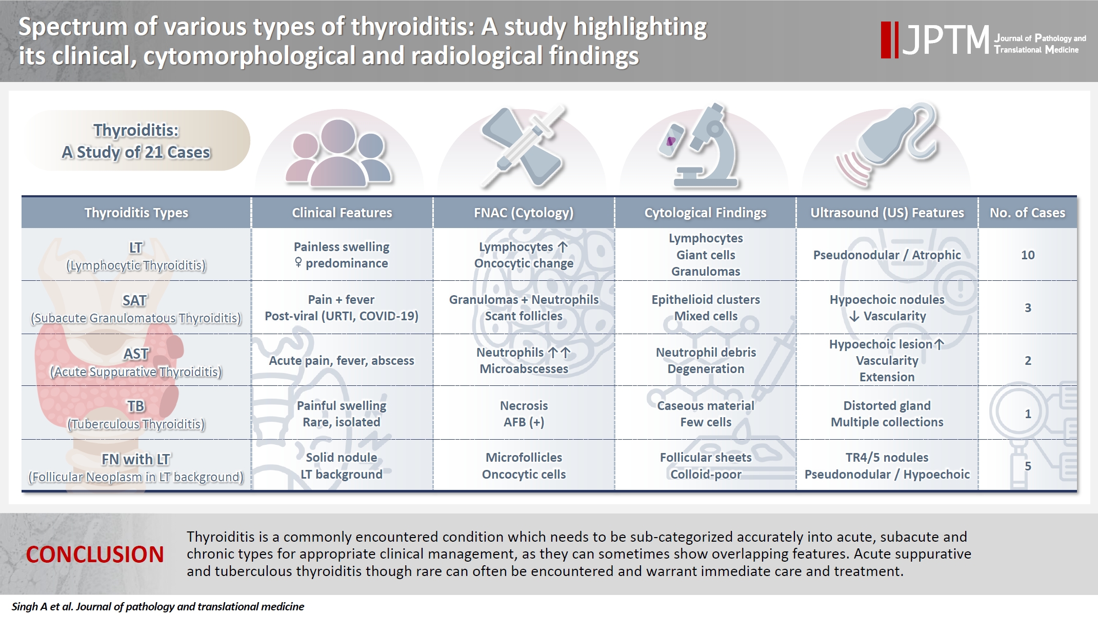
- Spectrum of thyroiditis types: clinical, cytomorphological, and radiological findings
- Anam Singh, Indrajeet Kundu
- J Pathol Transl Med. 2025;59(6):421-433. Published online November 6, 2025
- DOI: https://doi.org/10.4132/jptm.2025.08.13
- 3,824 View
- 183 Download
-
 Abstract
 PDF
PDF - Background
Thyroiditis encompasses a range of inflammatory conditions affecting the thyroid gland. Lymphocytic thyroiditis (LT) is a common form of thyroiditis, with acute suppuration of the thyroid, while tuberculous thyroiditis is relatively rare. Fine-needle aspiration cytology (FNAC) remains a safe and cost-effective tool for diagnosing thyroid-related diseases, especially when paired with ultrasound (US) and clinical examination. Methods: This is a cross-sectional study including 21 cases. The cases were reported as thyroiditis on US and FNAC, and the findings were correlated with patient clinical history, symptoms during presentation, and serological profiles. Results: The cases of thyroiditis encompassed the more common forms, LT and subacute granulomatous thyroiditis (SAT), as well as relatively rare forms like tuberculous thyroiditis and thyroid abscess. Cases of follicular neoplasms (FN) arising in the context of LT also are included in this study. The case of tuberculous thyroiditis presented as a bulky thyroid gland that appeared heterogeneous on US with extensive necrosis on FNAC. The cases of thyroid abscess and SAT presented with painful neck swellings, with granulomas in the latter cases. US features of LT showed an array of appearances ranging from pseudonodular to an atrophic thyroid gland. All cases of FN showed a lymphocytic background. Conclusions: Thyroiditis is a commonly encountered condition that needs to be sub-categorized accurately into acute, subacute, and chronic types for appropriate clinical management, as they can sometimes show overlapping features. Though rare, acute suppurative and tuberculous thyroiditis are often encountered and warrant immediate care and treatment.
Case Reports
- Alveolar Soft Part Sarcoma of the Lung Diagnosed by Fine Needle Aspiration Cytology: A Case Report .
- Dae Su Kim, Young Lyun Oh, Young Hyeh Ko
- J Pathol Transl Med. 1998;9(2):187-192.
- 1,806 View
- 12 Download
-
Abstract
PDF
- Alveolar soft part sarcoma(ASPS) is a rare malignant neoplasm with a distinct clinicopathologic entity of which fine needle aspiration(FNA) cytologic findings have been described in only a few reports. Although patients usually present with an isolated soft-tissue mass in the extremity, metastasis can occur in about 13 % of total cases and the most frequent metastatic site is the lung. We have recently experienced a FNA cytologic case of ASPS in the lung. A 23-year-old female patient was admitted to this hospital due to 2-month-history of cough. She had been good in health before the visit. Chest computed tomography revealed multiple, variable sized, bilateral pulmonary nodules. Physical examination and other staging work up revealed no other lesions except for pulmonary nodules. A percutaneous transthoracic FNA was performed from the pulmonary nodules. The smear was cellular and most cells were arranged singly. In addition, a few clusters lined by thin-walled vasculature with a pseudoalveolar pattern were present. Some of the tumor cells were large and polygonal to oval with abundant granular or vacuolated cytoplasm. Most cells were naked nuclei showing finely granular chromatin pattern with prominent central nucleoli.
- Fine Needle Aspiration Cytology of Glycogen-Rich Clear Cell Carcinoma of the Breast: A Report of Two Cases .
- Wan Seop Kim, Won Mi Lee, Eun Kyung Hong, Moon Hyang Park, Jung Dal Lee
- J Pathol Transl Med. 1998;9(2):213-219.
- 2,168 View
- 29 Download
-
Abstract
PDF
- Glycogen-rich clear cell carcinoma of the breast is an unusual variant of carcinoma with a recorded incidence of 1.4-3% of breast carcinomas. The cytologic characteristics have not been well described. We report two cases of glycogen-rich clear cell carcinoma with corresponding fine needle aspiration(FNA) cytologic findings and compare them to infiltrating ductal carcinoma and other clear cell malignancies with a review of literature. One was a 62-year-old woman exhibiting a palpable mass of the right breast. The smears showed atypical tight cell clusters and individually scattered single cells containing foamy or clear abundant cytoplasm with well defined cytoplasmic margins. Mild to moderate nuclear pleomorphism and a prominent nucleolus were present. The other was a 42-year-old woman who was admitted with a right breast mass. The smears showed moderately cellular, tightly cohesive tumor cells. The cytoplasmic outline was generally well demarcated. The tumor cells contained foamy to clear abundant cytoplasm with large and small vacuoles. The nuclear pleomorphism was marked. Both tumors resected by modified radical mastectomy, were diagnosed as glycogen-rich clear cell carcinoma. Histologically, the clear cell nature of tumor cells were not characteristic enough to predict this type of the tumor. Some cytologic features can be distinguished other clear cell breast cancer from glycogen-rich carcinoma. Recognition of these unusual patterns in a breast FNAC should raise the suspicion of a clear cell carcinoma including glycogen-rich subtype. Cytological localization of glycogen using PAS and D-PAS staining may permit the correct identification and differential diagnosis of this tumor.
- Cytologic Findings of Infectious Mononucleosis Lymphadenitis: A Report of Four Cases .
- Jin Hee Sohn, Eun Ha Jung, Hye Rim Park
- J Pathol Transl Med. 1998;9(2):227-232.
- 5,074 View
- 285 Download
-
Abstract
PDF
- Infectious mononucleosis(IM) is an acute self-limiting lymphoproliferative disorder associated with infection by the Epstein-Barr Virus(EBV), with the characteristic triad of fever, sore throat, and cervical or generalized lymphadenopathy. And also there are atypical lymphocytes in the peripheral blood. Cytological findings of IM lymphadenitis are characterized by a florid immunoblastic and atypical lymphoid cell proliferation. However, the small number of cases were studied by fineneedle aspiration cytology(FNAC) even though there was a complexity of lymph node pathology. It is important to recognize the reactive pattern of IM that would initiate EBV study and to avoid unnecessary biopsy. We studied findings of lymph node FNAC from 4 patients with EBV infection confirmed by EBV-specific serologic studies. All of the cases were positive for viral capsid antigen(VCA) and one case was positive for anti-EBV nuclear antigen(EBNA). Cytologically, all of the cases exhibited high cellularity and atypia with great numbers of large immunoblastic lymphocytes.
- Fine Needle Aspiration Cytology of Unusual Epidermoid Cyst with Diffuse Parakeratosis and Aggressive Growth: A Case Report.
- Hae Joo Nam
- J Pathol Transl Med. 1999;10(1):85-89.
- 2,427 View
- 24 Download
-
Abstract
PDF
- An extremely unusual case of epidermoid cyst showing diffuse parakeratosis and aggressive clinical behavior is presented. A destructive bone lesion with surrounding ill-defined soft tissue lesion was found by computed tomography in a 63 year-old man complaining of painful swelling of the right buttock. He had a history of surgical excision twice for epidermoid cysts of soft tissue of the right hip during recent one year. On aspiration cytology, the aspirate was highly cellular and mostly composed of desquamated nucleated squamous cells. Operation finding revealed that the iliac bone was irregularly destroyed and filled with gray-white cheesy material and necrotic bone debris. Adjacent gluteus muscle showed scattered gray-white lesions. The curettage specimen showed bone necrosis and desquamated squamous cells filling the marrow spaces. The lesion within muscle revealed epidermoid cyst with diffuse parakeratosis.
- Fine Needle Aspiration Cytology of Peripheral T Cell Lymphoma of the Lung: A Case Report .
- Ok Ran Shin, Eun Sun Jung, Youn Soo Lee, Chang Suk Kang, Byung Kee Kim, Sang In Shim
- J Pathol Transl Med. 1999;10(2):157-162.
- 2,009 View
- 16 Download
-
Abstract
PDF
- Primary non-Hodgkin's lymphoma of the lung is rare among extranodal lymphomas. The most common form is low grade B-cell type originated from the mucosa-associated lymphoid tissue (MALT) of the lung and primary peripheral T cell lymphoma of the lung is extremely rare. We recently experienced a case of fine needle aspiration cytology of primary peripheral T cell lymphoma of the lung in a 39-year-old male patient. The cytologic smears revealed some sheets of reactive epithelial cells, epithelioid histiocytes, and numerous polymorphous population of lymphoid cells composed of small and intermediate sized lymphoid cells and mature lymphocytes. Lymphoid cells were slightly larger than normal mature lymphocytes and showed significant irregularity of nuclear membrane. The internal nuclear structure was marked by chromatin clumping, clear parachromatin areas, and inconspicuous nucleoli. Histopathologically, atypical small lymphocytes infiltrated in the interstitium and alveolar sac. By the immunohistochemical study and molecular biologic study of gene rearrangement, the T cell clonality of atypical lymphoid cells was confirmed.
- Fine Needle Aspiration Cytology of Primary Malignant Lymphoma of the Thyroid Gland: A Case Report.
- Mi Seon Kwon, Seung Sook Lee, Jae Soo Koh, Jin Haeng Chung, Kyo Young Lee
- J Pathol Transl Med. 2001;12(1):67-71.
- 2,292 View
- 10 Download
-
Abstract
- Primary malignant lymphoma of the thyroid gland is uncommon malignancies. Its fine needle aspiration cytology (FNAC) findings are rarely described in the literature. This article highlights the FNAC diagnosis of primary malignant lymphoma of the thyroid gland. A 70-year-old female presented with a rapidly enlarging thyroid mass of five months' duration. FNAC smears showed low cellularity consisting of predominantly atypical enlarged lymphoid cells admixed with a few small lymphocytes, plasma cells, and oncocytic cells. Some disrupted lymphoid cells were also present. The tumor cells infiltrated into the thyroid follicular epithelium forming lymphoepithelial lesion. The cytologic appearance showed a diffuse mixture of cell types with only a few small, mature lymphocytes and many enlarged lymphoid cells. The enlarged lymphoid cells were atypical and pleomorphic with nuclear clefting and irregularities. Grossly, the left lobe of the thyroid was nearly replaced by a diffuse firm to soft solid mass with smooth tan fish-flesh homogeneous cut surface. Histological diagnosis was diffuse large B-cell lymphoma with areas of marginal zone B-cell lymphoma of MALT type.
- Fine Needle Aspiration Cytology of Solid Papillary Carcinoma of the Breast: Report of a case associated with mucinous carcinoma.
- Hee Kyung Kim, Dong Won Kim, So Young Jin, Dong Wha Lee
- J Pathol Transl Med. 2001;12(2):127-130.
- 2,392 View
- 38 Download
-
Abstract
PDF
- Solid papillary carcinoma of the breast is a distinctive form of intraductal papillary carcinoma frequently associated with both mucinous carcinoma and infiltrating ductal carcinoma, not otherwise specific. To our knowledge, this case is the first description of the cytologic aspects of solid papillary carcinoma of the breast in the Korean literature. We experienced a case of solid papillary carcinoma of the right breast diagnosed by fine needle aspiration cytology(FNAC) in a 70-year-old female. FNAC from the right breast showed high cellularity consisting of mostly tight clusters of tumor cells and a few scattered tumor cells. The nuclei were monotonously round to oval in shape with inconspicuous nucleoli. The cytoplasm was abundant and finely granular. Scant amount of mucinous material was present on the background. The diagnosis was confirmed histologically and immunohistochemically.
- Fine Needle Aspiration Cytology of Malignant Myoepithelioma of the Salivary Gland: A Case Report.
- Jae Hwa Lee, Jean Kyung Park, Bang Hur
- J Pathol Transl Med. 2002;13(1):28-32.
- 2,034 View
- 19 Download
-
Abstract
PDF
- Malignant myoepithelioma (myoepithelial carcinoma), is a very rare malignant epithelial neoplasm accounting for less than 1% of all salivary gland tumors and has an intermediate malignant potential. We report a case of malignant myoepithelioma arising in the left parotid gland in a 54-year-old man, which was difficult to differentiate from pleomorphic adenoma and other malignant salivary gland neoplasms. Fine needle aspiration cytology of the parotid gland showed cellular smear, composed of overlapped sheets and clusters or individually scattered tumor cells without any acinic or ductal structures. The tumor cells were rather uniform, with distinct cell borders and moderate amount of cytoplasm. The eccentrically located nuclei were oval to round and pleomorphic and showed prominent nucleoli. A few clear cells were noted in the cellular aggregates. Metachromatic matrix was seen between individual tumor cells in a lacelike fashion, resembling pleomorphic adenoma. According to the immunohistochemical staining, we recognized that the component cells are myoepithelial in nature, showing reactivity for the S-100 protein, vimentin, and actin.
Original Article
- Cytologic Features of Benign Phyllodes Tumors as Compared to Fibroadenomas of the Breast.
- Jae Hee Suh, Gyung Yub Gong, Jeong Mi park, Sei Hyun Ahn, On Ja Kim
- J Pathol Transl Med. 1996;7(2):151-156.
- 2,533 View
- 49 Download
-
Abstract
PDF
- Phyllodes tumor(PT) is a rare distinctive fibroepithelial breast tumor that occasionally shows unpredictable clinical behavior. Wide excision should be the primary treatment of PT and enucleation, the standard procedure for fibroadenoma(FA), is proscribed due to high frequency of local recurrence. Therefore an accurate preoperative diagnosis of PT is essential in order to ensure proper surgical treatment. However, the differentiation between benign PT and FA is often difficult on the basis of cytologic findings. In an attempt to better understand the cytologic features of benign PT and possibly to differentiate PT from FA on the findings of fine needle aspiration(FNA)smears, we reviewed cytologic smears from 22 histologically diagnosed cases each of benign PT and FA, respectively. The cytologic features assessed were cellularity and atypia of both epithelial and stromal components, and shape of epithelial cell clusters. Atypia of stromal cells was more frequent in PT, while blunt branching pattern of epithelial cells was more frequent in FA. The specific cytologic diagnosis of PT is not possible in many cases, but the abundance of stromal cells with moderate nuclear atypia in the correct clinical setting such as older age and larger size(>4cm) allows the diagnosis.
Case Report
- Fine Needle Aspiration Cytology of Extranodal Marginal Zone B cell Lymphoma with Abundant Plasma Cells and Eosinophilic Histiocytes in Parotid Gland.
- Youngseok Lee, Jungsuk An, Yang Seok Chae, Bom Woo Yeom, Jong Sang Choi, Chul Hwan Kim
- J Pathol Transl Med. 2007;18(2):165-169.
- 2,098 View
- 22 Download
-
Abstract
PDF
- The authors present the fine needle aspiration cytology (FNAC) cytologic findings of a case of extranodal marginal zone B cell lymphoma (MZBCL), which featured abundant plasma cells and eosinophilic histiocytes arising in both parotid glands. A 49-year-old female presented with palpable masses in both parotid glands. She had been suffering from systemic lupus erythematosus and rheumatoid arthritis. The lesions were evaluated by FNAC and smears showed a small number of clusters of oncocytic cells with abundant eosinophilic granular cytoplasm and small nuclei, intermixed with small to medium-sized lymphoid cells containing round to lobulated nuclei, which suggested Warthin's tumor. Some of lymphoid cells had a plasmacytoid appearance, and some scattered large cells contained a large amount of eosinophilic cytoplasm. Bilateral superficial parotidectomy was performed and a histopathologic study indicated MZBCL with abundant plasma cells, intermixed with eosinophilic histiocytes. The presence of oncocytic cells and a mixture of lymphoid and plasma cells indicates Warthin's tumor, but the cytologic features of a relatively monotonous small to medium-sized lymphoid infiltrate suggest the possibility of MZBCL in the clinical setting of an FNAC study performed on a patient suffering from a connective tissue disease.
Original Article
- Fine Needle Aspiration Cytology of the Breast Lesions: Application of the Masood's Scoring System.
- Ae Lee Kim, Hye Sun Kim, Han Kyeom Kim, Nam Hee Won, Mee Ja Park, Bum Hwan Koo
- J Pathol Transl Med. 1998;9(1):45-54.
- 2,212 View
- 33 Download
-
Abstract
PDF
- Fine needle aspiration cytology is considered as a useful diagnostic procedure in management of patients with breast lesions. This study was undertaken to evaluate the scoring system of Masood in the interpretation of breast aspirates, to establish the most useful cytologic criteria for the diagnosis of breast lesions, and to subclassify the benign breast diseases. To assess the feasibility of a cytologic grading system, 57 cases of benign breast disease, 61 cases of malignant breast disease were studied, respectively. The aspirates were evaluated for the cellular arrangement, the degree of cellular pleomorphism and anisonucleosis, and the presence of myoepithelial cells and nuceoli. Values ranging from 1 to 4 were assigned to each criterion and the sum of the individual values was made for each case. The presence of stroma, apocrine metaplasia, foamy histiocytes and inflammatory cells, background of the smear, and cellularity were also evaluated. Cut-off value of the scoring system of Masood between benign and malignant lesion was 15. Among the cytologic criteria, cellular arrangement, presence of myoepithelial cells, nucleoli, and stroma, status of chromatin pattern, and background of smear were useful criteria in the differentiation between benign and malignant lesions. Application of the scoring system of Masood does not always make the accurate diagnosis and the subclassification of benign breast disease.
Case Reports
- Multiple Angiomyolipoma of the Liver: Report of a Case with Diagnosis by Fine Needle Aspiration Cytology.
- Hyu Lee Yim, Kwang Wha Park, Kyi Beom Lee
- J Pathol Transl Med. 1998;9(1):79-84.
- 1,972 View
- 13 Download
-
Abstract
PDF
- Angiomyolipoma(AML) of the liver is a rare benign tumor; about 60 cases, almost solitary, have been reported. We present here a extremely rare case of multiple AML in the liver diagnosed by fine needle aspiration cytology (FNAC). Two large masses were found in a 51 year-old woman complaining of abdominal discomfort by computed tomography and several smaller masses are scattered in the liver. FNAC was performed, showing bundles of spindle shaped smooth muscle cells intermingled with mature fat cells. FNAC may be a valuable method for definitive diagnosis of hepatic AML. The diagnosis was further confirmed by histologic examination with immunohistochemical studies.
- Fine Needle Aspiration Cytology of Pulmonary Lesions in Wegener's Granulomatosis: A Case Report.
- Eun Joo Seo, Hi Jeong Kwon, Ki Ouk Min
- J Pathol Transl Med. 1998;9(1):85-88.
- 2,061 View
- 19 Download
-
Abstract
PDF
- We described the findings of fine needle aspiration cytology of the lung from a patient with Wegener's granulomatosis. Early diagnosis and prompt treatment of the patients with Wegener's granulomatosis is essential for a better prognosis. However, the variety of clinical presentations and nonspecific radiologic infiltrates of Wegener's granulomatosis frequently make the diagnosis difficult. Although an open lung biopsy is required for a firm diagnosis, fine needle aspiration cytology & biopsy preparation can also provide an adequate tissue sample, when the findings of fine needle aspiration are considered with clinical manifestations and ANCA value in the serum. The cytologic smears showed scattered necrotic tissue fragments entrapping many neutrophils and occasional epithelioid cells. Multinucleated giant cells were infrequently observed. Ziehl-Neelsen stain for acid fast bacilli was negative. All the cytologic features recapitulated the histopathologic findings of purulent and necrotizing granulomatous inflammation seen in Wegener's granulomatosis.
- Fine Needle Aspiration Cytology of Papillary Renal Cell Carcinoma: A Case Report.
- Yeong Ju Woo, Sung Sook Kim, Jong Wha Lee
- J Pathol Transl Med. 1998;9(1):95-98.
- 2,167 View
- 47 Download
-
Abstract
PDF
- Papillary renal cell carcinoma(RCC) is an uncommon subtype of RCC that has distinctive gross, histologic, and cytogenetic features. The cytologic features of FNA are abundant papillary clusters and relatively few single cells. The cells are usually small and contain uniform nuclei; numerous macrophages with foamy cytoplasm are often found in the background. We describe a case of papillary renal cell carcinoma evaluated by fine needle aspiration cytology(FNAC) in a 42 year-old man. The smear showed a few papillary clusters and numerous macrophages with foamy cytoplasm in the background. With adequate cellularity, papillary RCC can be distinguished reliably from non-papillary RCC by FNAC.
Original Article
- Fine Needle Aspiration Cytology of Adenomyoepithelioma of the Breast: Comparison with Typical Fibroadenoma.
- Hye Kyoung Yoon, Soo Jin Jung, Mi Seon Kang
- J Pathol Transl Med. 1998;9(1):105-110.
- 2,204 View
- 38 Download
-
Abstract
PDF
- Adenomyoepithelioma is an uncommon benign tumor of the breast. We present the fine needle aspiration cytologic features of adenomyoepithelioma in a 23 year-old Korean women, initially diagnosed as fibroadenoma. Aspiration cytologic findings of the left breast mass revealed high cellularity, small to medium sized, less cohesive epithelial clusters, rich naked cells and amorphous materials on background. The epithelial cells were round and uniform with no cytologic atypia or mitosis. Myoepithelial cells were conspicuous with peripheral rimming along the epithelial clusters. Small amount of fibrotic stromal tissues were observed. Distinguishing features from typical fibroadenoma are less tight epithelial clusters, dyscohesive epithelial cell aggregates, more abundant naked cells and scant stromal tissue fragments.
Case Report
- Fine Needle Aspiration Cytology of Metastatic Melanoma in the Breast: A Case Report.
- Young Lyun Oh, Young Hyeh Ko
- J Pathol Transl Med. 1998;9(1):111-116.
- 2,284 View
- 30 Download
-
Abstract
PDF
- Although the characteristic cytologic features of melanoma have been well described, the diagnosis of metastatic melanoma by fine needle aspiration cytology (FNAC) may be difficult in the case of amelanotic melanoma and in the absence of awareness of clinical history. Furthermore, when the breast is the site of initial presentation, it could simulate a primary breast carcinoma leading to misdiagnosis. The recognition of metastatic malignant melanoma in FNAC material is essential to avoid an unnecessary mastectomy and to ensure appropriate chemotherapy. We experienced a case of metastatic melanoma of breast which presented as solitary breast mass in a 56-year-old woman. She had a history of surgical excision of right foot for melanoma one year ago. The cytologic smears were composed of noncohesive epithelioid cells with round or eccentric nuclei, bi-or multi-nucleation, prominent nucleoli, fine chromatin, and intranuclear inclusions. The cytoplasm of tumor cells had scanty melanin pigment but were diffusely positive for S-100 protein.
Original Article
- Cytopathologic Analysis on Fine Needle Aspiration Cytologic Misdiagnoses of the Thyroid .
- Chan Pil Park, Joo Seob Keum, Won Mi Lee, Moon Hyang Park, Jung Dal Lee
- J Pathol Transl Med. 1998;9(2):169-180.
- 2,271 View
- 12 Download
-
Abstract
PDF
- Fine needle aspiration cytology(FNAC) has been used effectively as the initial modality in evaluating various thyroid lesions. We correlated cytologic and histopathologic features to investigate the diagnostic pitfalls of FNAC of the thyroid. A total of 1,593 FNACs of the thyroid were diagnosed at the Department of Pathology, Hanyang University Hospital, from January 1993 to December 1997. There were 963 cytologically benign cases(60.5%), 97 suspicious cases(6.1%), and 75 malignant cases(4.71%). The remaining 458 cases(28.8%) were unsatisfactory. Subsequent surgical resection was done in 192 cases. Seventy-two cases(37.5%) were cytologi cally diagnosed as benign, 45 cases(23.4%) suspicious, 56 cases(29.2%) malignant, and 19 cases(9.9%) unsatisfactory. Histopathologically, 101 cases were benign(11 thyroidites, 52 adenomatous hyperplasias, 34 follicular adenomas, and four Hurthle cell adenomas), and 91 cases malignant(72 papillary carcinomas, 16 follicular carcinomas, one medullary carcinoma, one anaplastic carcinoma, and one granular cell tumor). After excluding 19 unsatisfactory cases, 63 were misdiagnosed. They included 17 benign(three thyroidites and 14 adenomatous hyperplasias), 27 suspi cious(16 follicular adenomas, four Hurthle cell adenomas, and seven follicular carcinomas), and 19 malignant(16 papillary carcinoma, one medullary carcinoma, one anaplastic carcinoma, and one granular cell tumor) lesions. The accuracy rates in the benign, suspicious, and malignant categories were 54.9%, 49.8%, & 92.8%, respectively. The cytological pitfalls were as follows: (1) bloody background, (2) crowded follicular cell clusters indistinguishable between follicular neoplasia and adenomatous hyperplasia, (3) papillary structure, irregular nuclear membrane and pleomorphism mimicking those of papillary carcinoma, (4) indistinct eosinophilia in follicular epithelial cells, (5) unusual cellular components not commonly seen in FNACs of the thyroid.
 First
First Prev
Prev


