 E-submission
Search
- Page Path
- HOME > Search
Original Articles
- Progastrin, annexin A2, and tumor-associated macrophages in gastric adenocarcinoma
- Konstantinos Christofidis, Rodanthi Fioretzaki, Stylianos Mavropoulos Papoudas, Nikolaos Charalampakis, Nikolaos Kavantzas, Dimitrios Schizas, Stratigoula Sakellariou
- J Pathol Transl Med. 2026;60(2):263-279. Published online March 10, 2026
- DOI: https://doi.org/10.4132/jptm.2025.12.20
- 336 View
- 43 Download
-
 Abstract
 PDF
PDF Supplementary Material
 - Background
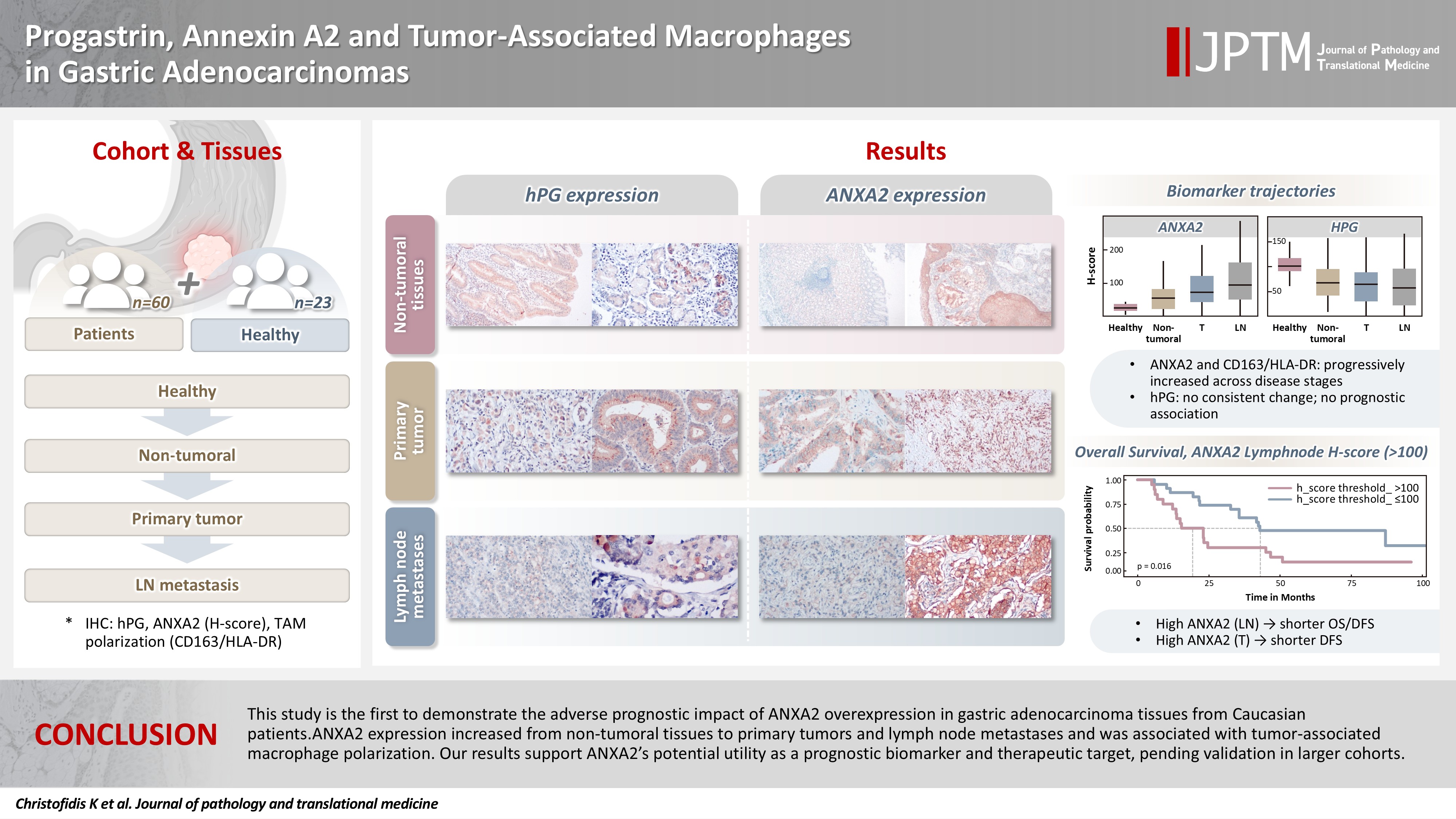
Gastric adenocarcinoma is a major cause of cancer mortality worldwide, and reliable biomarkers remain insufficient. This study investigates the immunohistochemical expression of progastrin (hPG) and annexin A2 (ANXA2) and the polarization of tumor-associated macrophages in gastric adenocarcinoma to explore their potential prognostic and biological significance. Methods: A retrospective analysis was conducted on formalin-fixed, paraffin-embedded tissue samples from 60 patients with gastric adenocarcinoma (primary tumors, lymph node metastases, and non-tumoral gastric mucosa) and gastric biopsies from 23 healthy controls. The expression of hPG and ANXA2 was quantified using the H-score, and the CD163/human leukocyte antigen–DR (HLA-DR) ratio was used to represent macrophage polarization (M2/M1). Statistical analyses included non-parametric tests, Spearman correlations, Kaplan-Meier survival curves, and Cox proportional-hazards models. Results: ANXA2 expression was significantly elevated in cancer cells from primary tumors and lymph node metastases, compared with the non-tumoral gastric mucosa tissues and gastric mucosa tissues from healthy controls. ANXA2 expression increased with the tumor grade. High ANXA2 levels were associated with shorter overall and disease-free survival, but they did not have independent prognostic value. Although hPG expression correlated positively with ANXA2, it showed no significant prognostic association. The CD163/HLA-DR ratio increased with tumor progression and negatively correlated with ANXA2, but it did not influence survival outcomes. Conclusions: This study is the first to demonstrate the adverse prognostic impact of ANXA2 overexpression in gastric adenocarcinoma tissues from Caucasian patients. Our results suggest that ANXA2 might have utility as a prognostic biomarker and therapeutic target, if further large-scale studies validate and expand our findings.
- Ultrastructural and Immunohistochemical Investigations of Exocrine and Endocrine Cells in Fetal Human Pancreas.
- Jung Ran Kim, Je G Chi, Jung Hee Cho
- Korean J Pathol. 1995;29(3):286-295.
- 2,050 View
- 24 Download
-
Abstract
PDF
- The pancreas consists of two types of tissue arising from same primitive cells, but with entirely different functions. Although the adult human pancreas and fetal islet tissue have been the subject of numerous electron microscopic studies, little is known of the ultrastructure of the developing human exocrine pancreas. The purpose of the current study is to investigate development of endo and exocrine of pancreas, especially during the middle trimester of human fetal life, which is the period of acinar cell maturation. Fresh autopsy specimens of pancreas, taken from 15 human fetuses at the 12th (n=2), 13-16th (n=5), 17-20th (n=4), 21-24th (n=2) and 25-28th (n=2) weeks of gestation, were studied electron microscopically, and immunohistochemically. Antisera against insulin, somatostatin, glucagon, pancreatic polypeptide and gastrin, were used for immunohistochemistry. By the 12th week, primitive exocrine acini were identified and these were matured rapidly in the next 6 weeks. At the 17th week stage, ultrastructural examination revealed atypical zymogen granules in the acinar cells. These became progressively less numerous in the 21-28 week period when classical zymogen granules increased upto the level of adult stage. All the endocrine cells were found at the 12th week, forming primitive or mature islets. The relative ratio of endocrine cells at the 12th week was about 35.4%, 24.9%, 39.8%, 0.5% for A, B, D & PP cell, respectively. But at the 25th to 28th week of development, the relative numbers of A and D cells decreased somewhat, whereas those of the B cells increased. The PP cells were constant. The G cells were found at the 12th week of fetal period, which appeared through out the on period.
 First
First Prev
Prev


