 E-submission
Search
- Page Path
- HOME > Search
Original Articles
- Immunohistochemical expression in idiopathic inflammatory myopathies at a single center in Vietnam
- Dat Quoc Ngo, Si Tri Le, Khanh Hoang Phuong Phan, Thao Thi Phuong Doan, Linh Ngoc Khanh Nguyen, Minh Hoang Dang, Thien Thanh Ly, Thu Dang Anh Phan
- J Pathol Transl Med. 2024;58(4):174-181. Published online June 25, 2024
- DOI: https://doi.org/10.4132/jptm.2024.05.02
- 5,481 View
- 276 Download
- 1 Web of Science
- 3 Crossref
-
 Abstract
 PDF
PDF - Background
The identification of idiopathic inflammatory myopathies (IIMs) requires a comprehensive analysis involving clinical manifestations and histological findings. This study aims to provide insights into the histopathological and immunohistochemical aspects of IIMs.
Methods
This retrospective case series involved 56 patients diagnosed with IIMs at the Department of Pathology, University of Medicine and Pharmacy at Ho Chi Minh City, from 2019 to 2023. The histology and immunohistochemical expression of HLA-ABC, HLA-DR, C5b-9, Mx1/2/3, and p62 were detected.
Results
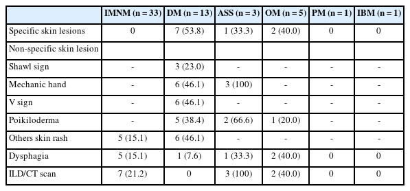
We examined six categories of inflammatory myopathy, including immunemediated necrotizing myopathy (58.9%), dermatomyositis (DM; 23.2%), overlap myositis (8.9%), antisynthetase syndrome (5.4%), inclusion body myositis (IBM; 1.8%), and polymyositis (1.8%). The average age of the patients was 49.7 ± 16.1 years, with a female-to-male ratio of 3:1. Inflammatory cell infiltration in the endomysium was present in 62.5% of cases, perifascicular atrophy was found in 17.8%, and fiber necrosis was observed in 42 cases (75.0%). Rimmed vacuoles were present in 100% of cases in the IBM group. Immunohistochemistry showed the following positivity rates: HLA-ABC (89.2%), HLA-DR (19.6%), C5b-9 (57.1%), and Mx1/2/3 (10.7%). Mx1/2/3 expression was high in DM cases. p62 vacuole deposits were noted in the IBM case. The combination of membrane attack complex and major histocompatibility complex I helped detect IIMs in 96% of cases.
Conclusions
The diagnosis of IIMs and their subtypes should be based on clinical features and histopathological characteristics. Immunohistochemistry plays a crucial role in the diagnosis and differentiation of these subgroups. -
Citations
Citations to this article as recorded by
- Evaluating the Diagnostic Potential of Myxovirus Resistance Protein 1 (MX1) and Myxovirus Resistance Protein 2 (MX2) As Biomarkers in Idiopathic Inflammatory Myopathies
Raghavee Neupane, Mustafa Haider, Perry Smith, Marc M Kesselman
Cureus.2026;[Epub] CrossRef - Rapidly Progressive Polymyositis With Vasculitis: The Pivotal Role of Histopathology in Diagnosis and Management
Amitha Venmanassery Karnalsingh, Arjun Karappilly Vijayan, Monica Roselin Edwin Peter, Dilan Davis
Cureus.2025;[Epub] CrossRef - Autoimmune Neuromuscular Disorders at a Molecular Crossroad: Linking Pathogenesis to Targeted Immunotherapy
Anca-Maria Florea, Dimela-Gabriela Luca, Eugenia Irene Davidescu, Bogdan-Ovidiu Popescu
International Journal of Molecular Sciences.2025; 26(23): 11736. CrossRef
- Evaluating the Diagnostic Potential of Myxovirus Resistance Protein 1 (MX1) and Myxovirus Resistance Protein 2 (MX2) As Biomarkers in Idiopathic Inflammatory Myopathies
- The Expression of C4d and HLA-DR in Renal Allografts with the Histologic Features of Antibody-Mediated Rejection.
- Young Soo Song, Moon Hyang Park
- Korean J Pathol. 2008;42(5):260-269.
- 2,224 View
- 28 Download
-
Abstract
PDF
- BACKGROUND
Deposition of C4d along the peritubular capillaries is generally associated with an antibody-mediated response. We evaluated, with performing C4d immunostaining, the diagnostic accuracy of the cases that were previously diagnosed as antibody-mediated rejection (ABMR) when based only on the histologic findings, and we examined possible correlation of C4d with HLA-DR.
METHODS
Forty-five renal transplantation biopsies, which showed ABMR-like histology, were obtained. The expressions of C4d and HLA-DR in the transplant rejection cases were investigated using immunofluorescent and/or immunohistochemical staining. RESULTS: There were 14 discordant cases among a total of 45 cases when C4d was used as a diagnostic marker and the original slides were reviewed. These total cases consisted of the C4d negative cases in two cases of hyperacute rejection and all the cases of ABMR and ABMR with chronic/sclerosing allograft nephropathy (CAN) and two C4d positive cases (one each of acute cellular rejection (ACR) and CAN according to their original diagnosis) and all these cases were then revised according to Banff 07. The expression of HLA-DR tended to be correlated with the log-transformed duration of grafts until three years after the transplantation. CONCLUSION: This study demonstrates that C4d together with the histologic findings should be used for making the diagnosis of ABMR. The tubular HLA-DR expression over time should be studied to further understand the mechanism of graft rejection.
- Immunohistochemical Analysis of HLA-DR and Secretory Component Expression in Gastric Adenocarcinoma.
- Ji Youn Bae, Soo Sang Sohn, Eun Sook Chang
- Korean J Pathol. 1996;30(4):293-300.
- 2,039 View
- 16 Download
-
Abstract
PDF
- Sixty one cases of gastric adenocarcinoma were studied immunohistochemically for expression of HLA-DR and secretory component(SC) in order to analyze the relationship between expression of these in gastric cancer cells and the adjacent mucosa. Immunostaining was detected within the cytoplasm and on the cell memgrane. The rate of HLA-DR and SC expressions in cancer cells were 59.0% and 49.2%, respectively, and 52.5%/52.5% and 31.2%/50.8% the mucosa in adjacent/remote from the site of to cancer. The SC expression in the adjacent mucosa was lower than that of the remote mucosa(p=0.027). The HLA-DR expression in the cancer cells in the intestinal type of gastric adenocarcinoma(73.9%) was higher than that of the diffuse type(14.3%) and it was statistically significant(p=0.02). The presence of an increased amount of lymphoid infiltration in the gastric mucosa was closely related to the expression of HLA-DR and SC. Decreased or absent expression of SC at the transitional mucosal cells was possibly a result of exposure to genotoxic agents due to the lack of protective function of SC-IgA. From these results, one can postulate that the expression of HLA-DR and SC may play an important role in atleration in microenvironment with lymphoid infiltration.
- Interdigitating Reticulum Cell Sarcoma of Lymph Node.
- Sung Suk Paeng, Yoon Ju Kim, Seong Eun Yang, Duck Hwan Kim, Hee Jin Chang, Jung Il Suh, Chu Woo Kim
- Korean J Pathol. 1996;30(7):635-642.
- 2,435 View
- 19 Download
-
Abstract
PDF
- We report a case of reticulum cell sarcoma in the right cervical lymph node of a 42-year-old male. It was a slowly growing, non-tender movable mass of 8 months duration. Microscopically, the lymph node was effaced by proliferating spindle cells arranged in broad sheets, bands, or fascicular patterns in paracortical area sparing of B-cell region. The tumor component was divided by fibrous band. The individual cells had oval to round or elongated nuclei, with inconspicuous nucleoli and moderate amounts of cytoplasms with indistinct cell borders. Pleomorphic large cells with binucleated, or multinucleated bizarre nuclei with prominent nucleoli, were partly admixed. In immunohistochemical stain, the tumor cell was strong positive for S-100 protein, HLA-DR, Mac387 and weakly positive for Leukocyte common antigen and equivocal for Vimentin. But it was negative for CD21, Ki-1, Desmin, Epithelial membrane antigen and Cytokeratin. These immunohistochemical findings suggested that the neoplastic cell was originated from the interdigitating reticulum cell of lymph node. The patient was treated by radiation therapy, and alive well at 37 months of follow-up.
 First
First Prev
Prev


