 E-submission
Search
- Page Path
- HOME > Search
Original Articles
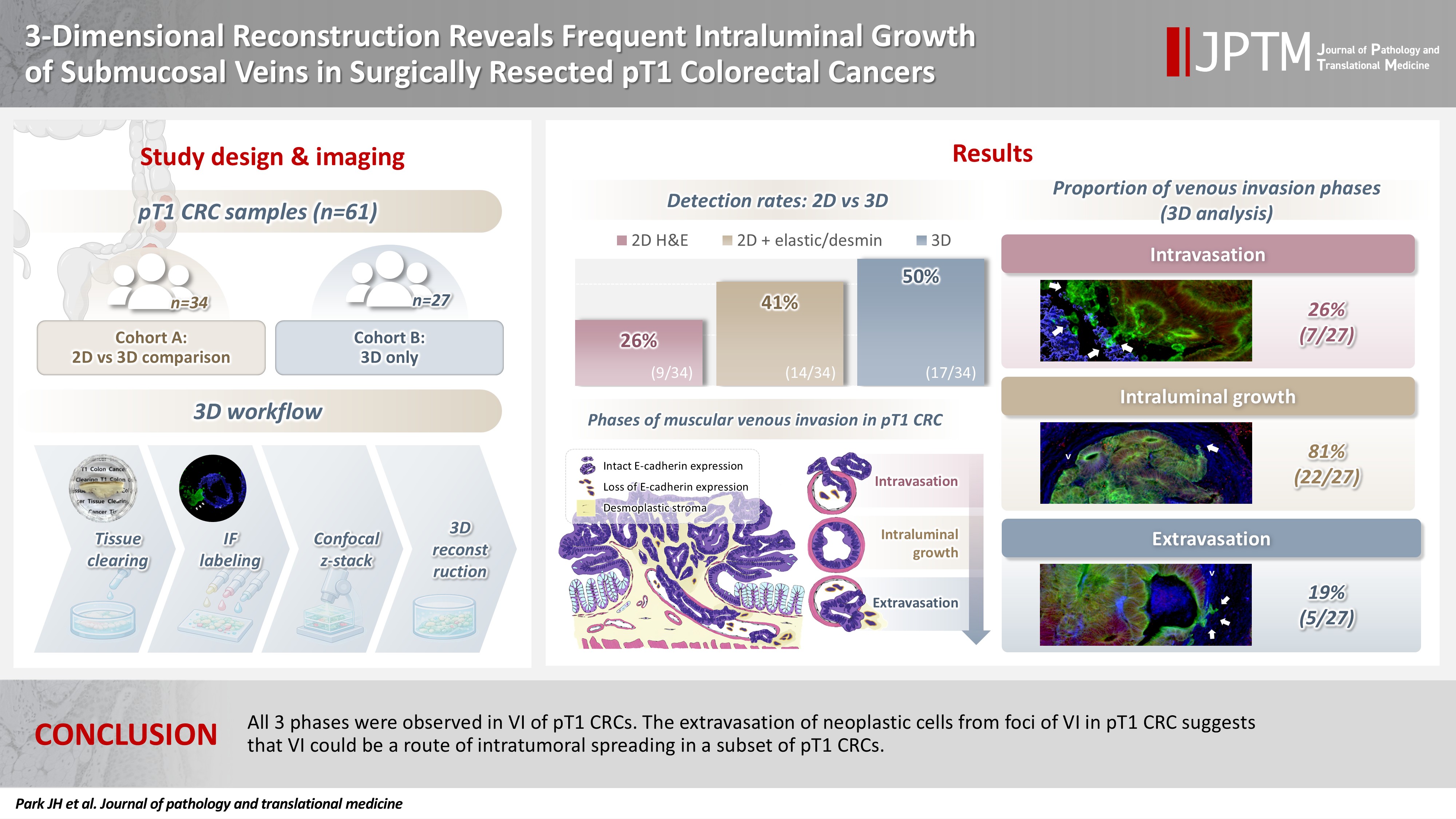
- 3-Dimensional reconstruction reveals frequent intraluminal growth of submucosal veins in surgically resected pT1 colorectal cancers
- Jihyun Park, Mi-Ju Kim, Yeon Wook Kim, Byong-Wook Lee, Junyoung Shin, Jinho Shin, Chan-Gi Pack, Dong-Hoon Yang, Jihun Kim, In Ja Park, Ralph H. Hruban, Seung-Mo Hong
- J Pathol Transl Med. 2026;60(2):246-262. Published online March 10, 2026
- DOI: https://doi.org/10.4132/jptm.2025.12.19
- 1,336 View
- 95 Download
-
 Abstract
 PDF
PDF - Background
Although venous invasion (VI) is associated with distant metastasis and observed in >50% of pT2–4 colorectal cancers (CRCs), the role of VI in pT1 CRCs is not well-defined. Methods: Thirty-four surgically resected pT1 CRCs were reevaluated for 2-dimensional (2D) VI using hematoxylin and eosin (H&E)–stained slides with additional elastic and desmin immunohistochemical staining (cohort A). Additionally, 27 pT1 CRCs without knowing VI status were selected for 3-dimensional (3D) VI evaluation only (cohort B). All 61 cases (cohorts A and B) were studied in 3D using tissue clearing. Results: VI was detected more commonly in 3D (17/34, 50.0%) than in 2D H&E slide evaluation (9/34, 26.5%, p = .047). When VI was identified in 3D (27/61, 44.3%), the most common phase was that of intraluminal growth (22/27, 81.5%), followed by intravasation (7/27, 25.9%) and extravasation (5/27, 18.5%). E-cadherin expression was characterized in 3D in foci of VI and varied in each phase of invasion. Conclusions: All three phases were observed in VI of pT1 CRCs. The extravasation of neoplastic cells from foci of VI in pT1 CRC suggests that VI could be a route of intratumoral spreading in a subset of pT1 CRCs.
- The Effects of Transforming Growth Factor beta1 on Apoptosis in Rat Hepatocellular Carcinoma.
- Young Euy Park, Young Hee Choi, Won Yo Lee, Jin Ja Park, Kyung Chan Choi, Hyung Shik Shin
- Korean J Pathol. 1999;33(2):71-79.
- 2,074 View
- 10 Download
-
Abstract
- Based upon the concept that carcinogenesis is associated with apoptosis, specific therapies designed to enhance the susceptibility of cancer cells to undergo apoptosis could be developed. Thus, in this paper, it was designed to investigate whether, using rat animal model with chemical-induced hepatocellular carcinoma, TGF-1 in vivo could induce apoptosis in cancer. The chemical hepatocarcinogenic procedure of Solt-Farber method was used on Sprague-Dawley rats. Experimental groups were divided into group A treated with the standard Solt-Farber regimen of diethylnitrosamine (DEN) and 2-Acetaminofluorene (AAF), group B TGF-, group C TGF-1, and group D adriamycin after hepatocellular carcinoma developed. For detection of apoptotic cells, apoptotic indices were examined by the in situ end DNA labelling method. The expression of proliferating cell nuclear antigen was examined by immunohistochemical staining. Apoptosis of rat hepatocellular carcinoma cells increased significantly to 4.92+/-2.32/HPF in the group C compared with the control group (A) (2.54+/-1.13/HPF; P<0.05). Two distinctly different populations of proliferating hepatocellular carcinoma cells were identified. The cells at G1/S boundary (weak granular staining) increased to 15.75+/-6.19/HPF and 6.45+/-2.93/HPF in the groups C and D, respectively, but decreased to 2.42+/-2.06/HPF in the group B compared with the control group (A) (6.38+/-2.18/HPF; p<0.05). The cells at S phase (strong granular staining) increased to 3.37+/-2.69/HPF in the group B but decreased to 0.32+/-0.47/HPF in the group D (p<0.05). In conclusion, these results indicate that the TGF-1 may be used as an effective anticancer agent.
Case Reports
- Malignant Eccrine Poroma of Abdomen Brief case report.
- Jin Ja Park, Young Hee Choi, Kyung Chan Choi, Young Euy Park
- Korean J Pathol. 1998;32(4):312-314.
- 2,232 View
- 21 Download
-
Abstract
PDF
- Eccrine porocarcinoma is a rare tumor of the skin. A case report of an eccrine porocarcinoma metastasizing to epidural space of spinal cord and inguinal area with a nine year follow up is described. The patient had a nodular growth of the abdomen with both inguinal lymphadenopathy three years before its first excision. After a follow up of nine years, he complained a weakness of lower extremities and back pain. Extradural mass of 10th thoracic vertebra and left inguinal mass were found. Subsequently, the masses histologically identical to the skin tumor were found.
- Polypoid Ganglioneuromatosis of Colon: A case report.
- Jin Ja Park, Kyung Chan Choi, Young Hee Choi, Young Euy Park
- Korean J Pathol. 1998;32(5):388-390.
- 2,168 View
- 10 Download
-
Abstract
- Gastrointestinal ganglioneuromatosis is an extremely rare lesion which typically occurs with a significant systemic syndrome. It is known to be a major component of multiple endocrine neoplasia, type 2b. We presented a case of polypoid ganglioneuromatosis of the colon in a 3-year-old female with abdominal pain and diarrhea. She had no clinical evidence of the systemic syndrome or von Recklinghausen's neurofibromatosis, conditions in which intestinal ganglioneuromatosis can occur. Gross examination showed diffuse polypoid masses in ascending and transverse colons with normal-appearing mucosa. Microscopic examination revealed a proliferation of spindle-shaped neuronal cells containing multiple clusters of mature ganglion cells in the mucosa, submucosa and proper muscle. We describe a case of colonic ganglioneuromatosis without any component of multiple endocrine neoplasia or family history.
 First
First Prev
Prev


