 E-submission
Search
- Page Path
- HOME > Search
Original Articles
- A single-institution demographic study of pathologically proven renal disease in kidney transplant recipients over the last 33 years
- Hyejin Noh, Jiyeon Kim, Yeong Jin Choi
- J Pathol Transl Med. 2026;60(4):398-412. Published online May 26, 2026
- DOI: https://doi.org/10.4132/jptm.2026.03.28
- 863 View
- 17 Download
-
 Abstract
 PDF
PDF Supplementary Material
 - Background
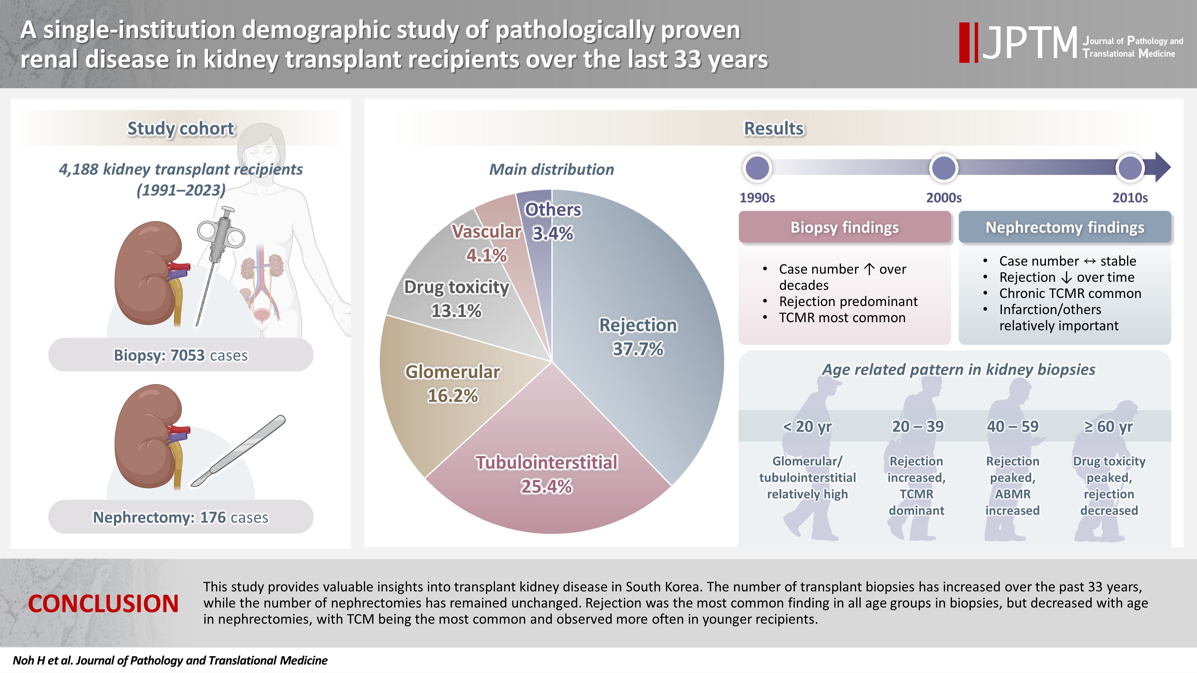
While the number of kidney transplants for end-stage renal disease (ESRD) is increasing, studies examining the long-term demographic analyses based on pathological diagnosis of transplant kidney remain limited. Methods: We conducted a retrospective analysis of 4,188 transplant recipients who underwent either biopsy or nephrectomy from 1991 to 2023 at Seoul St. Mary’s Hospital. Results: Among 7,229 pathologically confirmed cases, rejection was the most prevalent (37.7%), followed by tubulointerstitial (25.4%), glomerular, drug toxicity, and vascular diseases. In 7,053 transplant biopsies, rejection was predominant across all age groups, with T-cell mediated (TCM) category being the most common (60.1%), followed by antibody-mediated and mixed. Drug toxicity increased with age (p = .047), while glomerular and tubulointerstitial diseases were highest in recipients under 20 (p < .001). Among glomerular diseases, IgA-related glomerulonephritis (45.2%) was the most common. In 176 transplant nephrectomies, the most common diagnosis was rejection (33.5%), followed by renal infarction (19.9%), tubulointerstitial, vascular, glomerular disease, and drug toxicity. “Others” included infarction, ESRD, and lymphangiectasia, which increased with age (p = .011). In nephrectomy cases, rejection decreased over time, with chronic TCM rejection (40.7%) being the most frequent. Conclusions: This study provides valuable insights into transplant kidney disease in South Korea. The number of transplant biopsies has increased over the past 33 years, while the number of nephrectomies has remained unchanged. Rejection was the most common finding in all age groups in biopsies, but decreased with age in nephrectomies, with TCM being the most common and observed more often in younger recipients.
- A single-institution demographic study of pathologically proven kidney disease in South Korea over the last 33 years
- Hyejin Noh, Jiyeon Kim, Yeong Jin Choi
- J Pathol Transl Med. 2025;59(5):306-319. Published online September 10, 2025
- DOI: https://doi.org/10.4132/jptm.2025.06.18
- 3,479 View
- 100 Download
- 1 Web of Science
-
Abstract
PDFSupplementary Material
- Background
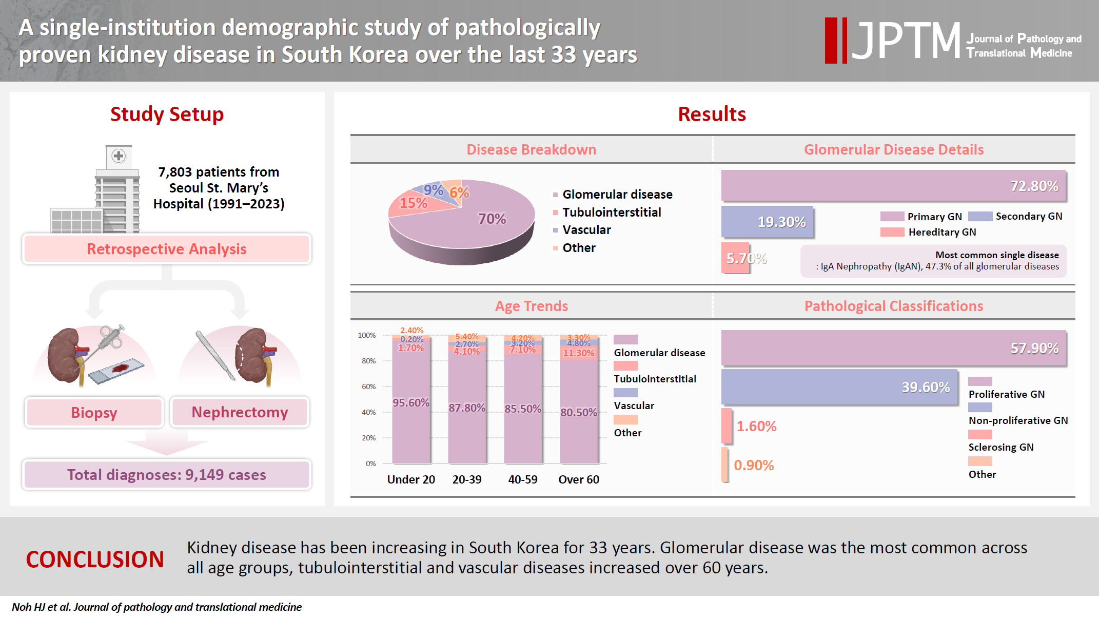
To date, epidemiological studies on the entire spectrum of kidney disease based on pathology have been rarely reported. Methods: A retrospective study was conducted on patients diagnosed with kidney disease at Seoul St. Mary's Hospital between 1991 and 2023. Results: Among 7,803 patients with native kidney disease, glomerular disease (70.3%) was the most common, followed by tubulointerstitial (15.1%) and vascular disease (8.8%). In kidney biopsy, glomerular disease (77.8%) showed the highest frequency, particularly in those under 20s (95.6%) (p = .013). Primary glomerulonephritis (GN) (72.8%) was the predominant glomerular disease, with IgA nephropathy (IgAN) (47.3%) being the most common one. Tubulointerstitial and vascular diseases increased with age, showing the highest prevalence in those over 60 years (p = .008 and p = .032, respectively). Glomerular disease was diagnosed at a younger age (39.7 ± 16.7 years) than tubulointerstitial (49.1 ± 16.2) and vascular (48.1 ± 15.3) diseases (p < .001). When glomerular diseases were classified morphologically, proliferative GN (57.9%) was the most common, followed by non-proliferative (39.6%) and sclerosing (1.6%). When classified by etiology, primary GN accounted for the most (72.8%), followed by secondary (19.3%) and hereditary GN (5.7%). In nephrectomy, tubulointerstitial disease (64.6%) was the most common. Those with a tubulointerstitial disease had a higher mean age than those with a glomerular disease (p < .001). In cases where nephrectomy was performed for glomerular diseases, IgAN (34.1%) was the most common diagnosis. Conclusions: Kidney disease has been increasing in South Korea for 33 years. Glomerular disease was the most common across all age groups, tubulointerstitial and vascular diseases increased over 60 years.
- Expression of Inducible Nitric Oxide Synthase and Nitric Oxide Mediated Apoptosis in Neuronal PC12 Cells after Lipopolysaccharide/Tumor Necrosis Factor-/Interferon- Treatment.
- Jiyeon Kim, Jiyoung Kim, Kuseong Kang, Eunkyoung Kwak, Jiyoung Park, Taein Park, Yoonkyung Sohn
- Korean J Pathol. 2002;36(4):249-256.
- 2,149 View
- 20 Download
-
Abstract
PDF
- BACKGROUND
Inducible nitric oxide synthase (iNOS) has been detected in a number of pathologic conditions in the central nervous system. This study was investigated the patterns of iNOS expression in the neuronal PC12 cell and the effects of nitric oxide on the apoptosis of PC12 cells.
METHODS
The stimulating agents for induction of iNOS expression in PC12 cells were bacterial lipopolysaccharide (LPS), tumor necrosis factor-alpha (TNF-), and interferon-gamma (IFN-).
RESULTS
The expression iNOS mRNA and protein in PC12 cells stimulated with LPS/TNF-/IFN- were profoundly increased. The expression of iNOS mRNA arose at 6 hours, peaked at 12 hours, and declined to 48 hours after LPS/TNF-/ IFN- treatment. iNOS protein was increased up to 24 hours in LPS/TNF-/IFN- treated PC12 cells while the expression of nNOS was unaffected. Accumulation of NO derivatives in the culture media was markedly increased at least at up to 48 hours after LPS/TNF-/IFN- treatment. The induction of iNOS expression and NO production in differentiated PC12 cells was correlated with apoptotic cell death judged by transmission electron microscopy and DNA fragmentation from the results of the Terminal deoxynucleotidyl-transferase-mediated dUDP biotin nick end-labeling (TUNEL) method. After treatment with NOS inhibitor, N-monomethylarginine (NMMA), a profound decrease in NO production by LPS/TNF-/IFN- treated PC12 cells was noted. And the LPS/TNF-/IFN- induced apoptosis was prevented by the NMMA treatment.
CONCLUSIONS
From the above results it is concluded that the expression of iNOS in differentiated PC12 cells is induced by the combined application of LPS, TNF-, and IFN-. And the apoptosis of cultured PC12 cells is mediated by iNOS-derived NO.
 First
First Prev
Prev


