 E-submission
Search
- Page Path
- HOME > Search
Original Articles
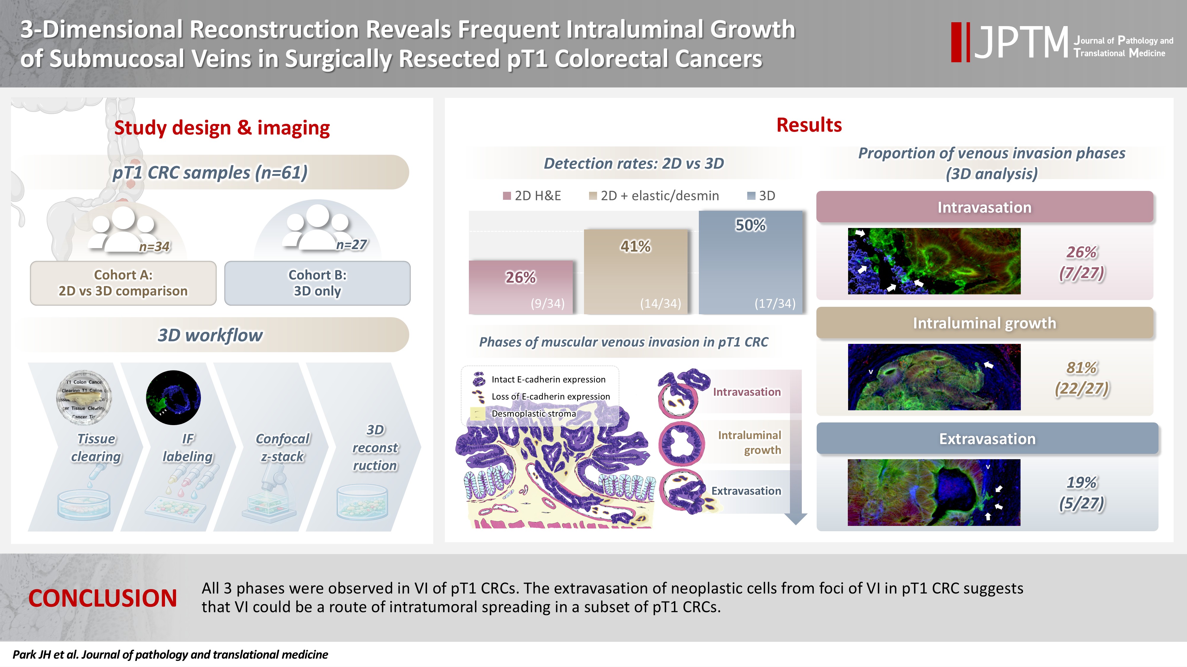
- 3-Dimensional reconstruction reveals frequent intraluminal growth of submucosal veins in surgically resected pT1 colorectal cancers
- Jihyun Park, Mi-Ju Kim, Yeon Wook Kim, Byong-Wook Lee, Junyoung Shin, Jinho Shin, Chan-Gi Pack, Dong-Hoon Yang, Jihun Kim, In Ja Park, Ralph H. Hruban, Seung-Mo Hong
- J Pathol Transl Med. 2026;60(2):246-262. Published online March 10, 2026
- DOI: https://doi.org/10.4132/jptm.2025.12.19
- 1,336 View
- 95 Download
-
 Abstract
 PDF
PDF - Background
Although venous invasion (VI) is associated with distant metastasis and observed in >50% of pT2–4 colorectal cancers (CRCs), the role of VI in pT1 CRCs is not well-defined. Methods: Thirty-four surgically resected pT1 CRCs were reevaluated for 2-dimensional (2D) VI using hematoxylin and eosin (H&E)–stained slides with additional elastic and desmin immunohistochemical staining (cohort A). Additionally, 27 pT1 CRCs without knowing VI status were selected for 3-dimensional (3D) VI evaluation only (cohort B). All 61 cases (cohorts A and B) were studied in 3D using tissue clearing. Results: VI was detected more commonly in 3D (17/34, 50.0%) than in 2D H&E slide evaluation (9/34, 26.5%, p = .047). When VI was identified in 3D (27/61, 44.3%), the most common phase was that of intraluminal growth (22/27, 81.5%), followed by intravasation (7/27, 25.9%) and extravasation (5/27, 18.5%). E-cadherin expression was characterized in 3D in foci of VI and varied in each phase of invasion. Conclusions: All three phases were observed in VI of pT1 CRCs. The extravasation of neoplastic cells from foci of VI in pT1 CRC suggests that VI could be a route of intratumoral spreading in a subset of pT1 CRCs.
- Diagnostic value of cytology in detecting human papillomavirus–independent cervical malignancies: a nation-wide study in Korea
- Hye-Ra Jung, Junyoung Shin, Chong Woo Yoo, Eun Na Kim, Cheol Lee, Kyeongmin Kim, Ho-chang Lee, Yonghee Lee, Ji Hye Kim, Soo Jin Jung, Yumin Chung, Joo Yeon Kim, Hye Eun Park, Tae Hoen Kim, Wonae Lee, Min-Sun Cho, Ran Hong, Yoon Jung Choi, Younghee Choi, Young Sub Lee, Sang-Ryung Lee, Myunghee Kang, Young Jin Seo, Seung-Sook Lee, Yoon-Jung Hwang, Hyun-Jung Kim
- J Pathol Transl Med. 2025;59(6):444-452. Published online November 11, 2025
- DOI: https://doi.org/10.4132/jptm.2025.10.21

- 5,328 View
- 164 Download
-
Abstract
PDF
- Background
Human papillomavirus (HPV) independent cervical malignancies (HPV-IDCMs) have recently been classified by the World Health Organization (WHO) 5th edition. These malignancies have historically received limited attention due to their rarity and the potential for evasion of HPV-based screening.
Methods
We retrospectively reviewed 5,854 biopsy-confirmed cervical malignancies from 22 institutions over 3 years (July 2020–June 2023). Histologic classification followed the WHO guidelines. HPV independence was confirmed by dual negativity for p16 and HPV; discordant cases (p16-positive/HPV-negative) underwent additional HPV testing using paraffin-embedded tissue. Cytological results were matched sequentially to histological confirmation.
Results
The prevalence of HPV-IDCM was 4.4% (257/5,854) overall and was 3.6% (208/5,805 cases) among primary cervical malignancy. Patient age of HPV-IDCM was 29 to 89 years (median, 57.79). Its histologic subtypes included primary adenocarcinoma (n = 116), endometrial adenocarcinoma (n = 35), squamous cell carcinoma (n = 72), metastatic carcinoma (n = 14), carcinoma, not otherwise specified (n = 10), neuroendocrine carcinoma (n = 3), and others (n = 7). Among 155 cytology-histological matched cases, the overall and primary Pap test detection rates were 85.2% (132/155) and 83.2% (104/125), respectively. The interval between cytology and histologic confirmation extended up to 38 months.
Conclusions
HPV-IDCMs comprised 3.6% of primary cervical malignancies with a high detection rate via cytology (83.2%). These findings affirm the value of cytological screening, particularly in patients with limited screening history or at risk for HPV-independent lesions, and may guide future screening protocols.
- Primary Rhabdomyosarcoma of the Breast: Study of Three Cases at One Institution with a Review of Primary Breast Sarcomas
- Junyoung Shin, Hee Jeong Kim, Dae-Yeon Kim, Gyungyub Gong, Kyung-Ja Cho
- J Pathol Transl Med. 2019;53(5):308-316. Published online August 2, 2019
- DOI: https://doi.org/10.4132/jptm.2019.07.22
- 8,586 View
- 141 Download
- 7 Web of Science
- 9 Crossref
-
Abstract
PDF
- Background
Primary breast sarcoma (PBS) is rare, comprising approximately 1% of breast malignancies. Rhabdomyosarcoma (RMS) accounts for an extremely small proportion of PBSs, often leading to delayed histologic confirmation.
Methods
Upon reviewing Asan Medical Center’s pathology database between 2000 and 2018, 41 PBS cases were retrieved, including three cases of primary RMS of the breast. Their clinicopathological features were analyzed, and the literature related to PBS and primary RMS of the breast was reviewed.
Results
We identified three primary breast RMS cases from our institution database, comprising 7.3% of PBS: one case each of spindle cell/sclerosing RMS (ssRMS), alveolar RMS (aRMS), and embryonal RMS (eRMS). All cases involved adolescents or young adults (14, 16, and 25 years, respectively) who underwent mastectomy or radiotherapy and were confirmed using immunohistochemical testing for myogenin, desmin, and myogenic differentiation. The ssRMS patient experienced recurrence at the operation site 4 months post-surgery despite undergoing concurrent chemoradiotherapy. The aRMS patient had multiple metastases at diagnosis and showed FAX3-FOXO1 fusion transcripts; she died 22 months after the diagnosis. The eRMS patient had enlarged axillary lymph nodes; post-radiotherapy, the lesion recurred as multiple metastases to the bone and lung. She died 18 months post-diagnosis.
Conclusions
Our experience on RMS cases suggests that spindle cell or small round cell malignancy in breasts of young female should raise suspicion for the possibility of primary or secondary RMS. To our knowledge, this is the second report of primary breast ssRMS and it may help clinicians who encounter this rare disease in the future. -
Citations
Citations to this article as recorded by
- Primary Ewing sarcoma of the breast in a male adolescent: A case report
Joie Sheen A. Bastian, Willie T. Hao, Lawrence Faith A. Lucañas-Yap
Journal of Pediatric Surgery Case Reports.2026; 127: 103205. CrossRef - Clinicopathological and molecular features of breast metastases in alveolar rhabdomyosarcoma: A series of 3 cases
Wanni Xu, Li Yang, Yuxin Hui, Peizhuo Yao, Yiwei Jia, Xinyu Wei, Shuqun Zhang
Annals of Diagnostic Pathology.2026; 83: 152627. CrossRef - Management of Pediatric Breast Masses for the Pediatric Surgeon: Expert Consensus Recommendations From the APSA Cancer Committee
Dana Schwartz, Elisabeth T. Tracy, Bindi Naik-Mathuria, Richard D. Glick, Stephanie F. Polites, Peter Mattei, David Rodeberg, Andres F. Espinoza, Sara A. Mansfield, Dave R. Lal, Meera Kotagal, Timothy Lautz, Jennifer Aldrink, Barrie S. Rich
Journal of Pediatric Surgery.2025; 60(2): 161916. CrossRef - Differential diagnosis of primary mesenchymal neoplasms of the breast
Mine Ozsen, Seyit Ali Volkan Polatkan, Ulviye Yalcınkaya, Sahsine Tolunay, Mustafa Sehsuvar Gokgoz
Clinical and Translational Oncology.2024; 27(1): 223. CrossRef - Primary breast rhabdomyosarcoma in a 17-year-old girl
Laxmi Singotia, V.S. Haritha
Journal of Cancer Research and Therapeutics.2023; 19(7): 2070. CrossRef - High-Grade Spindle Cell Lesions of the Breast
Esther Yoon, Qingqing Ding, Kelly Hunt, Aysegul Sahin
Surgical Pathology Clinics.2022; 15(1): 77. CrossRef - Primary Small Cell Malignancies of the Breast: Are They Rare Malignancies?
Kemal Behzatoğlu, Fernando Schmitt
Acta Cytologica.2022; 66(4): 347. CrossRef - Recurrent malignant phyllodes tumor of the breast: An extremely rare case of recurrence with only rhabdomyosarcoma components
Jia Han, Shuice Liu, Akihoro Shioya, Motona Kumagai, Emi Morioka, Miki Noguchi, Masafumi Inokuchi, Sohsuke Yamada
SAGE Open Medical Case Reports.2022;[Epub] CrossRef - Primary rhabdomyosarcoma: An extremely rare and aggressive variant of male breast cancer
Cătălin Bogdan Satală, Ioan Jung, Tivadar Jr Bara, Patricia Simu, Iunius Simu, Madalina Vlad, Rita Szodorai, Simona Gurzu
World Journal of Clinical Cases.2020; 8(19): 4466. CrossRef
- Primary Ewing sarcoma of the breast in a male adolescent: A case report
Case Study
- WITHDRAWN:Primary Rhabdomyosarcoma of the Breast: A Report of Two Cases and Literature Review
- Junyoung Shin, Hee Jeong Kim, Dae-Yeon Kim, Gyungyub Gong, Kyung-Ja Cho
- Received August 6, 2018 Accepted September 13, 2018 Published online October 4, 2018
- DOI: https://doi.org/10.4132/jptm.2018.09.14
- 4,525 View
- 62 Download
- 1 Crossref
-
Citations
Citations to this article as recorded by- Primary Alveolar Rhabdomyosarcoma of the Breast in an Adult: An Extremely Rare Case
Helen J. Trihia, Natasa Novkovic, Ioannis Provatas, Anastasios Mavrogiorgis, Evangelos Lianos
Case Reports in Pathology.2019; 2019: 1. CrossRef
- Primary Alveolar Rhabdomyosarcoma of the Breast in an Adult: An Extremely Rare Case
 First
First Prev
Prev


