 E-submission
Search
- Page Path
- HOME > Search
Review Article
- The evolving role of TRPS1 in dermatopathology: insights from the past 4 years
- Mokhtar H. Abdelhammed, Woo Cheal Cho
- J Pathol Transl Med. 2026;60(2):129-143. Published online January 29, 2026
- DOI: https://doi.org/10.4132/jptm.2025.11.25
- 3,412 View
- 230 Download
-
 Abstract
 PDF
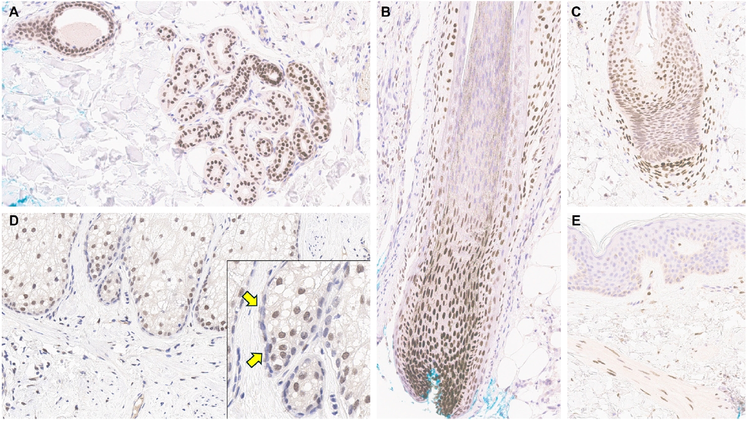
PDF - Over the past 4 years, trichorhinophalangeal syndrome type 1 (TRPS1) has rapidly gained attention among practicing pathologists, with numerous studies emerging that both support and question its diagnostic utility. Initially regarded as a highly specific marker for tumors of mammary origin, TRPS1 is now recognized to have broader expression patterns, including in a variety of cutaneous neoplasms. This is likely due to embryologic parallels between breast tissue and skin adnexal structures, an overlap that was underappreciated in early investigations. Although TRPS1 lacks absolute specificity—even among cutaneous neoplasms—it can still offer meaningful diagnostic value when interpreted alongside conventional immunohistochemical markers and within the appropriate morphologic context. Noteworthy diagnostic applications include mammary Paget disease, primary extramammary Paget disease, rare adnexal neoplasms such as endocrine mucin-producing sweat gland carcinoma and primary cutaneous NUT adnexal carcinoma, and cutaneous metastases from breast carcinoma. In this review, we present the most comprehensive and up-to-date evaluation of the utility and limitations of TRPS1 immunohistochemistry in dermatopathology. Our aim is to deepen understanding of this emerging marker and provide practical guidance on its optimal integration with established immunohistochemical panels to enhance diagnostic accuracy in routine practice.
Case Reports
- Malignant Melanoma Arising in Giant Congenital Melanocytic Nevus: A case report.
- Jung Sun Kim, Sang Yong Song, Kye Yong Song, Je G Chi
- Korean J Pathol. 1993;27(6):650-655.
- 2,367 View
- 39 Download
-
Abstract
PDF
- Giant congenital melanocytic nevus is found in 0.1% of live born infants. If present, this lesion has a 6.3% chance to develop malignant melanoma. We report such a case in a 22-year-old woman who had multiple pigmented skin lesions since birth. Rapidly growing masses were recently detected in the 19 cm-sized occipital pigmented lesion. Removed scalp lesion revealed yellowish white lobulated soft nodules in the background of pigmented nevus. Microscopically, the nodules consisted of epithelioid cells with prominent nucleoli, and pleomorphic cells including signetring cells. These cells seldom contained melanin pigment. There were metastatic aggregates of tumor cells in the cervical lymph node, which were reminiscent of germinal centers of lymph nodes. S-100 protein immunostaining was helpful to distinguish them. Incidentally, focally scattered pigmented spindle cells were seen in the capsule of a lymph node
- Congenital Melanocytic Schwannoma in Ankle Joint Potentially Malignant: A case report.
- Jong Tae Park, Chang Soo Park, Sang Woo Juhng, Kyu Hyuk Cho
- Korean J Pathol. 1987;21(4):308-316.
- 2,138 View
- 10 Download
-
Abstract
PDF
- Congenital malignant melanocytic schwannoma in ankle joint was not reported on literature and it was a very interesting case. Light microscopically, melaninladen cells were mixed in abundant wavy spindle cells, some mitotic cells were also observed. Ultrastructurally, melanosomes in variable stages of development were scattered in the cytoplasm which had basal lamina. Collagen bundles were abundant in the intercellular connective tissue. It was histologically malignant tumor and clinically recurred. But in non-congenital potentially malignant melanocytic schwannoma which had been reported, reccurrence or distant metastasis were not noted. So, further clinical survey may be necessary for evaluation of the malignant behavior of this neoplasm.
 First
First Prev
Prev


