Search
- Page Path
- HOME > Search
Original Articles
- Unraveling the crucial role of CCL3 in nasopharyngeal carcinoma: bioinformatics and immunohistochemical insights
- Xiaopeng Guo, Zhen Sun, Ya Liang, Aoshuang Chang, Junjun Ling, Houyu Zhao, Xianlu Zhuo
- J Pathol Transl Med. 2025;59(5):281-290. Published online September 8, 2025
- DOI: https://doi.org/10.4132/jptm.2025.05.23
- 1,536 View
- 137 Download
-
 Abstract
 PDF
PDF - Background
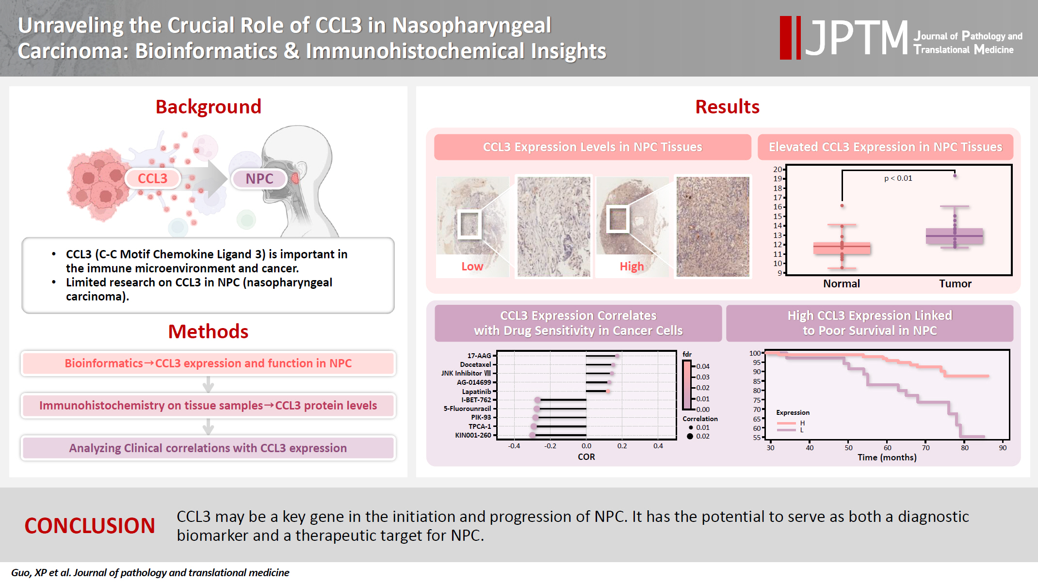
C-C motif chemokine ligand 3 (CCL3) is a crucial chemokine that plays a fundamental role in the immune microenvironment and is closely linked to the development of various cancers. Despite its importance, there is limited research regarding the expression and function of CCL3 in nasopharyngeal carcinoma (NPC). Therefore, this study seeks to examine the expression of CCL3 and assess its clinical significance in NPC using bioinformatics analysis and experiments. Methods: The bioinformatics approach was employed to assess the expression and function of CCL3 in NPC. Subsequently, protein expression of CCL3 was detected in an NPC cohort using immunohistochemistry based on a tissue microarray. The relationship between CCL3 expression and clinical features was then investigated. Results: A total of 20 CCL3-related genes and 14 possible target genes were identified through bioinformatics analysis, many of which play crucial roles in pathways such as chemokine signaling pathway and transcriptional misregulation in cancer signaling pathways. CCL3 was found to be associated with drug resistance and various immune cell infiltrations. In NPC, CCL3 expression was significantly higher than normal controls, and high expression of CCL3 correlated with cervical lymph node metastasis, tumor recurrence, advanced clinical stage, and poor prognosis. Conclusions: CCL3 may be a key gene in the initiation and progression of NPC. It has the potential to serve as both a diagnostic biomarker and a therapeutic target for NPC.
- Tissue Microarray Analysis of the Expression of p53, c-kit and CD34 in Sarcomas.
- Jinyoung Yoo, Kyung Shin Park, Seok Jin Kang, Chang Suk Kang
- Korean J Pathol. 2004;38(4):221-227.
- 2,111 View
- 15 Download
-
 Abstract
 PDF
PDF - BACKGROUND
Our objectives in this study were to (1) evaluate the possible role of p53, c-kit and CD34 proteins in sarcomas and to determine their potential relationship; (2) use a tissue microarray to compare the immunohistochemical staining results on both the tissue microarrays and the corresponding whole tissue sections.
METHODS
Whole sections from 85 sarcomas were studied for the immunohistochemical expression of p53, c-kit and CD34. Tissue microarrays consisting of triplicate 2 mm cores from the corresponding blocks were constructed and stained according to the same protocols as those used for the whole sections.
RESULTS
On whole section analysis, p53 protein was expressed in 25 cases (29.4%). Expression of c-kit was observed in 31 specimens (36.5%), whereas CD34 expression was noted in 11 tumors (12.9%). The overall concordance between triplicates was 96% (217/226). The consensus score from the combined triplicates agreed with the results on the whole sections at 91.4% (233/255). The correlations between p53 and CD34, and between c-kit and CD34, were statistically significant (p=.028 and p=.010 respectively).
CONCLUSIONS
p53 and c-kit express relatively frequently in sarcomas. Tissue microarrays are an effective alternative to whole sections; however, the presence of triplicate punches seems to improve the yield but not the concordance of data.
- Expression of CD44 Isoforms and Its Significance in Renal Cell Carcinoma.
- Ghil Suk Yoon, Hee Yeon Hong, Tae Sook Kim
- Korean J Pathol. 2005;39(4):251-257.
- 2,124 View
- 30 Download
-
 Abstract
 PDF
PDF - Background
: CD44 is a transmembranous glycoprotein that participates in cell-cell and cell-matrix interactions, and it also contributes to cell migration. In vitro studies have suggested that the expression of CD44 isoforms is associated with tumor metastasis. Since it is not clear whether the CD44 isoforms play a role in the tumorigenesis, differentiation, progression or metastasis of renal cell carcinomas (RCCs). Methods : We performed immunohistochemistry with primary antibodies for the standard CD44 (CD44s) and the CD44 variant exon 6 (CD44v6) on the archival paraffin-embedded tissue microarray (TMA) specimens from 51 RCC patients. Results : In the normal kidney, the expressions of both CD44s and CD44v6 were negligible. The CD44s expression was increased in accordance with the tumor size (p<0.01), but it was not related to the microvessel density (MVD). No CD44v6 expression was observed in all RCC cases. Univariate analysis indicated that stage, tumor size, lymph node metastasis and distant organ metastasis were the statistically significant prognostic factors for disease free survival (DFS) (p<0.01), and the multivariate analysis proved that stage (p<0.01) and tumor size (p<0.05) were the independent prognostic factors for DFS. Conclusions : Our results suggest that CD44s, but not CD44v6, plays a role in tumor progression and it could be a potential prognostic factor for patients with RCCs.
- Analysis of Gene Expression in Renal Cell Carcinomas Using cDNA Microarray: Reduced Expression of Decorin in Renal Cell Carcinomas.
- Jin Sook Lee, Kang Suek Suh, Kyung Un Choi, Jee Yeun Kim, Do Youn Park
- Korean J Pathol. 2003;37(4):232-238.
- 1,887 View
- 14 Download
-
 Abstract
 PDF
PDF - BACKGROUND
Identification of the genes expressed differentially in renal cell carcinoma (RCC)but not in the non-cancerous kidney is important for understanding the molecular basis ofrenal cell carcinoma and for defining possible prognostic value and therapeutic intervention.We investigated the changes in gene expression accompanying the development and progression of kidney cancer by cDNA microarrays.
METHODS
To identify molecular alterations in renal cell carcinoma, we measured expression profiles for paired neoplastic and noncancerouskidney samples from an individual by means of a cDNA microarry representing 7, 500genes. Of the differentially expressed genes, we assessed the decorin gene at the proteinlevel using immunohistochemistry.
RESULTS
The 60 genes were noted to have more than a fivefold change in expression (either increased or decreased) in RCC compared to the noncancerouskidney. The changed genes are those associated with signal transduction, metabolizingenzymes, the cytoskeleton, cell adhesion, cell cycle control, modulation of transcription, the tumor suppressor gene and tumor antigens. Under immunohistochemistry, the expressionof decorin was significantly decreased in the tumor than in the non-cancerous kidney.The expression rate of decorin was not associated with the patient's sex, age, histologic type, Fuhrmann nuclear grade and T stage.
CONCLUSION
The author predicted that these geneexpression profiling experiments will lead to improvements in the basic understanding of renaltumor pathogenesis and will promote the discovery of novel molecular markers for renal tumordiagnosis and therapy.
- Reduced Expression of Claudin-7 Correlates with Invasiveness and Nuclear Grade of Breast Carcinomas.
- Sang Hee Seok, Su Hwan Kang, Soo Jung Lee, Tae Yoon Hwang, Young Kyung Bae
- Korean J Pathol. 2007;41(3):158-164.
- 2,056 View
- 20 Download
-
 Abstract
 PDF
PDF - Background
: Claudins are important components of the tight junctions in the intercellular barriers and cell polarity. Among them, claudin-7 is down-regulated in breast cancers compared with the normal breast epithelium. The aim of this study was to determine the expression pattern and prognostic value of claudin-7 in breast carcinomas.
Methods
: Claudin-7 expression was evaluated immunohistochemically in 42 cases of ductal carcinoma in situ (DCIS) and in 142 cases of invasive breast carcinoma (IBC) using a tissue microarray (TMA).
Results
: Claudin- 7 was strongly expressed in the normal luminal epithelial cells in the breast lobule. The level of claudin-7 expression was significantly lower or absent in 45.2% (19/42) of DCIS and 72.5% (103/142) of IBC. A loss or reduced expression of claudin-7 correlated with the invasiveness (p=0.001) of breast carcinomas and a high nuclear grade (p=0.013) in IBC.
Conclusion
Claudin-7 is an important tight junction protein in the breast and a loss of expression may assist in the dissociation and invasion of tumor cells.

 E-submission

 First
First Prev
Prev


