 E-submission
Search
- Page Path
- HOME > Search
Case Study
- Multidimensional analysis of concurrent proximal bronchiolar adenoma and lung carcinoma
- Lu-Yao Li, Gong-Ming Dong, Yun-Peng Zhang, Ting-Ting Wang, Fu-Quan Jia, Guan-Jun Zhang
- J Pathol Transl Med. 2026;60(3):356-363. Published online March 23, 2026
- DOI: https://doi.org/10.4132/jptm.2025.12.31
- 1,896 View
- 85 Download
-
 Abstract
 PDF
PDF Supplementary Material
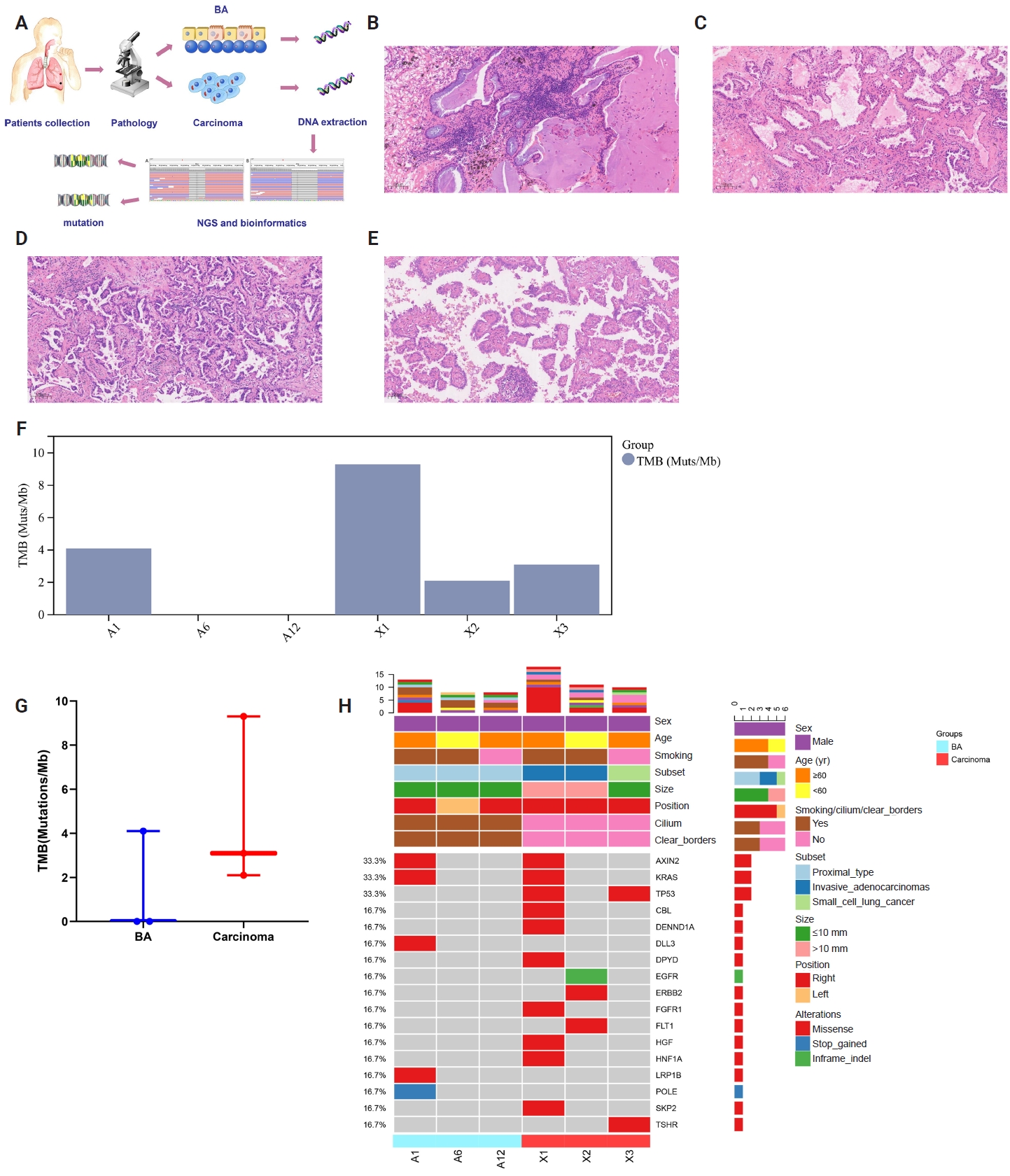
 - Bronchiolar adenoma (BA) is a rare type of lung tumor characterized by bilayered epithelial cells having a continuous basal layer and a luminal layer. It resembles mucinous adenocarcinoma (MA) on frozen section, with difficulty in distinguishing the basal layer. Immunohistochemistry is the best choice for verifying the diagnosis. This study aimed to comprehensively characterize three cases of BA-combined carcinoma using clinical, histopathological, and genetic features. BA and carcinoma sections were subjected to next-generation sequencing, respectively. It was hypothesized that while different mutation forms matched different regions, BA and lung adenocarcinoma shared the same gene mutation when they co-occurred in the same location. BA with extensive carcinoma is extremely rare and presents diagnostic challenges due to its overlap with conditions such as MA. Because of its distinctive morphological characteristics, BA may be regarded as a low-grade malignancy, particularly during a confusing evaluation. A multifaceted examination of clinical, radiological, immunohistochemical, and genetic data is necessary for an accurate diagnosis.
Case Report
- Morphological Features of Metastatic Gastrointestinal Stromal Tumors after Gleevec Treatment: Two Cases Report.
- Joon Hyuk Choi, Young Kyung Bae, Sun Kyo Song, Hong Jin Kim, Min Chul Shim, Kyung Hee Lee
- Korean J Pathol. 2009;43(4):368-373.
- DOI: https://doi.org/10.4132/KoreanJPathol.2009.43.4.368
- 3,961 View
- 29 Download
-
Abstract
PDF
- We report two patients with metastatic gastrointestinal stromal tumors (GISTs) with a focus on the morphological features related to Gleevec treatment. In case 1, a 50-year-old woman presented with a 1.8 cm metastatic GIST in the liver after resection of a gastric GIST. Majority of the metastatic tumor showed fibrosis and hyalinization after 8 weeks of Gleevec treatment. CD117-positive cells were present in approximately 1% of the overall tumor. In case 2, a 2 cm and 14 cm metastatic liver masses were found in a 54-year-old man who had a rectal GIST. After 4 weeks of Gleevec treatment, metastatic tumors showed a decrease in size on CT scan. The metastatic tumors showed a decrease in number of tumor cells. The hemorrhage, cystic changes, necrosis, and fibrosis made up approximately 90% of the tumor. The morphological features related to Gleevec treatment are important for correct diagnosis and evaluation of tumor response and prognosis.
Original Article
- Prenatal Development of Sebaceous Gland: Morphologic and Morphometric Observation.
- Im Joong Yoon, Je Geun Chi, Kye Yong Song
- Korean J Pathol. 1998;32(4):273-282.
- 2,376 View
- 22 Download
-
Abstract
PDF
- This study was conducted to illustrate the histological and morphometric features of the sebaceous gland of human fetal skin. For this purpose, we studied 12 human embryos and 60 fetuses from the 4 th to 38 th week of gestation. In each case, we sampled eight different areas of skin, i.e., scalp, forehead, face, chest, abdomen, back, extremity, and palm and sole. Through routine tissue processing, hematoxylin and eosin preparations were made for morphology and morphometric analysis. The sebaceous gland anlagen is noted in the face and scalp by the 14th week of gestation, being subsequently generalized in other parts of the body, namely by 16th week of gestation. The lobation of the sebaceous gland subsequently differentiated into multilobular appearance in the face and scalp by the 17th week of gestation and in the chest and abdomen by the 26th week of gestation. The sebaceous ducts were seen by the 21th week of gestation in face and scalp, and in the chest and abdomen by the 27th week of gestation. In morphometric observation, the number and diameter of sebaceous gland were reached its peak during the 21st to 24th week of gestation, and then decreased gradually until it became constant in later days of the gestational period. In general, cephalic portion of the body had more sebaceous glands and also was larger in diameter. This difference became negligible as fetuses reached the term.
 First
First Prev
Prev


