 E-submission
Search
- Page Path
- HOME > Search
Original Articles
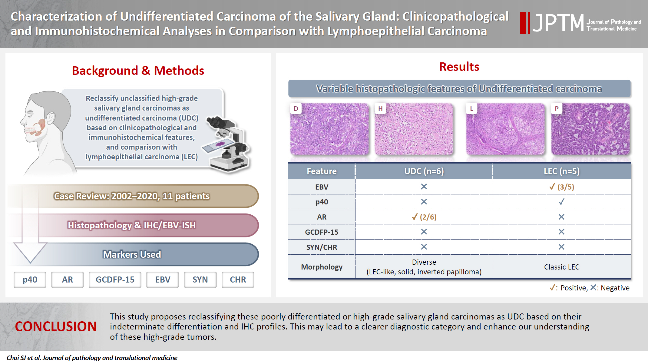
- Characterization of undifferentiated carcinoma of the salivary gland: clinicopathological and immunohistochemical analyses in comparison with lymphoepithelial carcinoma
- Sangjoon Choi, Gyuheon Choi, Hee Jin Lee, Joon Seon Song, Yoon Se Lee, Seung-Ho Choi, Kyung-Ja Cho
- J Pathol Transl Med. 2025;59(6):361-370. Published online September 8, 2025
- DOI: https://doi.org/10.4132/jptm.2025.07.07
- 5,132 View
- 314 Download
-
 Abstract
 PDF
PDF - Background
This study aimed to reclassify a subset of poorly differentiated salivary gland carcinoma that do not conform to any entities of the current World Health Organization (WHO) classification into the category of undifferentiated carcinoma (UDC) because they lack specific histologic differentiation or immunophenotype. Methods: Cases of salivary gland carcinomas from Asan Medical Center (2002–2020) that did not fit any existing WHO classification criteria and were diagnosed as poorly differentiated carcinoma, high-grade carcinoma, or UDC, were retrospectively reviewed. Immunohistochemical (IHC) staining for p40, neuroendocrine markers, androgen receptor (AR), and gross cystic disease fluid protein 15 (GCDFP-15) and Epstein-Barr virus (EBV) in situ hybridization (ISH) were performed. Clinical data were collected from the electronic medical records. Results: Six salivary gland carcinomas did not align with any specific entities and lacked distinct differentiation. Two of six cases displayed lymphoepithelial carcinoma (LEC)-like morphology but were negative or showed negligible immunoreactivity for p40 and EBV ISH, distinguishing them from LEC of the salivary gland. Two cases showed strong AR positivity, suggesting a potential overlap with salivary duct carcinoma (SDC) but lacked classic SDC morphologies and GCDFP-15 expression. No cases expressed neuroendocrine markers. Conclusions: This study proposes reclassifying these poorly differentiated or high-grade salivary gland carcinomas as UDC based on their indeterminate differentiation and IHC profiles. This may lead to a clearer diagnostic category and enhance our understanding of these high-grade tumors.
- Contribution of cytologic examination to diagnosis of poorly differentiated thyroid carcinoma
- Na Rae Kim, Jae Yeon Seok, Yoo Seung Chung, Joon Hyop Lee, Dong Hae Chung
- J Pathol Transl Med. 2020;54(2):171-178. Published online February 5, 2020
- DOI: https://doi.org/10.4132/jptm.2019.12.03

- 10,108 View
- 210 Download
- 6 Web of Science
- 7 Crossref
-
Abstract
PDF
- Background
The cytologic diagnosis of poorly differentiated thyroid carcinoma (PDTC) is difficult because it lacks salient cytologic findings and shares cytologic features with more commonly encountered neoplasms. Due to diverse cytologic findings and paucicellularity of PDTC, standardization of cytologic diagnostic criteria is limited. The purpose of this study is to investigate and recognize diverse thyroid findings of fine needle aspiration (FNA) cytology and frozen smear cytology in diagnosis of this rare but aggressive carcinoma.
Methods
The present study included six cases of FNA cytology and frozen smears of histologically diagnosed PDTCs.
Results
PDTC showed cytologic overlap with well-differentiated thyroid carcinomas (WDTCs). Five of six cases showed dedifferentiation arising from well differentiated thyroid carcinomas. Only one de novo PDTC showed highly cellular smears composed of discohesive small cells, high nuclear/cytoplasmic (N/C) ratio, prominent micronucleoli, and irregular nuclei. Retrospectively reviewed, these findings are highly suspicious for PDTC. Cytologic findings of nuclear atypia, pleomorphism, and irregularity were frequently found, whereas scattered small cells were seen only in the de novo case.
Conclusions
Heterogeneous cytologic findings of PDTCs are shared with those of WDTCs and contribute to difficult preoperative cytologic diagnoses. Most PDTCs show dedifferentiation from WDTCs. Albeit rare, de novo PDTC should be considered with cytology showing discohesive small cells with high N/C ratio. This will enable precise diagnosis and prompt treatment of this aggressive malignancy -
Citations
Citations to this article as recorded by
- Practical and challenging issue in thyroid cytopathology
Qianqian Zhang, Belen Padial Urtueta, Elisabetta Merenda, Gabriele Rotondaro, Noemi Morelli, Alessia Piermattei, Patrizia Straccia, Federica Cianfrini, Angela Feraco, Alessia Granitto, Antonino Mule, Esther Diana Rossi
Human Pathology.2026; 169: 106019. CrossRef - Plasma cells and plasmacytoid features in thyroid lesions
Qianqian Zhang, Angela Feraco, Belen Padial Urtueta, Elisabetta Merenda, Luisa Cioni, Alessia Piermattei, Patrizia Straccia, Federica Cianfrini, Antonino Mule, Liron Pantanowitz, Esther Diana Rossi
Virchows Archiv.2026;[Epub] CrossRef - Non-papillary thyroid carcinoma diagnoses in The Bethesda System for Reporting Thyroid Cytopathology categories V and VI: An institutional experience
Myunghee Kang, Na Rae Kim, Jae Yeon Seok
Annals of Diagnostic Pathology.2024; 71: 152263. CrossRef - Cytologic features of differentiated high‐grade thyroid carcinoma: A multi‐institutional study of 40 cases
Vanda F. Torous, Tikamporn Jitpasutham, Zubair Baloch, Richard L. Cantley, Darcy A. Kerr, Xiaoying Liu, Zahra Maleki, Ross Merkin, Vania Nosé, Liron Pantanowitz, Isabella Tondi Resta, Esther D. Rossi, William C. Faquin
Cancer Cytopathology.2024; 132(8): 525. CrossRef - An Unexpected Finding of Poorly Differentiated Thyroid Carcinoma in a Toxic Thyroid Nodule
Kimberly Yuang, Huda Al-Bahadili, Alan Chang
JCEM Case Reports.2023;[Epub] CrossRef - Revisiting the cytomorphological features of poorly differentiated thyroid carcinoma: a comparative analysis with indeterminate thyroid fine-needle aspiration samples
Yazeed Alwelaie, Ali Howaidi, Mohammed Tashkandi, Ahmad Almotairi, Hisham Saied, Moammar Muzzaffar, Doaa Alghamdi
Journal of the American Society of Cytopathology.2023; 12(5): 331. CrossRef - Characterization of the genomic alterations in poorly differentiated thyroid cancer
Yeeun Lee, SeongRyeol Moon, Jae Yeon Seok, Joon-Hyop Lee, Seungyoon Nam, Yoo Seung Chung
Scientific Reports.2023;[Epub] CrossRef
- Practical and challenging issue in thyroid cytopathology
Case Reports
- Sertoli-Leydig Cell Tumor of Hemangiopericytoma Pattern: A case report.
- Hye Jin Lee, Young im Han, Hyeon Ok Kim, Kang Suek Suh, Sun Kyung Lee
- Korean J Pathol. 1995;29(6):815-818.
- 2,207 View
- 17 Download
-
Abstract
PDF
- The Sertoli-Leydig cell tumor is a gonadal tumor of sex-cord stromal type, similar to that seen in of the various phases of testicular development in the male. This tumor is exceedingly rare, accounting for only 0.1% to 0.5% of all primary ovarian neoplasms. It occurs predominantly in the second and third decades(mean age about 25 years), less than 10% after menopause. We investigated a case of poorly differentiated Sertoli-Leydig cell tumor of right ovary, occured in a 76-year-old woman. Grossly, the tumor measured 2, 100 gm in weight and 25 x 19 x 8 cm in dimensions. The outer surface was smooth and glistening without rupture of the capsule. Cut sections revealed a multilobulated brown solid mass with multiple cystic change. Microscopically, it showed the typical findings o a Sertoli-Leydig cell tumor. The characteristic feature is hemangiopericytoma paftem of sarcomatoid spindle cells. Therefore, we present it with a brief review of the literature.
- Aspiration Cytology of Insular Carcinoma of Thyroid: A Case Report.
- Young Il Yang, Chan Hawn Kim, Shin Kwang Khang
- J Pathol Transl Med. 1994;5(1):46-51.
- 2,144 View
- 12 Download
-
Abstract
PDF
- Fine needle aspiration cytologic features of a case of insular carcinoma of the thyroid in a 23-year-old woman who presented a palpable neck mass is described. The aspirate showed cellular smear arranged in trabeculae, solid or loose clusters, and microfollicles in necrotic background. The tumor cells had uniform, small round, hyperchromatic nuclei. The chromatin was finely granular, and nuclear membrane was smooth. Nucleoli were not discernible. Nuclear pleomorphism was minimal. The cytoplasm was usually scanty, pale, poorly outlined, and almostly amphophilic. Sometimes paranuclear cytoplasmic vacuoles were noted. Final diagnosis was confirmed by total thyroidectomy as insular carcinoma.
- Fine Needle Aspiration Cytoloy of Poorly Differentiated "Insular" Carcinoma of the Thyroid: A Case Report.
- Hee Jung Lee, Kyung Shin Park, Young Shin Kim, Kyo Young Lee, Chang Suck Kang, Sang In Shim
- J Pathol Transl Med. 1998;9(1):117-122.
- 1,986 View
- 11 Download
-
Abstract
PDF
- Cytologic features of a poorly differentiated "insular" carcinoma of the thyroid are presented. In fine needle aspiration cytology, the aspirates were highly cellular and tumor cells were arranged in loose clusters or singly dispersed on focally necrotic background. Occasional microfollicles were evident. The tumor cells had poorly defined, scanty cytoplasm and most of the nuclei were fairly uniform with coarse chromatin pattern. A few large pleomorphic cells were also noted. The cytologic findings of the present case were correlated well with the histologic findings, which showed typical insular pattern and the presence of uniform cells with occasional pleomorphism.
 First
First Prev
Prev


