Search
- Page Path
- HOME > Search
Review
- Clinical management of abnormal Pap tests: differences between US and Korean guidelines
- Seyeon Won, Mi Kyoung Kim, Seok Ju Seong
- J Pathol Transl Med. 2020;54(3):213-219. Published online April 15, 2020
- DOI: https://doi.org/10.4132/jptm.2020.03.11
- 7,017 View
- 155 Download
- 2 Web of Science
- 2 Crossref
-
 Abstract
 PDF
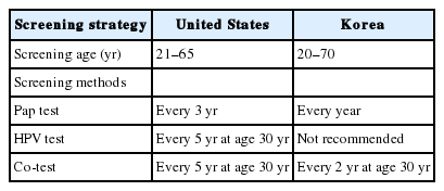
PDF - Cervical cancer has been the most common gynecological cancer in Korea but has become a preventable disease with regular screening and proper vaccination. If regular screening is provided, cervical cancer does not progress to more than carcinoma in situ, due to its comparatively long precancerous duration (years to decades). In 2012, the American Society for Colposcopy and Cervical Pathology published guidelines to aid clinicians in managing women with abnormal Papanicolaou (Pap) tests, and they soon became the standard in the United States. Not long thereafter, the Korean Society of Gynecologic Oncology and the Korean Society for Cytopathology published practical guidelines to reflect the specific situation in Korea. The detailed screening guidelines and management options in the case of abnormal Pap test results are sometimes the same and sometimes different in the United States and Korean guidelines. In this article, we summarize the differences between the United States and Korean guidelines in order to facilitate physicians’ proper management of abnormal Pap test results.
-
Citations
Citations to this article as recorded by- Analysis of HR-HPV Infection Concordance Rates in Cervical and Urine Specimens; Proposal of Additional Cervical Screening Process for Women Who Refuse Invasive Cervical Sampling
Dong Hyeok Kim, Hyunwoo Jin, Kyung Eun Lee
Journal of Personalized Medicine.2022; 12(12): 1949. CrossRef - Analysis of HR-HPV Prevalence among Unvaccinated Busan Women
Dong Hyeok Kim, Kyung Eun Lee
Biomedical Science Letters.2022; 28(4): 229. CrossRef
- Analysis of HR-HPV Infection Concordance Rates in Cervical and Urine Specimens; Proposal of Additional Cervical Screening Process for Women Who Refuse Invasive Cervical Sampling
Case Report
- A Case of Ovarian Microinvasive Mucinous Carcinoma and Co-existent Angiosarcoma.
- Jin Hyung Heo, Yoon Hee Lee, Gwang Il Kim, Tae Heon Kim, Haeyoun Kang, Hee Jung An, Bo Sung Yoon, Seok Ju Seong, Hyun Park, Ji Young Kim
- Korean J Pathol. 2011;45(1):96-100.
- DOI: https://doi.org/10.4132/KoreanJPathol.2011.45.1.96
- 3,331 View
- 24 Download
- 5 Crossref
-
 Abstract
 PDF
PDF - Primary ovarian angiosarcoma is very rare with only 27 cases reported so far in the medical literature. We report here on a rare case of ovarian microinvasive mucinous carcinoma that was coexistent with angiosarcoma in a 54-year-old woman. The tumor was a 26x19x10 cm-sized multilocular cystic mass with a 4x3 cm-sized solid hematoma-like nodule in the center. Microscopically, it was composed mostly of mucinous tumor of various grades from borderline to microinvasive carcinoma. The hematoma-like area turned out to be an angiosarcoma, composed of pleomorphic cells that formed slit-like spaces, spindle cells that formed short fascicles and anastomosing vascular channels with atypical endothelial cells. All these cells were positive for CD31, CD34 and factor VIII-related antigen. The patient developed peritoneal and pleural metastases, which were angiosarcoma and mucinous carcinoma, respectively. We believe this case is only the fourth example of an ovarian collision tumor of angiosarcoma and surface epithelial tumor.
-
Citations
Citations to this article as recorded by- Ovarian angiosarcoma: A systematic review of literature and survival analysis
Shafi Rehman, Arya Harikrishna, Amisha Silwal, B.R. Sumie, Safdar Mohamed, Nisha Kolhe, Meghana Maddi, Linh Huynh, Jesus Gutierrez, Yoshita Rao Annepu, Ameer Mustafa Farrukh
Annals of Diagnostic Pathology.2024; 73: 152331. CrossRef - Tumor to Tumor Metastasis: A Case Report of Metastatic Angiosarcoma to an Ovarian Brenner Tumor and Review of the Literature
Bilge Dundar, Audai Alrwashdeh, Laila Dahmoush
International Journal of Gynecological Pathology.2023; 42(2): 176. CrossRef - Collision Tumors in Ovary: Case Series and Literature Review
Borges A, Loddo A, Martins A, Peiretti M, Fanni D, Djokovic D
Journal of Surgical Oncology.2019; : 1. CrossRef - Angiosarcoma Arising in Ovarian Mucinous Tumor: A Challenge in Intraoperative Frozen Section Diagnosis
Surapan Khunamornpong, Jongkolnee Settakorn, Kornkanok Sukpan, Tip Pongsuvareeyakul, Sumalee Siriaunkgul
Case Reports in Pathology.2016; 2016: 1. CrossRef - Impact of body burden of pesticide residues on the reproductive tract of buffalo
KARANPREET KAUR, SARVPREET SINGH GHUMAN, OPINDER SINGH, JASBIR SINGH BEDI, JATINDER PAUL SINGH GILL
The Indian Journal of Animal Sciences.2016;[Epub] CrossRef
- Ovarian angiosarcoma: A systematic review of literature and survival analysis
Original Article
- Pregnancy-Related Cytologic Changes In Cervicovaginal Smears.
- Yi Kyeong Chun, Hoi Sook Jang, Hye Sun Kim, Sung Ran Hong, Jong Sun Choi, Ji Young Park, Jung Sook Cho, Seok Ju Seong, Jae Hyug Yang, Hy Sook Kim
- Korean J Cytopathol. 2004;15(2):92-100.
- 2,425 View
- 51 Download
-
 Abstract
 PDF
PDF - Due to insufficient clinical information, most cervicovaginal smears from pregnant or postpartum women have been screened without regard to pregnancy-related cytological changes. Here, we have reviewed 116 abnormal cervicovaginal smears from 103 pregnant and postpartum women. Initial cytological diagnoses revealed the following: 9 cases of high-grade squamous intraepithelial lesions (HSIL), 8 cases of low-grade squamous intraepithelial lesions (LSIL), 85 cases of atypical squamous cells of undetermined significance (ASCUS), and 14 cases involving atypical glandular cells of undetermined significance (AGUS). 31 cases, upon review, involved pregnancy-related cytological changes, comprising 25 cases of decidua cells, 4 cases of Arias-Stella reaction, and 2 cases of decidual cells coupled with Arias-Stella reaction. Interpretation errors were detected in 14 cases: 13 cases of decidual cells interpreted as either ASCUS favor reactive or ASCUS ruled out HSIL, and one case of Arias-Stella reaction was interpreted as ASCUS ruled out HSIL. Decidual cells and degenerated glandular cells with Arias-Stella reaction can result in diagnostic mistakes. In order to avoid misdiagnosis and unnecessary surgeries, both clinicians and pathologists must be aware of the pregnancy-related cytological changes. The clinician should also always inform the pathologist on the pregnancy status of the patient.

 E-submission


 First
First Prev
Prev


