 E-submission
Search
- Page Path
- HOME > Search
Original Article
- 3-Dimensional reconstruction reveals frequent intraluminal growth of submucosal veins in surgically resected pT1 colorectal cancers
- Jihyun Park, Mi-Ju Kim, Yeon Wook Kim, Byong-Wook Lee, Junyoung Shin, Jinho Shin, Chan-Gi Pack, Dong-Hoon Yang, Jihun Kim, In Ja Park, Ralph H. Hruban, Seung-Mo Hong
- J Pathol Transl Med. 2026;60(2):246-262. Published online March 10, 2026
- DOI: https://doi.org/10.4132/jptm.2025.12.19
- 1,327 View
- 95 Download
-
 Abstract
 PDF
PDF - Background
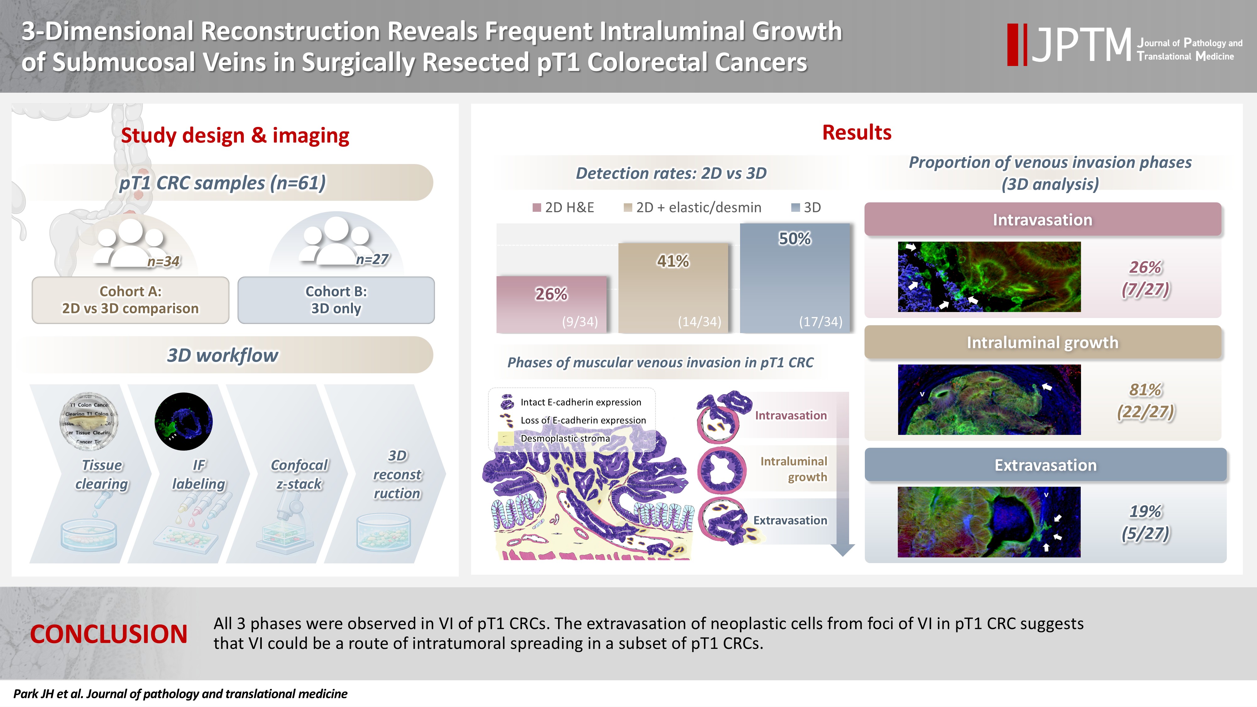
Although venous invasion (VI) is associated with distant metastasis and observed in >50% of pT2–4 colorectal cancers (CRCs), the role of VI in pT1 CRCs is not well-defined. Methods: Thirty-four surgically resected pT1 CRCs were reevaluated for 2-dimensional (2D) VI using hematoxylin and eosin (H&E)–stained slides with additional elastic and desmin immunohistochemical staining (cohort A). Additionally, 27 pT1 CRCs without knowing VI status were selected for 3-dimensional (3D) VI evaluation only (cohort B). All 61 cases (cohorts A and B) were studied in 3D using tissue clearing. Results: VI was detected more commonly in 3D (17/34, 50.0%) than in 2D H&E slide evaluation (9/34, 26.5%, p = .047). When VI was identified in 3D (27/61, 44.3%), the most common phase was that of intraluminal growth (22/27, 81.5%), followed by intravasation (7/27, 25.9%) and extravasation (5/27, 18.5%). E-cadherin expression was characterized in 3D in foci of VI and varied in each phase of invasion. Conclusions: All three phases were observed in VI of pT1 CRCs. The extravasation of neoplastic cells from foci of VI in pT1 CRC suggests that VI could be a route of intratumoral spreading in a subset of pT1 CRCs.
Case Study
- A Case of Giant Colonic Muco-submucosal Elongated Polyps Associated with Intussusception
- Joo Heon Kim, Seung Yun Lee, Je Ho Jang, Hyun Young Han, Dong Wook Kang
- J Pathol Transl Med. 2016;50(6):474-478. Published online May 23, 2016
- DOI: https://doi.org/10.4132/jptm.2016.04.27
- 12,563 View
- 134 Download
- 6 Web of Science
- 8 Crossref
-
Abstract
PDF
- Colonic muco-submucosal elongated polyp (CMSEP), a newly categorized non-neoplastic colorectal polyp, is a pedunculated and elongated polyp composed of normal mucosal and submucosal layers without any proper muscle layer. We herein report a giant variant of CMSEP associated with intussusception in the rectosigmoid colon, with a review of the literature. A 48-year-old woman underwent a laparoscopic low anterior resection due to multiple large submucosal polypoid masses associated with intussusception. Grossly, the colonic masses were multiple pedunculated polyps with a long stalk and branches ranging in size from a few millimeters to 14.0 cm in length. Microscopically, there was no evidence of hyperplasia, atypia, or active inflammation in the mucosa. The submucosal layers were composed of edematous and fibrotic stroma with fat tissue, dilated vessels, and lymphoid follicles.
-
Citations
Citations to this article as recorded by
- Unusually rapid growth of a duodenal muco-submucosal elongated polyp: A case report
Yi Yang, Ding-Fu Zhong
World Journal of Gastrointestinal Surgery.2025;[Epub] CrossRef - Multiple enteric muco-submucosal elongated polyps causing intussusception
Atsuki Taniguchi, Izuru Endo, Takeyoshi Nishiyama, Nobuyuki Watanabe, Osamu Yoshida, Hiroaki Asano, Masatoshi Kubo, Tetsunobu Udaka
Clinical Journal of Gastroenterology.2024; 17(1): 41. CrossRef - Intussusception due to a Muco-submucosal Elongated Polyp in the Small Intestine—A Case Report—
Hiroki ISHIGE, Ken IMAIZUMI, Takumu FUKASAWA, Keiichiro ITO, Hiroyuki KASAJIMA, Satoru MUNAKATA, Norihiko SHIMOYAMA, Kazuaki NAKANISHI
Nihon Rinsho Geka Gakkai Zasshi (Journal of Japan Surgical Association).2024; 85(6): 744. CrossRef - Jejunal Intussusception Caused by Enteric Muco-submucosal Elongated Polyp: A Case Report
Young Min Jo
Soonchunhyang Medical Science.2024; 30(2): 60. CrossRef - Jejunal intussusception and perforation due to enteric muco-submucosal elongated polyp: a case report and literature review
Ryosuke Kikuchi, Shigenobu Emoto, Hiroaki Nozawa, Kazuhito Sasaki, Koji Murono, Shinya Abe, Hirofumi Sonoda, Aya Shinozaki-Ushiku, Soichiro Ishihara
Surgical Case Reports.2023;[Epub] CrossRef - A stalk with no polyp—A muco‐submucosal elongated polyp in the duodenum
Neil O’Morain, Ciaran McCloskey, Sinead Flanagan, Glen Doherty
United European Gastroenterology Journal.2023; 11(4): 392. CrossRef - Duodenal Worm-Like Polyp
Pan Pan, Guoshan Zhang, Xiao Cui, Liang Liu
Digestive Diseases and Sciences.2023; 68(12): 4275. CrossRef - Colonic Mucosubmucosal Elongated Polyp in the Sigmoid Colon on Surveillance Colonoscopy
Xiaowen Fan, Melissa Hershman, Gabriel Levi, Ilan Weisberg
ACG Case Reports Journal.2019; 6(6): e00110. CrossRef
- Unusually rapid growth of a duodenal muco-submucosal elongated polyp: A case report
Original Article
- Distribution of Smooth Muscles in Hemorrhoids.
- Jae Gul Chung, Ghee Young Choe, Gyung Yub Gong, Eun Sil Yu, Jin Cheon Kim, In Chul Lee
- Korean J Pathol. 1994;28(2):154-159.
- 2,176 View
- 13 Download
-
Abstract
PDF
- Hemorrhoids are one of the commonest disorders specific to the human. However, the pathogenesis is not well understood so far. Anal submucosa is largely composed of blood vessels, loose connective tissue and smooth muscles, forming muscular network around the venous plexuses. We analyzed the distribution of smooth muscles in the hemorrhoidal tissues. Immunohistochemical stainings for desmin, vimentin, and Factor VIII related antigen were performed using six freshly frozen hemorrhoidal tissues. All of them were diagnosed as external hemorrhoids. Four anal tissues from Miles' operation specimen without hemorrhoids were used as normal controls. In all six cases, venous plexuses were variably dilated and smooth muscle cells were unevenly distributed. In minimally involved areas, there were relatively sufficient amount of perivascular smooth muscles which were arranged in their bundles. In contrast, only single scattered cells or very small amount of smooth muscle bundles were noted around the dilated vascular plexuses in severely affected areas. In two severe hemorrhoidal tissue samples, vascular plexuses were markedly dilated and only single scattered smooth muscle cells were seen. In conclusion, the total amount of smooth muscles in the submucosa of hemorrhoid tissue was reduced than those of the normal controls. The degree of hemorrhoidal dilation was inversely related to the amount of smooth muscles. However, causal relation between diminution of submucosal smooth muscles and venous dilation remains to be clarified.
Case Report
- Acute Gastric Anisakiasis: A case report.
- So Young Jin, Soon Hee Jung, Tai Seung Kim
- Korean J Pathol. 1989;23(1):149-153.
- 2,041 View
- 14 Download
-
Abstract
PDF
- We report a case of a 41-year-old female patient who suffered from the acute abdominal pain for several hours after eating raw sea-fishes. After the fibergastroscopy and the abdominal C-T scan, the clinicians found a gastric submucosal mass and performed the explolaparotomy to get an wedge of stomach. Sections revealed a larva surrounded by phlegmonous inflammation with intense eosinophilic infiltration in the widened gastic submucosa. The larva presented the characteristics of the Anisakis: two lateral chords with renette cell, thich smooth cuticle and well developed musculature.
 First
First Prev
Prev


