
 E-submission

Search
- Page Path
- HOME > Search
Original Articles
- Concordance of flow cytometric E6/E7 mRNA detection with human papillomavirus DNA testing and cervical cytology in a cervical cancer screening cohort
- Dat Quoc Ngo, Trang Kieu Ha, Mi Ngoc Thi Tran, Anh Mai Thi Nguyen, Truong Nhat Trinh, Thien Thanh Ly, Linh Ngoc Khanh Nguyen, Thu Dang Anh Phan
- Received January 13, 2026 Accepted September 9, 2026 Published online November 10, 2026
- DOI: https://doi.org/10.4132/jptm.2026.09.09 [Epub ahead of print]
- 60 View
- 1 Download
-
 Abstract
 PDF
- Background
Overexpression of viral oncogenes E6 and E7 of high-risk (HR)–human papillomavirus (HPV) plays a key role in the progression to cervical intraepithelial neoplasia (CIN) and cancer. Detection of E6/E7 mRNA may help distinguish between transient and transforming infections, thus improving cervical cancer screening accuracy. This study aimed to evaluate the association of E6/E7 mRNA expression with HR-HPV infection, cervical cytology, and histologically confirmed cervical lesions and to compare its agreement with HPV DNA testing and cytology. Methods: A total of 526 women undergoing cervical cancer screening between January 2023 and May 2025 were enrolled. E6/E7 mRNA expression was assessed using flow cytometry with the HPV OncoTect 3Dx E6/E7 mRNA assay and compared with HR-HPV DNA testing, cervical cytology, and available histopathologic findings from biopsy specimens. Results: Among 526 samples, eight (1.5%) tested positive for E6/E7 mRNA, compared to 30 (5.7%) HR-HPV DNA–positive and 17 (3.2%) abnormal cytology results. Median E6/E7 mRNA expression was higher in HR-HPV– positive cases and in cytologically abnormal cases. Almost perfect agreement was observed between E6/E7 mRNA results, HR-HPV DNA testing, and cytology. Histopathologic follow-up was available for nine women with cytological abnormalities ≥low-grade squamous intraepithelial lesion, including four CIN2 and five CIN1 cases. E6/E7 mRNA expression was markedly elevated in one CIN2 case associated with HPV16 infection (37.51%), while two CIN1 cases demonstrated high mRNA expression despite negative HPV DNA results. Conclusions: Flow-cytometric E6/E7 mRNA detection showed high overall agreement with HPV DNA testing and cytology in this screening cohort. Larger prospective studies with comprehensive histologic follow-up are needed to determine its clinical utility.
- Immunohistochemical expression in idiopathic inflammatory myopathies at a single center in Vietnam
- Dat Quoc Ngo, Si Tri Le, Khanh Hoang Phuong Phan, Thao Thi Phuong Doan, Linh Ngoc Khanh Nguyen, Minh Hoang Dang, Thien Thanh Ly, Thu Dang Anh Phan
- J Pathol Transl Med. 2024;58(4):174-181. Published online June 25, 2024
- DOI: https://doi.org/10.4132/jptm.2024.05.02
- 7,712 View
- 286 Download
- 1 Web of Science
- 3 Crossref
-
Abstract
PDF
- Background
The identification of idiopathic inflammatory myopathies (IIMs) requires a comprehensive analysis involving clinical manifestations and histological findings. This study aims to provide insights into the histopathological and immunohistochemical aspects of IIMs.
Methods
This retrospective case series involved 56 patients diagnosed with IIMs at the Department of Pathology, University of Medicine and Pharmacy at Ho Chi Minh City, from 2019 to 2023. The histology and immunohistochemical expression of HLA-ABC, HLA-DR, C5b-9, Mx1/2/3, and p62 were detected.
Results
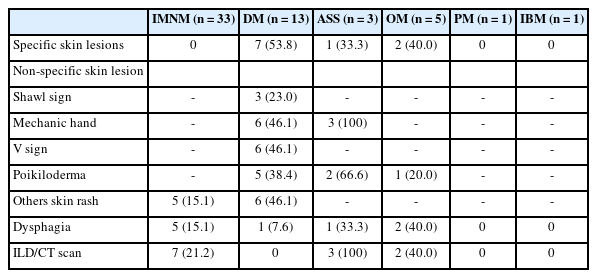
We examined six categories of inflammatory myopathy, including immunemediated necrotizing myopathy (58.9%), dermatomyositis (DM; 23.2%), overlap myositis (8.9%), antisynthetase syndrome (5.4%), inclusion body myositis (IBM; 1.8%), and polymyositis (1.8%). The average age of the patients was 49.7 ± 16.1 years, with a female-to-male ratio of 3:1. Inflammatory cell infiltration in the endomysium was present in 62.5% of cases, perifascicular atrophy was found in 17.8%, and fiber necrosis was observed in 42 cases (75.0%). Rimmed vacuoles were present in 100% of cases in the IBM group. Immunohistochemistry showed the following positivity rates: HLA-ABC (89.2%), HLA-DR (19.6%), C5b-9 (57.1%), and Mx1/2/3 (10.7%). Mx1/2/3 expression was high in DM cases. p62 vacuole deposits were noted in the IBM case. The combination of membrane attack complex and major histocompatibility complex I helped detect IIMs in 96% of cases.
Conclusions
The diagnosis of IIMs and their subtypes should be based on clinical features and histopathological characteristics. Immunohistochemistry plays a crucial role in the diagnosis and differentiation of these subgroups. -
Citations
Citations to this article as recorded by
- Evaluating the Diagnostic Potential of Myxovirus Resistance Protein 1 (MX1) and Myxovirus Resistance Protein 2 (MX2) As Biomarkers in Idiopathic Inflammatory Myopathies
Raghavee Neupane, Mustafa Haider, Perry Smith, Marc M Kesselman
Cureus.2026;[Epub] CrossRef - Rapidly Progressive Polymyositis With Vasculitis: The Pivotal Role of Histopathology in Diagnosis and Management
Amitha Venmanassery Karnalsingh, Arjun Karappilly Vijayan, Monica Roselin Edwin Peter, Dilan Davis
Cureus.2025;[Epub] CrossRef - Autoimmune Neuromuscular Disorders at a Molecular Crossroad: Linking Pathogenesis to Targeted Immunotherapy
Anca-Maria Florea, Dimela-Gabriela Luca, Eugenia Irene Davidescu, Bogdan-Ovidiu Popescu
International Journal of Molecular Sciences.2025; 26(23): 11736. CrossRef
- Evaluating the Diagnostic Potential of Myxovirus Resistance Protein 1 (MX1) and Myxovirus Resistance Protein 2 (MX2) As Biomarkers in Idiopathic Inflammatory Myopathies
 First
 Prev




