Search
- Page Path
- HOME > Search
Case Study
- Fallopian Metaplastic Papillary Tumour: An Atypical Transdifferentiation of the Tubal Epithelium?
- Miguel Fdo. Salazar, Isaías Estrada Moscoso, Lorena Troncoso Vázquez, Nubia Leticia López García, Paola Andrea Escalante Abril
- J Pathol Transl Med. 2015;49(2):148-155. Published online March 12, 2015
- DOI: https://doi.org/10.4132/jptm.2014.10.15
- 10,263 View
- 60 Download
- 3 Web of Science
- 3 Crossref
-
 Abstract
 PDF
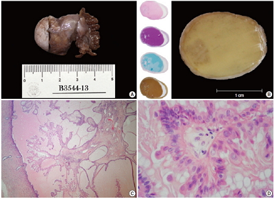
PDF - A metaplastic papillary tumor of the Fallopian tube is an extremely uncommon condition, with odd and confusing features that make it difficult to categorize as benign or borderline. Here, we summarize all the published cases to date and document the case of a 41-year-old woman diagnosed with this alteration after her last childbirth and ensuing tubal ligation. One of the tubes was bulky and filled with a caramel-like substance encircling a blurry spot. Light microscopy detailed a slender stalk covered by eosinophilic, columnar plump cells, showing atypical nuclei and focal budding. Mitotic figures were absent. The immunohistochemistry panel was positive for pan-cytokeratin, epithelial membrane antigen, cyclin D1, and hormone receptors. Additionally, a proliferation index of less than 5% was rated using Ki-67. The true nature of this tumor (reactive vs neoplastic) is uncertain. Nonetheless, its association with pregnancy suggests an adaptive change, likely similar to the atypical transdifferentiation proposed for Arias-Stella reaction.
-
Citations
Citations to this article as recorded by- Ungewöhnliche Proliferation des Eileiters
Angelina Vlaški, Vanessa Neukunft, Andrea Maria Gassel, Frederick Klauschen, Doris Mayr
Die Pathologie.2025; 46(1): 56. CrossRef - Fallopian tube papilloma
Shashank Mishra, Prerna Guleria
Indian Journal of Pathology and Microbiology.2021; 64(3): 608. CrossRef - Metaplastic papillary tumour of the fallopian tube, a rare entity, analysed by next‐generation sequencing
Sandra Sunitsch, Julia Reisinger, Luca Abete, Karl Kashofer, Peter Regitnig
Histopathology.2020; 76(6): 923. CrossRef
- Ungewöhnliche Proliferation des Eileiters
Case Report
- Early Gastric Carcinoma with Hepatoid Differentiation: Report of a case with histotopographic analysis.
- Gyeong Hoon Kang, Chong Jai Kim, Yong Il Kim
- Korean J Pathol. 1991;25(6):594-600.
- 2,083 View
- 14 Download
-
 Abstract
 PDF
PDF - A 56-year-old man received subtotal gastrectomy for an early gastric carcinoma type IIa+IIc with submucosal invasion. The tumor was made up of mixed papillo-tubular adenocarcinoma and solid carcinomatous portion, the latter comprising approximately four-fifths of the total tumor mass. The solid portion was confined within the submucosa and revealed a mixture of trabecular, compact and pelioid patterns of large polyhedra cells, resembling hepatocellular carcinoma of the liver(Edmondson-Steiner grade 2). Sinusoid-like vascular stroma of classical trabecular hepatocellular carcinoma intervened the tumor cell nests but was not associated with endothelial-cell lining. Immunohistochemical stainings with alpha-fetoprotein and alpha1-antitrypsin gave a strong reactivity in those areas of hepatoid differentiation and in the adjacent minute portion of adenocarcinoma. The findings suggest that a portion of gastric carcinoma may transdifferentiate into cells with hepatoid features along the line of endodermal lineage.

 E-submission


 First
First Prev
Prev


