Search
- Page Path
- HOME > Search
Original Articles
- Characterization of undifferentiated carcinoma of the salivary gland: clinicopathological and immunohistochemical analyses in comparison with lymphoepithelial carcinoma
- Sangjoon Choi, Gyuheon Choi, Hee Jin Lee, Joon Seon Song, Yoon Se Lee, Seung-Ho Choi, Kyung-Ja Cho
- J Pathol Transl Med. 2025;59(6):361-370. Published online September 8, 2025
- DOI: https://doi.org/10.4132/jptm.2025.07.07
- 2,746 View
- 253 Download
-
 Abstract
 PDF
PDF - Background
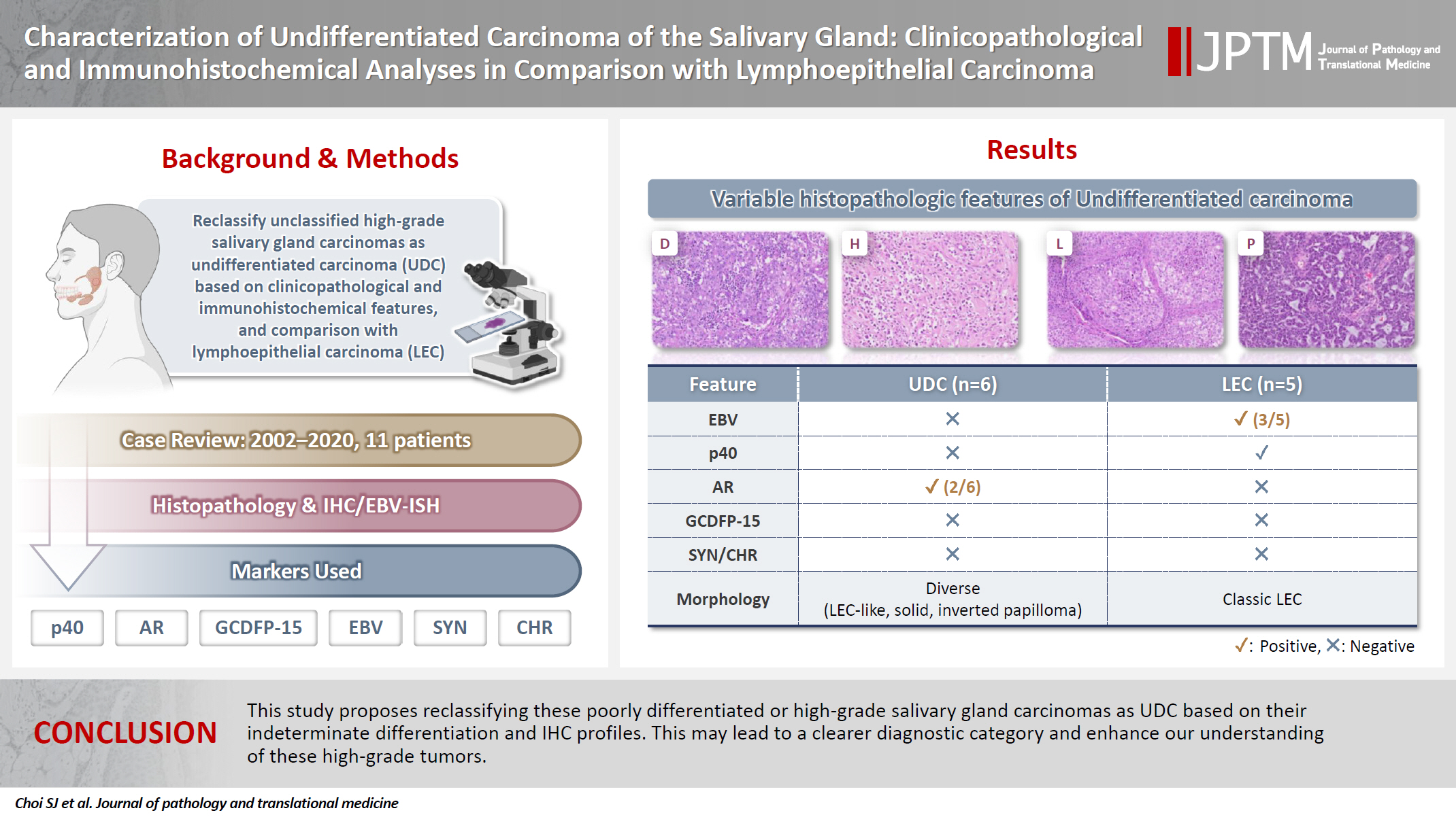
This study aimed to reclassify a subset of poorly differentiated salivary gland carcinoma that do not conform to any entities of the current World Health Organization (WHO) classification into the category of undifferentiated carcinoma (UDC) because they lack specific histologic differentiation or immunophenotype. Methods: Cases of salivary gland carcinomas from Asan Medical Center (2002–2020) that did not fit any existing WHO classification criteria and were diagnosed as poorly differentiated carcinoma, high-grade carcinoma, or UDC, were retrospectively reviewed. Immunohistochemical (IHC) staining for p40, neuroendocrine markers, androgen receptor (AR), and gross cystic disease fluid protein 15 (GCDFP-15) and Epstein-Barr virus (EBV) in situ hybridization (ISH) were performed. Clinical data were collected from the electronic medical records. Results: Six salivary gland carcinomas did not align with any specific entities and lacked distinct differentiation. Two of six cases displayed lymphoepithelial carcinoma (LEC)-like morphology but were negative or showed negligible immunoreactivity for p40 and EBV ISH, distinguishing them from LEC of the salivary gland. Two cases showed strong AR positivity, suggesting a potential overlap with salivary duct carcinoma (SDC) but lacked classic SDC morphologies and GCDFP-15 expression. No cases expressed neuroendocrine markers. Conclusions: This study proposes reclassifying these poorly differentiated or high-grade salivary gland carcinomas as UDC based on their indeterminate differentiation and IHC profiles. This may lead to a clearer diagnostic category and enhance our understanding of these high-grade tumors.
- Primary Undifferentiated Carcinoma of the Endometrium with Small Cell and Trophoblastic Differentiation.
- Chul Hwan Kim, Seoung Hye Park, In Sun Kim, Seung Yong Paik
- Korean J Pathol. 1990;24(1):58-64.
- 2,112 View
- 19 Download
-
 Abstract
 PDF
PDF - This report describes a very rare case of primary undifferentiated carcinoma of the endometrium with small cell and trophoblastic differentiation. The patient was 54-year-old woman with complaints of vaginal bleeding and palpable lower abdominal mass. The light microscopic findings revealed predominantly small cells with round nuclei, spindle cells, and large cells with hyperchromatic bizarre nuclei. Foci of syncytiotrophoblastic giant cells are scattered, especially in the hemorrhagic areas. Immunohistochemical stainging for neuron specific enolase and beta-hCG showed positive reactions to small cells and syncytiotrophoblastic giant cells, respectively. Argentaffin and argyrophil stains, however, showed negative reactions to small cells. The histogenesis of small cell undifferentiated carcinoma of the endometrium remains unclear; however, it may arise from epithelial precursors instead of neuroendocrine cells, and syncytiotrophoblastic cells may be differentiated or dedifferentiated from the undifferentiated carcinoma cells.
- Immunohistochemical and Ultrastructural Studies on the Histogenesis of Thyroid Undifferentiated Carcinoma.
- Myoung Ja Jeong, Woo Sung Moon, Young Hye Lee, Myoung Jae Kang, Ho Yeul Choi, Sang Ho Kim, Dong Geun Lee
- Korean J Pathol. 1995;29(6):756-765.
- 1,991 View
- 15 Download
-
 Abstract
 PDF
PDF - Histologic, immunohistochemical and ultrastructural studies were performed on 6 cases of undifferentiated thyroid carcinoma to study the histogenesis of the undifferentiated thyroid carcinoma, to determine the most useful markers for diagnosing these tumors and to investigate the nature of osteoclast-like giant cells rarely observed in these tumors. For the immuno-histochemical study, a panel of antibodies to epithelial (cocktailed keratin, low molecular weight keratin, CEA), mesenchymal(vimentin, desmin, actin, FVIIIRAg) endocrine(calcitonin, chromogranin), lymphocytic(LCA), histiocytic(alpha-l-ACT, alpha-1-AT, lysozyme, CD68), and Schwann cell(S-100 protein) markers were used. The following results were obtained; 1) Well differentiated carcinoma was associated with 2 cases of spindle cell type and 1 case of giant cell type of undifferentiated thyroid carcinoma and a transitional zone between the well differentiated and undifferentiated lesions was observed. 2) All of the examined cases expressed keratin, and 3 tumors expressed CEA. 3) All the mesenchymal markers, LCA, S-100 protein, calcitonin, and chromogranin were not expressed. Vimentin was coexpressed with keratin in 4 cases. 4) Osteoclast-like giant cells were observed in 1 case of spindle cell type. They expressed CD68 but not keratin. 5) Ultrastructural study revealed the desmosomes between the tumor cells and non-neoplastic, follicular, thyroid epithelial cells. The above results indicate that undifferentiated thyroid carcinoma originates from follicular epithelial cell, keratin is the most useful marker for diagnosis of this tumor, and the osteoclast-like giant cells are histiocytic in nature and reactive, rather than neoplastic.
- Immunohistochemical Study of Primary Large Cell Undifferentiated Carcinoma of the Lung.
- Hye Seung Han, Jeong Wook Seo, Eui Keun Ham
- Korean J Pathol. 1996;30(5):417-426.
- 2,197 View
- 26 Download
-
 Abstract
 PDF
PDF - We performed a histopathologic and immunohistochemical study of 23 cases of surgically resected large cell undifferentiated carcinoma(LCUC) of the lung. The relative incidence of LCUC was 7.6% among the total resected cases of primary lung cancer over 7 years(1987-1993). The mean age of the patients was 56 years and 21 cases were male. The mean size of the mass was 5 cm and 11 cases were located peripherally. According to the histologic features, LCUC could be divided into three groups: squamous cell carcinoma-like(6 cases), adenocarcinoma-like(13 cases), and small cell carcinoma-like(4 cases) groups. The histologic differences were related to the variations of the immunohistochemical properties, but there were no differences in prognosis among these groups. Immunoreactivity to cytokeratin(CAM 5.2) was demonstrated in 22/23(96%). Carcinoembryonic antigen was positive in 13/23(57%). Neuron specific enolase and chromogranin were positive in 11/23(48%) and 5/23(22%), respectively. Vimentin was seen in 11/23(48%). From these observations, we could subclassify them by their immunologic phenotypes; exocrine features in 6/23(26%), neuroendocrine(NE) features in 4/23(17%), both exocrine and NE phenotypes in 7/23(30%), and 6 cases(26%) showed neither phenotype. The group with NE features showed a worse prognosis(P<0.05) and immunoreactivity for vimentin was also related to a worse prognosis(P<0.05). These findings imply that the immunohistochemical properties of LCUC are closely related to the histopathologic features. The groups, subdivided by histology and immunoreactivity, showed no prognostic difference except for the NE differentiation and reaction for vimentin.
Case Report
- Sinonasal Undifferentiated Carcinoma: A Case Report .
- Mi Kyung Shin, Yang Seok Chae
- J Pathol Transl Med. 1997;8(1):98-102.
- 2,066 View
- 32 Download
-
 Abstract
 PDF
PDF - Sinonasal undifferentiated carcinoma(SNUC) is a distinct, relatively rare neoplasm arising in the nasal cavity and paranasal sinuses composed of undifferentiated epithelial cells and clinically characterized by a fulminant course. We report a case of SNUC in a 56-year old man who have had bilateral neck masses since one month ago before coming to our hospital. The paranasal computed tomography showed soft mass density in the left maxillary sinus and the nasal cavity with bone destruction in the anterior medial and the inferior maxillary sinus wall. This mass was extruded into the left orbital wall. Biopsy of the nasal mass and fine needle aspiration(FNA) of the neck mass were done. FNA revealed medium-sized neoplastic cells forming clusters or individually dispersed. Nuclei were round to oval, slightly to moderately pleomorphic, and hyperchromatic. Chromatin was finely granular, but occasionally was coarsely granular. Nucleoli varied from large to inconspicuous and the cytoplasm was scanty.

 E-submission

 First
First Prev
Prev


