 E-submission
Search
- Page Path
- HOME > Search
Case Studies
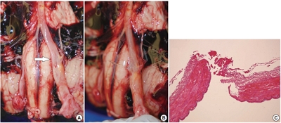
- Inconspicuous longitudinal tears of the intracranial vertebral artery in traumatic basal subarachnoid hemorrhage
- Seongho Kim
- J Pathol Transl Med. 2020;54(2):179-183. Published online November 8, 2019
- DOI: https://doi.org/10.4132/jptm.2019.10.15
- 11,446 View
- 211 Download
- 1 Web of Science
- 2 Crossref
-
 Abstract
 PDF
PDF - Blunt force trauma to the head or neck region can cause traumatic basal subarachnoid hemorrhage (TBSAH), which can result in rapid loss of consciousness and death; however, detecting such a vascular injury is difficult. Posterior neck dissection was performed to investigate the bleeding focus in TBSAH cases 2018 and 2019. In all four cases, autopsies revealed a longitudinal tear in the midsection of the vertebral artery’s intracranial portion. The midportion of the intracranial vertebral artery appears to be most vulnerable to TBSAH. Interestingly, three of the cases showed only a vaguely visible longitudinal fissure in the artery without a grossly apparent tear; rupture was confirmed by microscopic examination. Longitudinal fissures of the intracranial vertebral artery, which are difficult to identify without detailed examination, may be overlooked in some cases of TBSAH. Thus, careful gross and microscopic examination of the vertebral artery is recommended in cases of TBSAH.
-
Citations
Citations to this article as recorded by
- Traumatic vertebrobasilar pseudoaneurysms: diagnostic pitfalls on CT angiography with forensic implications — two case reports
Numfon Tweeatsani, Kana Unuma, Yukiko Uemura, Hirotaro Iwase, Yohsuke Makino
Emergency Radiology.2025; 33(1): 189. CrossRef - Effect of Ginseng Extract Ginsenoside Rg1 on Mice with Intracerebral Injury
Zixin Zhuang, Jinman Chen, Hao Xu, Yongjun Wang, Qianqian Liang
Chinese Medicine and Culture.2023;[Epub] CrossRef
- Traumatic vertebrobasilar pseudoaneurysms: diagnostic pitfalls on CT angiography with forensic implications — two case reports
- Erdheim-Chester Disease Involving Lymph Nodes and Liver Clinically Mimicking Lymphoma: A Case Report
- Yeoun Eun Sung, Yoon Seo Lee, Jieun Lee, Kyo Young Lee
- J Pathol Transl Med. 2018;52(3):183-190. Published online December 27, 2017
- DOI: https://doi.org/10.4132/jptm.2017.10.16
- 11,148 View
- 234 Download
- 11 Web of Science
- 12 Crossref
-
Abstract
PDF
- Erdheim-Chester disease (ECD) is a rare non-Langerhans cell histiocytosis and multisystem disease. First described in 1930, there are no more than 750 cases reported. The etiology remains unknown, but a majority of cases of ECD and Langerhans cell histiocytosis were found to have clonal mutations involving genes of the mitogen-activated protein kinase pathway. We recently encountered a 53-year-old male patient with extensive ECD involving the systemic lymph nodes, pleura, liver, and long bones clinically mimicking malignant lymphoma. Biopsies were performed at multiple sites, including a pleural mass, an external iliac lymph node, bone marrow, and the liver. Based on histopathological and immunohistochemical findings of positivity for CD68 and negativity for CD1a and S-100, the patient was diagnosed with ECD. Interferon-α was administered as the first-line treatment, but the patient rapidly progressed to hepatic failure after 2 months of treatment. We report this rare case of ECD clinically mimicking malignant lymphoma and diagnosed by careful pathological review.
-
Citations
Citations to this article as recorded by- Pleuropulmonary Involvement in Erdheim–Chester Disease
Steven Tessier, Aldo A. Acosta-Medina, Brandon T. Larsen, Jason R. Young, Ronald S. Go, Jay H. Ryu
Mayo Clinic Proceedings.2026; 101(6): 971. CrossRef - Case Report: Erdheim–Chester disease with liver involvement successfully treated with trametinib
Long Chang, Kai-ni Shen, Ya-Ping Luo, Xin-xin Cao
Frontiers in Oncology.2026;[Epub] CrossRef - Progress and challenges towards high-density magnetic random access memory: evolution of high-performance perpendicularly magnetized magnetic tunnel junctions with elaborate cell structure design
Toshihiko Nagase, Hisanori Aikawa, Masatoshi Yoshikawa, Masahiko Nakayama
Journal of Physics D: Applied Physics.2026; 59(21): 213001. CrossRef - Pathologic characteristics of histiocytic and dendritic cell neoplasms
Sun Och Yoon
Blood Research.2024;[Epub] CrossRef - Erdheim Chester Disease Mimicking Lymphoma: A Case Report
Philipp Moritz Wunschel, Wolfgang Voss, Marc Keberle
RöFo - Fortschritte auf dem Gebiet der Röntgenstrahlen und der bildgebenden Verfahren.2022; 194(03): 310. CrossRef - Erdheim–Chester Disease with Isolated CNS Involvement: A Systematic Review of the Literature
Anam Haque, Carlos A. Pérez, Thejasvi A. Reddy, Rajesh K. Gupta
Neurology International.2022; 14(3): 716. CrossRef - Exploring Erdheim-Chester Disease: A Histopathological Insight into a Rare Disorder
Banyameen Iqbal, Indranil Dey, Iqra Mushtaq
Journal of Medical Sciences and Interdisciplinary Research.2022; 2(2): 26. CrossRef - Erdheim‐Chester disease with long‐standing diabetes insipidus and generalized edema
Faezeh Sadat Naji, Minoo Sadat Hajmiri, Zahra Mazari, Faeze Salahshour, Vahid Soleimani, Manouchehr Nakhjavani, Mahboobeh Hemmatabadi
Clinical Case Reports.2021;[Epub] CrossRef - Morbus Erdheim-Chester
J. Knitza, E. Kampylafka, J. Wacker, G. Schett, B. Manger
Zeitschrift für Rheumatologie.2019; 78(1): 66. CrossRef - New causes of hypophysitis
Kevin C.J. Yuen, Vera Popovic, Peter J. Trainer
Best Practice & Research Clinical Endocrinology & Metabolism.2019; 33(2): 101276. CrossRef - Primary Necrobiotic Xanthogranulomatous Sialadenitis with Submandibular Gland Localization without Skin Involvement
Myunghee Kang, Na Rae Kim, Dong Hae Chung, Jae Yeon Seok, Dong Young Kim
Journal of Pathology and Translational Medicine.2019; 53(4): 261. CrossRef - Interferon-α
Reactions Weekly.2018; 1709(1): 128. CrossRef
- Pleuropulmonary Involvement in Erdheim–Chester Disease
Case Reports
- Chondroblastoma of the Lumbar Spine: A Case Report and Review of the Literature.
- Sun A Kim, Kyung Ja Cho, Yong Koo Park, Jong Seok Lee, Heon Ju Kwon, Hyewon Chung, Mi Jung Kim
- Korean J Pathol. 2011;45(5):532-536.
- DOI: https://doi.org/10.4132/KoreanJPathol.2011.45.5.532
- 5,404 View
- 39 Download
- 9 Crossref
-
Abstract
PDF
- We report a case of chondroblastoma arising in the lumbar spine in a 25-year-old man who presented with low back pain of 5 years duration. Plain radiography and computed tomography revealed a well-defined osteolytic mass surrounded by marginal sclerosis in the third lumbar vertebra. The mass encroached on the left neural foramen on magnetic resonance imaging. Histologically, the tumor consisted of round to oval cells with eosinophilic cytoplasm and randomly scattered osteoclastic type giant cells. There were characteristic chicken-wire calcification and aneurysmal bone cyst-like changes. Chondroblastomas of the lumbar spine are extremely rare, and only nine cases have been reported. Spinal chondroblastoma should be distinguished from other benign bone tumors, because it tends to show aggressive biological behavior with high recurrence and mortality rates.
-
Citations
Citations to this article as recorded by- Unveiling Primary Bone Tumors of the Spine: A Review of Essential Imaging Clues
Noah Tregobov, Michal Krolikowski, Ryan Dragoman, Benjamin Brakel, Peter L. Munk, Manraj K. S. Heran
Diagnostics.2025; 15(23): 2970. CrossRef - Chondroblastoma of Thoracic Vertebrae: a Case Report and Review of the Literature
Alireza Tabibkhooei, Parisa Javadnia
Indian Journal of Surgical Oncology.2024; 15(S1): 22. CrossRef - Clinicopathological characteristics and prognostic factors in axial chondroblastomas: a retrospective analysis of 61 cases and comparison with extra-axial chondroblastomas
Bo-Wen Zheng, Bo-Yv Zheng, Hua-Qing Niu, Ming-Xiang Zou, Hai-Lin Wu, Ming Wang, Xue-Lin Li
World Journal of Surgical Oncology.2023;[Epub] CrossRef - Commentary on Letter to the Editor concerning “Management of cranial chondroblastoma in adults; a pooled analysis” by Amr Muhammed et al.
Amr Muhammed
American Journal of Otolaryngology.2021; 42(1): 102749. CrossRef - Prognostic Significance of Tumor-Associated Macrophages in Chondroblastoma and Their Association with Response to Adjuvant Radiotherapy
Bo-Wen Zheng, Min-Liang Yang, Wei Huang, Bo-Yv Zheng, Tao-Lan Zhang, Jing Li, Guo-Hua Lv, Yi-Guo Yan, Ming-Xiang Zou
Journal of Inflammation Research.2021; Volume 14: 1991. CrossRef - Sacral chondroblastoma — a rare location, a rare pathology: A case report and review of literature
Bo-Wen Zheng, Hua-Qing Niu, Xiao-Bin Wang, Jing Li
World Journal of Clinical Cases.2021; 9(20): 5709. CrossRef - Intraoperative crush smear cytology of vertebral chondroblastoma: A diagnostic challenge
Shilpa P. Tathe, Sanjay N. Parate, Kirti N. Jaiswal, Archana A. Randale
Diagnostic Cytopathology.2018; 46(1): 79. CrossRef - Clinical features, treatments and long-term follow-up outcomes of spinal chondroblastoma: report of 13 clinical cases in a single center
Qi Jia, Chao Liu, Jian Yang, Yong Ji, Haifeng Wei, Tielong Liu, Xinghai Yang, Cheng Yang, Jianru Xiao
Journal of Neuro-Oncology.2018; 140(1): 99. CrossRef - Chondroblastoma of the thoracic spine: a rare location. Case report with radiologic-pathologic correlation
A. Venkatasamy, M. P. Chenard, G. Massard, J.-P. Steib, G. Bierry
Skeletal Radiology.2017; 46(3): 367. CrossRef
- Unveiling Primary Bone Tumors of the Spine: A Review of Essential Imaging Clues
- Cytohistologic Features of Chordoma Arising in Thoracic Spine: A Case Report.
- Seung Yeon Ha, Insun Kim, Sung Hye Park, Heum Rye Park
- J Pathol Transl Med. 1995;6(2):199-203.
- 2,421 View
- 15 Download
-
Abstract
PDF
- Chordoma is relatively uncommon tumor comprising 1~4% of primary malignant bone tumors, and believed to arise from the remnants of notochordal tissue. Because of its occurrence in the thoracic spine, we report a case of chordoma in volving the thoracic spine. A 45-year-old male was suffered from chest pain radiating to the back. Chest CT showed a well marginated, round huge mass with multiseptated enhancement at the thoracic spine from T5 to T8 level, After percutaneous needle aspiration, piecemeal resection of the tumor was done. On cytologic smears. two types of neoplastic cells were arranged in sheets and cords in mucinous background. One type of cells consisted of medium sized cells with pink cytoplasm and round nuclei. The other type had voluminous bubbly or clear cytoplasm divided by intracytoplasmic septae imparting a feathery or basket-like appearance. Histologically, the tumor showed lobulated feature divided by fibrous septae and the tumor cells were pink eosinophilic or physaliphorous in morphology. Immunohistochemically, the tumor cells revealed strong positivity for low(AE1) and high (AE3) molecular weight cytokeratins.
- Malignant Eccrine Poroma of Abdomen Brief case report.
- Jin Ja Park, Young Hee Choi, Kyung Chan Choi, Young Euy Park
- Korean J Pathol. 1998;32(4):312-314.
- 2,276 View
- 21 Download
-
Abstract
PDF
- Eccrine porocarcinoma is a rare tumor of the skin. A case report of an eccrine porocarcinoma metastasizing to epidural space of spinal cord and inguinal area with a nine year follow up is described. The patient had a nodular growth of the abdomen with both inguinal lymphadenopathy three years before its first excision. After a follow up of nine years, he complained a weakness of lower extremities and back pain. Extradural mass of 10th thoracic vertebra and left inguinal mass were found. Subsequently, the masses histologically identical to the skin tumor were found.
Original Article
- Cytologic Features of Plasmacytoma of the Ovary and Breast Occurred in a Patient with Solitary Plasmacytoma of Vertebra .
- Mi OK Park, Hoon Kyu Oh, Yong Jin Kim, Jae Bok Park
- J Pathol Transl Med. 1997;8(2):164-169.
- 2,201 View
- 21 Download
-
Abstract
PDF
- A case of plasmacytoma of the ovary and breast, which developed in a patient with a solitary plasmacytoma in the lumbar vertebra for nine months, was diagnosed cytologically and histologically. Enlargement of the right ovary and multiple palpable masses in the right and left breast were already present at six months after the diagnosis of vertebral solitary plasmacytoma. At eight months, plasma cell leukemia developed, and nine months the enlarged both ovaries, replaced by yellowish-gray solid tumors showed infiltration of immature plasma cells. The cytologic features of the ovarian tumors were same with those of the breast tumor. The tumor cells were of predominantly immature plasma cells with one or more nuclei. Some mature plasma cell had an eccentric nucleus with single nucleolus and peripherally clumped chromatin. Binucleated or multinucleated giant cells were often present. Histologically, sheets of poorly differentiated plasmacytoid tumor cells were separated by strands of hyaline fibrous tissue. On immunohistochemical stains, the tumor cells showed strong reactivity for lambda-light chain but no reaction for kappa-light chain, cytokeratin, or leukocyte common antigen.
 First
First Prev
Prev


