 E-submission
Search
- Page Path
- HOME > Search
- Clinicopathological characteristics of digestive system angioleiomyomas: case report and literature review
- Georgios Kalliopitsas, Christos Topalidis, Constantine Halkias, Theodora Gkeka, Konstantinos Sapalidis, Triantafyllia Koletsa
- J Pathol Transl Med. 2025;59(6):453-459. Published online October 28, 2025
- DOI: https://doi.org/10.4132/jptm.2025.08.04
- 4,022 View
- 119 Download
-
 Abstract
 PDF
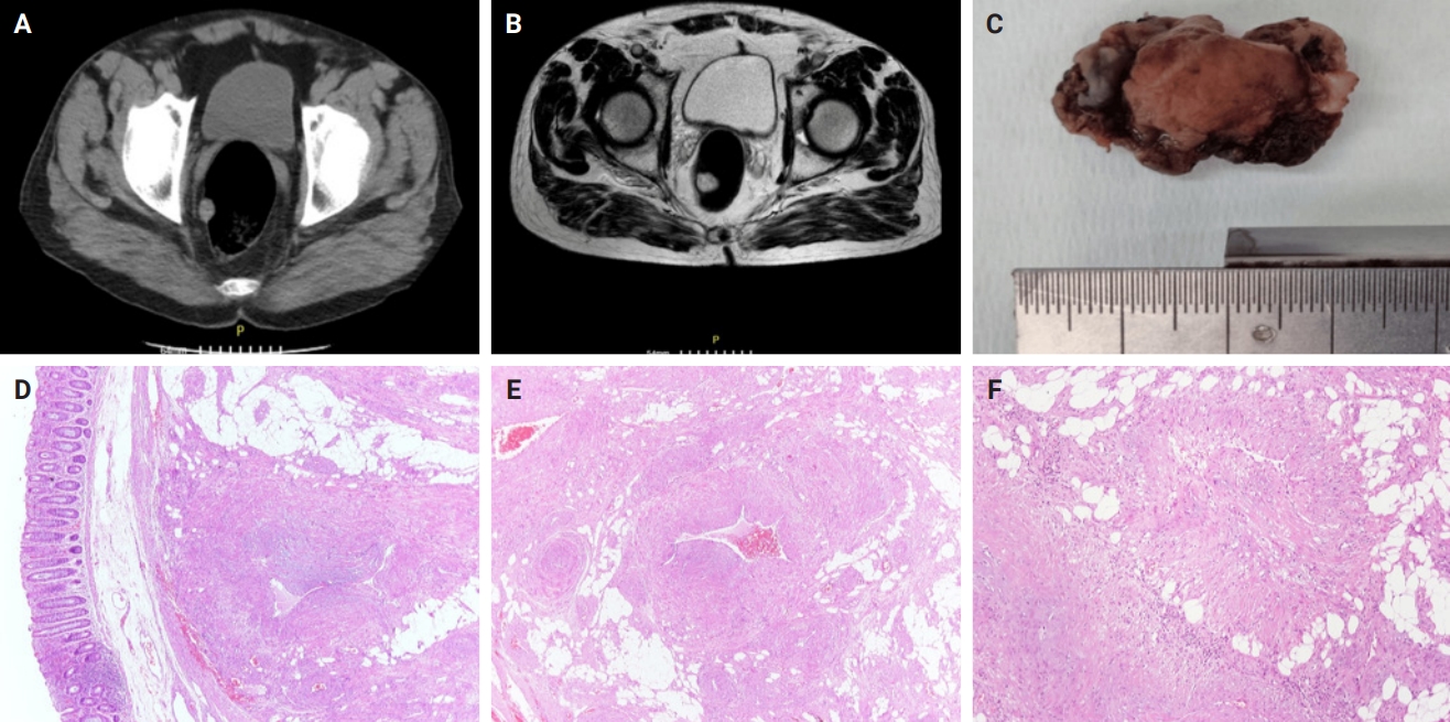
PDF - Angioleiomyomas are benign soft tissue tumors originating from the vascular wall. Although angioleiomyomas mainly occur in extremities, followed by head, neck, and trunk, they can also be found throughout the digestive system and especially in the oral cavity. Herein, the fourth case of a rectal angioleiomyoma in the English literature is reported and the clinicopathological features of digestive system angioleiomyomas were investigated. In contrast to their soft tissue counterparts, digestive system angioleiomyomas mainly affect males at a slightly younger age. Angioleiomyomas are mainly asymptomatic and only rarely elicit pain. Clinicians consider angioleiomyomas infrequently and instead include more common soft tissue or epithelial tumors in their differential diagnosis. To prevent angiomyolipoma misdiagnosis, pathologists should exercise caution when examining an angioleiomyoma composed of adipose tissue, smooth muscle, and blood vessels. Pathologists, radiologists, and surgeons should be aware that angioleiomyomas can occur in the digestive system.
- Adenomatoid odontogenic tumor: clinicopathological analysis of 34 cases from Karachi, Pakistan
- Summaya Zafar, Sehar Sulaiman, Madeeha Nisar, Poonum Khan, Nasir Ud Din
- J Pathol Transl Med. 2025;59(6):390-397. Published online October 16, 2025
- DOI: https://doi.org/10.4132/jptm.2025.07.11

- 5,292 View
- 182 Download
- 1 Web of Science
- 1 Crossref
-
Abstract
PDF
- Background
Adenomatoid odontogenic tumor (AOT) is a benign slow-growing neoplasm of odontogenic epithelial origin that is relatively uncommon. Only a few studies have described its histological features. Hence, we aimed to describe the clinicopathological features of AOT in a cohort of patients. Methods: AOT cases diagnosed between 2009 and 2024 were searched electronically. Glass slides were retrieved from archives and were reviewed by two pathologists to record the associated morphological features. Other data including patient demographics and tumor site were collected by reviewing histopathology reports. Results: The age of patients ranged from 9 to 44 years (mean, 17.7 years), and most were female (55.9%). The maxilla (44.1%) was the most common tumor site. Histologically, a predominantly solid growth pattern (n = 34) accompanied by ducts with a cuboidal/columnar epithelial lining (n = 31), eosinophilic secretions (n = 31), calcifications (n = 31), lattice work pattern (n = 30), and cystic areas (n = 20) were observed. Less frequent features included calcifying epithelial odontogenic tumor (CEOT)–like areas (n = 13), osteodentin (n = 6), association with impacted tooth (n = 3), mucin in tubules (n = 7), fibrocollagenous stroma (n = 6), mucin in ducts (n = 3) and ossifying fibroma-like areas (n = 6). The association of ducts with a cuboidal/columnar epithelial lining, lattice work pattern, calcifications, and eosinophilic secretions with gingival tumors was statistically significant (p ≤ .05). Additionally, tooth tumors were significantly associated with CEOT-like areas (p = .03). Conclusions: Our study confirms the trends in the clinicopathological features of AOT in previous case reports. Our results suggest that AOTs usually exhibit a predominantly solid pattern with duct-like spaces. Only a few cases with CEOT-like and ossifying fibroma-like areas were observed, similar to infrequent cases reported in the past. -
Citations
Citations to this article as recorded by
- Intraosseous lesions of the jaw: a clinicohistological study
Hadeel Odeh, Esra Nsour, Muna A. Salameh, Zayed M. Al-Zu’bi, Ali Al Khader
BMC Oral Health.2026;[Epub] CrossRef
- Intraosseous lesions of the jaw: a clinicohistological study
- Evaluation of potential prognostic significance of JUNB in human prostate cancer: a bioinformatic and histopathological study
- Noha R. Noufal, Einas M. Yousef, Mohamed Taha
- J Pathol Transl Med. 2025;59(5):291-305. Published online September 8, 2025
- DOI: https://doi.org/10.4132/jptm.2025.06.06
- 3,777 View
- 132 Download
-
Abstract
PDF
 Supplementary Material
 - Background
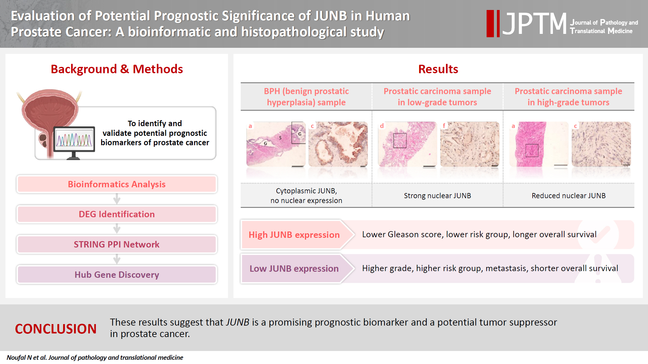
Prostate cancer is one of the most common malignancies in males worldwide. Serum prostate-specific antigen is a frequently employed biomarker in the diagnosis and risk stratification of prostate cancer; however, it is known for its low predictive accuracy for disease progression. New prognostic biomarkers are needed to distinguish aggressive prostate cancer from low-risk disease. This study aimed to identify and validate potential prognostic biomarkers of prostate cancer. Methods: Two prostate cancer datasets from the Gene Expression Omnibus were analyzed to identify differentially expressed genes between benign prostatic hyperplasia (BPH) and prostatic carcinoma. Immunohistochemistry was used to evaluate the JUNB proto-oncogene, a subunit of the AP-1 transcription factor (JUNB), in 70 prostate cancer patients and 10 BPH samples. Results: Our findings showed that JUNB was significantly enriched in prostate cancer-related pathways and biological processes. JUNB expression was considerably higher in prostatic adenocarcinoma patients than in BPH patients. Regarding JUNB expression in prostate cancer cases, lower levels of JUNB expression were associated with higher grades of prostatic adenocarcinoma. Lower JUNB expression was associated with a higher risk of prostatic adenocarcinoma progression and shorter overall survival. Conclusions: These results suggest that JUNB is a promising prognostic biomarker and a potential tumor suppressor in prostate cancer.
- Benign Triton Tumor: A Rare Entity in Head and Neck Region
- Krishnappa Amita, S. Vijay Shankar, Kuchangi C. Nischal, Haleuoor B. Basavaraj
- Korean J Pathol. 2013;47(1):74-76. Published online February 25, 2013
- DOI: https://doi.org/10.4132/KoreanJPathol.2013.47.1.74
- 9,920 View
- 58 Download
- 12 Crossref
-
Abstract
PDF
Benign triton tumors (BTT) are very rare lesions composed of mature skeletal muscle and neural tissue. We report a case of a 14-year-old boy who presented with asymptomatic swelling of the chin over an 18-month duration which increased gradually to involve the left side of the lower lip. Clinically, a diagnosis of neurofibroma was made. Excisional biopsy confirmed the diagnosis of a BTT. Having an affinity for large nerve trunks like the brachial and sciatic, these tumors rarely occur in the head and neck region. When they do, they may involve the large central cranial nerve trunk and present as intracranial masses or involve the smaller peripheral nerve twigs and present as asymptomatic skin nodules, of which only four cases involving peripheral nerves are reported in the English literature. Here, we report the fifth documented case of a BTT involving the mental branch of the trigeminal nerve. A brief review of the literature is also provided.
-
Citations
Citations to this article as recorded by- Pediatric Benign Nasal Triton Tumor: Its Diagnosis and Treatment
Marco Berlucchi, Iara Comincini, Luca O. Redaelli de Zinis, Iacopo Ghini, Maria P. Bondioni
Annals of Otology, Rhinology & Laryngology.2026; 135(4): 313. CrossRef - Update on MR Imaging of Soft Tissue Tumors of Head and Neck
Justin D. Rodriguez, A. Morgan Selleck, Ahmed Abdel Khalek Abdel Razek, Benjamin Y. Huang
Magnetic Resonance Imaging Clinics of North America.2022; 30(1): 151. CrossRef - Pediatric benign triton tumor of trigeminal nerve: a case report and literature review
Sophia Peng, Mandana Behbahani, Shelly Sharma, Stacy Speck, Nitin R. Wadhwani, Jeff C. Rastatter, Tord D. Alden
Child's Nervous System.2022; 38(11): 2055. CrossRef - Rhabdomyomatous mesenchymal hamartoma presenting as a chin nodule in a 15‐year‐old male
Lisa M. Marinelli, Wendi E. Wohltmann, Kevin D. Myers, Geoffrey T. Sasaki
Journal of Cutaneous Pathology.2021; 48(2): 322. CrossRef - Pediatric Benign Tumors With a Skeletal Muscle Component: Myogenin Expression, Diagnostic Pitfalls, and New Molecular Insights
Lara Berklite, John Ozolek, Larry Wang, Luisa Santoro, Vittoria Donofrio, Alessandra Stracuzzi, Ivy John, Rita Alaggio
Pediatric and Developmental Pathology.2021; 24(3): 213. CrossRef - Incidental Hamartoma in an elderly patient: a case report
Tae-Sung Joo, Hyejee Kim, In-Ki Park, Jae-Ho Shin
BMC Ophthalmology.2020;[Epub] CrossRef - Peripheral Nerve Sheath Tumors of Head and Neck: Imaging-Based Review of World Health Organization Classification
Ahmed Abdel Khalek Abdel Razek, Omneya A. Gamaleldin, Nermeen A. Elsebaie
Journal of Computer Assisted Tomography.2020; 44(6): 928. CrossRef - Benign peripheral nerve tumors
Zinon T. Kokkalis, Nikolaos A. Stavropoulos, Andreas F. Mavrogenis, Andreas Panagopoulos, Panayotis N. Soucacos
Injury.2019; 50: S77. CrossRef - Tumor de tritón benigno: reporte de un caso en órbita
Tatiana Urrea Victoria, Luis Alberto Ruíz Robles, Ana María Vanegas Monroy, Humberto Quintana Muñoz
Universitas Médica.2017;[Epub] CrossRef - Benign Triton Tumor: Multidisciplinary Approach to Diagnosis and Treatment
Raj Thakrar, Caroline D. Robson, Sara O. Vargas, John G. Meara, Reza Rahbar, Edward R. Smith
Pediatric and Developmental Pathology.2014; 17(5): 400. CrossRef - Ectomesenchymoma with Embryonal Rhabdomyosarcoma and Ganglioneuroma, Arising in Association with Benign Triton Tumor of the Tongue
Katherine A. VandenHeuvel, David F. Carpentieri, Jie Chen, Kar-Ming Fung, David M. Parham
Pediatric and Developmental Pathology.2014; 17(3): 226. CrossRef - Adulthood Benign Triton Tumor Developed in the Orbit
Dong Hyeon Bae, Choong Hyun Kim, Jin Hwan Cheong, Jae Min Kim
Journal of Korean Neurosurgical Society.2014; 56(2): 146. CrossRef
- Pediatric Benign Nasal Triton Tumor: Its Diagnosis and Treatment
- Diagnostic Difficulties in Fine Needle Aspiration of Benign Salivary Glandular Lesions
- Hye Jung Jo, Hyo Jung Ahn, Soojin Jung, Hye-Kyoung Yoon
- Korean J Pathol. 2012;46(6):569-575. Published online December 26, 2012
- DOI: https://doi.org/10.4132/KoreanJPathol.2012.46.6.569
- 10,842 View
- 63 Download
- 12 Crossref
-
Abstract
PDF
Background The diagnostic accuracy of fine needle aspiration cytology (FNAC) of salivary lesions is relatively high, but cytologic interpretation might be confusing if the sample is lacking typical cytologic features.
Methods There were 77 cases of benign salivary lesions, consisting of pleomorphic adenoma (PA) in 61 cases, Warthin's tumor (WT) in 12 cases, and other benign lesions in 4 cases. The causes of the discrepancies between the FNAC and the histologic diagnoses were evaluated.
Results Major discrepancies were noted in 4 of the 61 PA cases, and in 1 of 12 WT cases. The causes of the major discrepancies were a mislabeled site in 1 PA and 1 WT case, and an interpretation error in 3 PA cases. Minor discrepancies were more common in the WT cases (7 of 12 cases) than in the PA cases (11 of 61 cases). The causes of the minor discrepancies were a mislabeled site in 1 PA and 1 WT case, an inadequate sample in 7 PA and 2 WT cases, a lack of typical cytomorphology in 2 PA and 2 WT cases, and an interpretation error in 1 PA and 2 WT cases.
Conclusions To increase the diagnostic accuracy in the benign salivary lesions, recognition of both characteristic and less typical cytomorphology is needed.
-
Citations
Citations to this article as recorded by- Deep Learning Based on Ultrasound Images Differentiates Parotid Gland Pleomorphic Adenomas and Warthin Tumors
Yajuan Li, Mingchi Zou, Xiaogang Zhou, Xia Long, Xue Liu, Yanfeng Yao
Ultrasonic Imaging.2025; 47(3-4): 107. CrossRef - Clinical decision making when cytology indicates a Warthin tumor
Minna Sirviö, Katri Aro, Mira Naukkarinen, Antti Mäkitie, Jussi Tarkkanen, Jetta Kelppe, Timo Atula
Scientific Reports.2024;[Epub] CrossRef - Different MRI-based radiomics models for differentiating misdiagnosed or ambiguous pleomorphic adenoma and Warthin tumor of the parotid gland: a multicenter study
Jing Yang, Qiu Bi, Yiren Jin, Yong Yang, Ji Du, Hongjiang Zhang, Kunhua Wu
Frontiers in Oncology.2024;[Epub] CrossRef - Diagnostic Accuracy of Fine Needle Aspiration Cytology (FNAC) in Salivary Gland Lesions with Histopathological Examination (HPE) Correlation in a Tertiary Care Centre in Southern India
Banumathi Kasinathan, Babu Manohar, H. Ganapathy
Indian Journal of Otolaryngology and Head & Neck Surgery.2023; 75(2): 871. CrossRef - Diagnostic performance of qualitative and radiomics approach to parotid gland tumors: which is the added benefit of texture analysis?
Federica Vernuccio, Federica Arnone, Roberto Cannella, Barbara Verro, Albert Comelli, Francesco Agnello, Alessandro Stefano, Rosalia Gargano, Vito Rodolico, Giuseppe Salvaggio, Roberto Lagalla, Massimo Midiri, Antonio Lo Casto
The British Journal of Radiology.2021;[Epub] CrossRef - Improving the diagnosis of common parotid tumors via the combination of CT image biomarkers and clinical parameters
Dan Zhang, Xiaojiao Li, Liang Lv, Jiayi Yu, Chao Yang, Hua Xiong, Ruikun Liao, Bi Zhou, Xianlong Huang, Xiaoshuang Liu, Zhuoyue Tang
BMC Medical Imaging.2020;[Epub] CrossRef Evaluation of Salivary Gland Lesions by Fine Needle Aspiration Cytology at a Tertiary Care Hospital, Western Nepal
Anuj Poudel, Bigya Shrestha, Sudeep Regmi
Pathology and Laboratory Medicine International.2020; Volume 12: 9. CrossRef- Ultrasound-guided fine-needle capillary cytology of parotid gland masses coupled with a rapid-on-site evaluation improves results
R. Barats, S. Evrard, L. Collin, S. Vergez, S. Gellée, M. Courtade-Saïdi
Morphologie.2018; 102(336): 25. CrossRef - The Value of Ultrasound-Guided Fine-Needle Aspiration Cytology by Cytopathologists in the Diagnosis of Major Salivary Gland Tumors
Shahrzad Negahban, Sadegh Shirian, Bijan Khademi, Ahmad Oryan, Roshanak Sadoughifar, Mohammadian-Panah Mohammad, Azita Aledavood, Khosrow Daneshbod, Yayha Daneshbod
Journal of Diagnostic Medical Sonography.2016; 32(2): 92. CrossRef - Salivary Gland Tumors: A Diagnostic Dilemma!
Ranjit Kumar Peravali, H. Hari Kishore Bhat, Varsha H. Upadya, Anmol Agarwal, Sushma Naag
Journal of Maxillofacial and Oral Surgery.2015; 14(S1): 438. CrossRef - Diagnostic problems of salivary gland tumors
Ruchita Tyagi, Pranab Dey
Diagnostic Cytopathology.2015; 43(6): 495. CrossRef - Pleomorphic Adenoma of the Accessory Parotid Gland: Case Report and Reappraisal of Intraoral Extracapsular Dissection for Management
Tibebu M. Tsegga, Jennifer D. Britt, Aragon R. Ellwanger
Journal of Oral and Maxillofacial Surgery.2015; 73(3): 564. CrossRef
- Deep Learning Based on Ultrasound Images Differentiates Parotid Gland Pleomorphic Adenomas and Warthin Tumors
- Chondroblastoma-like Extraskeletal Chondroma: A case report.
- Jung Won Lee, Dae Su Kim, Mi Kyung Kim, Yeon Lim Suh
- Korean J Pathol. 1999;33(1):55-58.
- 2,376 View
- 49 Download
-
Abstract
PDF
- Extraskeletal chondromas are relatively uncommon benign cartilaginous tumors of the soft tissue and well known to pose a considerable diagnostic problem because of histological variations including the immature appearance of their tumor cells. Recently, we have experienced a case of extraskeletal chondroma mimicking benign chondroblastoma. The patient was a 47-year-old woman who complained of a painful subcutaneous swelling on the radial aspect of 4th proximal interphalangeal (PIP) joint in the left hand for 6 months. Radiologic examination of the 4th finger revealed a 1cm-sized soft tissue mass. Histologically, the tumor was characterized by a lobulated mass which was composed of dense proliferation of chondroblast-like cells admixed with a few multinucleated giant cells of osteoclastic type. However, there were focal areas of typical chondroma which showed lace-like intense calcification around the differentiated chondrocytes.
- Subcutaneous Neuromuscular Hamartoma: A case report.
- Dong Hoon Kim, Eun Kyung Hong, Jung Dal Lee
- Korean J Pathol. 1999;33(1):62-64.
- 2,556 View
- 42 Download
-
Abstract
PDF
- Subcutaneous form of neuromuscular hamartoma is extremely rare and histologically different from the conventional neuromuscular hamartoma of the peripheral nerve or benign Triton tumor by an indistinct nodular growth with ill-defined margin and marked collagen interposition. It is usually not associated with a major nerve. We report a case of subcutaneous neuromuscular hamartoma developed in the forehead of 24-year-old man. The tumor showed proliferation of dense, hyalinized fibrous tissue, in which single or group of mature skeletal muscle fibers and nerve fibers were haphazardly intermixed. Recognition of abnormally arranged muscle and nerve fibers is important not to miss this lesion.
- Benign Cystic Mesothelioma.
- Sung Chul Lim, You Kyung Jeong, Mi Sook Lee, Yun Shin Kim, Hyun Jong Park, Sang Joon Choi
- Korean J Pathol. 1997;31(6):595-597.
- 2,314 View
- 16 Download
-
Abstract
PDF
- Benign cystic mesothelioma (BCM) is a rare mesothelial lesion that forms multicystic masses in the upper abdomen, pelvis and retroperitoneum. Although it is categorized as a benign lesion, it has a tendency to recur. It is uncertain whether the nature of this lesion is reactive or neoplastic, but many articles support the conclusion that it is reactive rather than neoplastic. The majority of cases were associated with a history of a previous abdominal or pelvic operation, or an evidence of endometriosis or a pelvic inflammatory disease, or a combination of these findings. In a 26-year-old woman we experienced a case of BCM which was incidentally discovered at cesarean delivery revealing multilocular thin and translucent walled cysts in the pelvic cavity. Microscopic examination revealed a thin cyst wall that was composed of fibrous connective tissue and lined by internal stratified and external nonstratified single cuboidal epithelia.
- Urinary Cytologic Findings of Urothelial Lesions.
- Yoon Jung Choi, Kwang Gil Lee
- J Pathol Transl Med. 1994;5(2):130-136.
- 2,275 View
- 17 Download
-
Abstract
PDF
- Urinary cytology is increasingly accepted as a diagnostic tool in the detection and follow-up of patients with bladder cancer. However, its value is reduced by several limitations, especially by the tack of cytologic criteria specifically reflecting the morphology of low-grade urothelial neoplasm. We reviewed histologically proven 50 cases of urine cytology with emphasis on cytologic findings of benign atypia and differential findings of urothelial neoplasm according to the grade. The diagnoses included 17 benign lesions (including 5 cases of urine calculi) and 33 malignant lesions(including 28 transitional cell carcinomas. 3 squamous cell carcinomas, 1 adenocarcinoma and 1 prostate adenocarcinoma). Diagnostic accuracy was 92%. Important cytodiagnostic criteria for benign atypia and low grade malignancy were cellularity, number of cell clusters, and morphology and arrangement of urothelial cells. The cytologic findings of urothelial neoplasms according to histologic grade were relatively well correlated with the histologic findings. However, the cytologic criteria were not sufficient to readily distinguish grade I from grade II. In view of this, we think that cytologic nomenclature "low-grade" and "high-grade" is a more reliable criterion. Recognition of subtle cellular morphologic features specific for urothelial lesions(including benign or malignancy) and proper fixation, processing and staining of specimen can expand the role of urinary cytology in detection and follow-up of patients.
- Benign Lymphoepithelial Cyst: A case report.
- Jin Haeng Chung, Gyeong Hoon Kang, Je G Chi
- Korean J Pathol. 1996;30(6):551-553.
- 2,712 View
- 20 Download
-
Abstract
PDF
- An intraparotid benign lymphoepithelial cyst is a rare disease characterized by unilateral painless swelling of parotid region. The histogenesis is controversial. Surgical excision is recommended for diagnosis and curative treatment. We present a case of benign lymphoepithelial cyst arising in a patient with neurofibromatosis. A 46-year-old woman presented with a slowly growing multilocular cystic mass in the left cheek. The cystic mass measured 4 cm in maximal outer diameter and the cystic wall was thick and yellowish pale to gray, soft with well circumscribed margin. Microscopically, the multilocular cyst was lined by stratified squamous epithelium for the most part and underlying lymphoid tissue aggregates with follicles and sharply demarcated from adjacent salivary parenchyma which is of normal appearance and without lymphoid aggregates. Since this lesion is absolutely benign, it is important to separate this benign cyst from cystic salivary gland tumors.
- Squamous Cell Carcinoma and Struma Ovarii Arising in Benign Cystic Teratoma.
- Eun Sook Nam, Young Seek Kim, Yang Seok Chae, Kap No Lee, Seung Yong Paik
- Korean J Pathol. 1991;25(5):462-466.
- 3,620 View
- 10 Download
-
Abstract
- Malignant tumor is found in 1-2% of ovarian benign cystic teratomas. Among these malignant neoplasms, squamous cell carcinoma is by far the most common malignancy, whereas the incidence of struma ovarii is less than 5% in mature teratoma. As far as concerned the struma ovarii, a very small percentage is associated with carcinoid, mucinous or serous cystadenoma, or Brenner tumor. However, any reports of struma ovarii associated with squamous cell carcinoma in the same ovary could not be found in English literature. Recently we have experienced a case of squamous cell carcinoma and struma ovarii arising in an ovarian benign cystic teratoma in 72 year old female patient.
- Differential Diagnosis of Fine Needle Aspiration Cytology of Benign Lymphadenopathy.
- Eun Mee Han, Dong Eun Song, Dae Un Eom, Hye Jeong Choi, Hee Jeong Cha, Jooryung Huh
- J Pathol Transl Med. 2006;17(2):99-107.
- 3,169 View
- 39 Download
-
Abstract
PDF
- In the investigation of superficial lymphadenopathy of unknown cause, fine needle aspiration (FNA) cytology plays an invaluable role. It enables the differentiation of benign lymphadenopathy from lymphoid and non-lymphoid malignancies, obviating the need for open biopsy, and allowing the triage of patients. Cytopathologists should be familiar with the typical FNA patterns of benign lymphadenopathy, and recognize and differentiate among categories. In a minority of cases of benign lymphadenopathy, FNA can render a specific diagnosis. Benign lymphadenopathies are generally categorized into reactive lymphoid hyperplasia (RLH), inflammatory or infectious processes, and benign lymphoproliferative disorders. RLH characteristically presents with a heterogeneous and polymorphous smear composed of normal cellular constituents of lymph nodes, in contrast with the homogeneous or monomorphic smear of most lymphomas. The caveat is that various malignant disorders may also present with polymorphous populations. It is also important to recognize thatbenign lymphoid smears may sometimes contain atypical cells that raise the suspicion of malignancy. Clinical information should always be the integral part of the diagnostic criteria in FNA of lymphadenopathy. If there is any doubt about the benign nature of the smear, it is prudent to suggest biopsy and ancillary studies.
- Benign Epithelial Changes of Endometrium: Based on 450 hysterectomy specimens obtained from Jan. 1994 to Dec. 1994.
- Hye Kyung Lee, Myung Jin Joo, Kwang Min Lee, Dong Kyu Chung
- Korean J Pathol. 1996;30(11):966-971.
- 2,416 View
- 14 Download
-
Abstract
PDF
- To evaluate the incidence and clinico-pathologic correlation of benign epithelial changes of endometrium, we tried to classify the changes into squamous cell change, ciliary change, eosinophilic cell change, papillary surface epithelial change, and mucinous cell change by the criteria of Hendrickson. Based on the 450 hysterectomy specimens obtained from Jan. 1994 to Dec. 1994 in PMC, the incidence of the cell changes was as follows: squamous cell change: 1.1%, eosinophilic cell change: 6.8%, mucinous cell change: 6.6%, ciliary change: 10.4%, papillary surface epithelial change: 16.4%. Squamous cell change was noted in severe endometritis or endometrial hyperplasia and papillary surface epithelial proliferation was mainly associated with plasma cell infiltration in adenomyosis or leiomyoma. Eosinophilic change and ciliary change were sometimes concomitantly found in dilated glands of the basal layer or in the invaded glands of adenomyosis. The results of this study suggested a correlation of benign epithelial changes with endometritis, adenomyosis, leiomyoma and dysfunctional uterine bleeding.
- Benign Fibrous Histiocytoma of Spinal Cord.
- Hae Joo Nam, Won Hee Choi, Tae Sook Lee
- Korean J Pathol. 1988;22(4):510-514.
- 2,371 View
- 34 Download
-
Abstract
PDF
- Fibrous histiocytoma composed of fibroblasts and histiocytes is quite variable in histologic pattern. The biologic behavior is unpredictable by histologic ground. This tumor is well-known in subcutaneous tissue and deep soft tissue, but quite rare in central nervous system. We experienced a case of the fibrous histiocytoma involving the dura mater of spinal cord in a 26 year old female patient. In gross findings, the mass was a well demarcated, ovoid mass attached to the dura matter, and measured 2.5x1.5 cm in diameter. The cut surface was rubbery, solid, gray-white or yellow. Microscopically, the lesion consisted of polyhedral cells with round or oval nuclei and faintly eosinophilic or vaculoated cytoplasm, and multinucleated giant cells. Some giant cells were Touton-type. Composing cells were bland-looking. Mitotic figures were average 3 per 10 high power fields.
 First
First Prev
Prev


