 E-submission
Search
- Page Path
- HOME > Search
Original Article
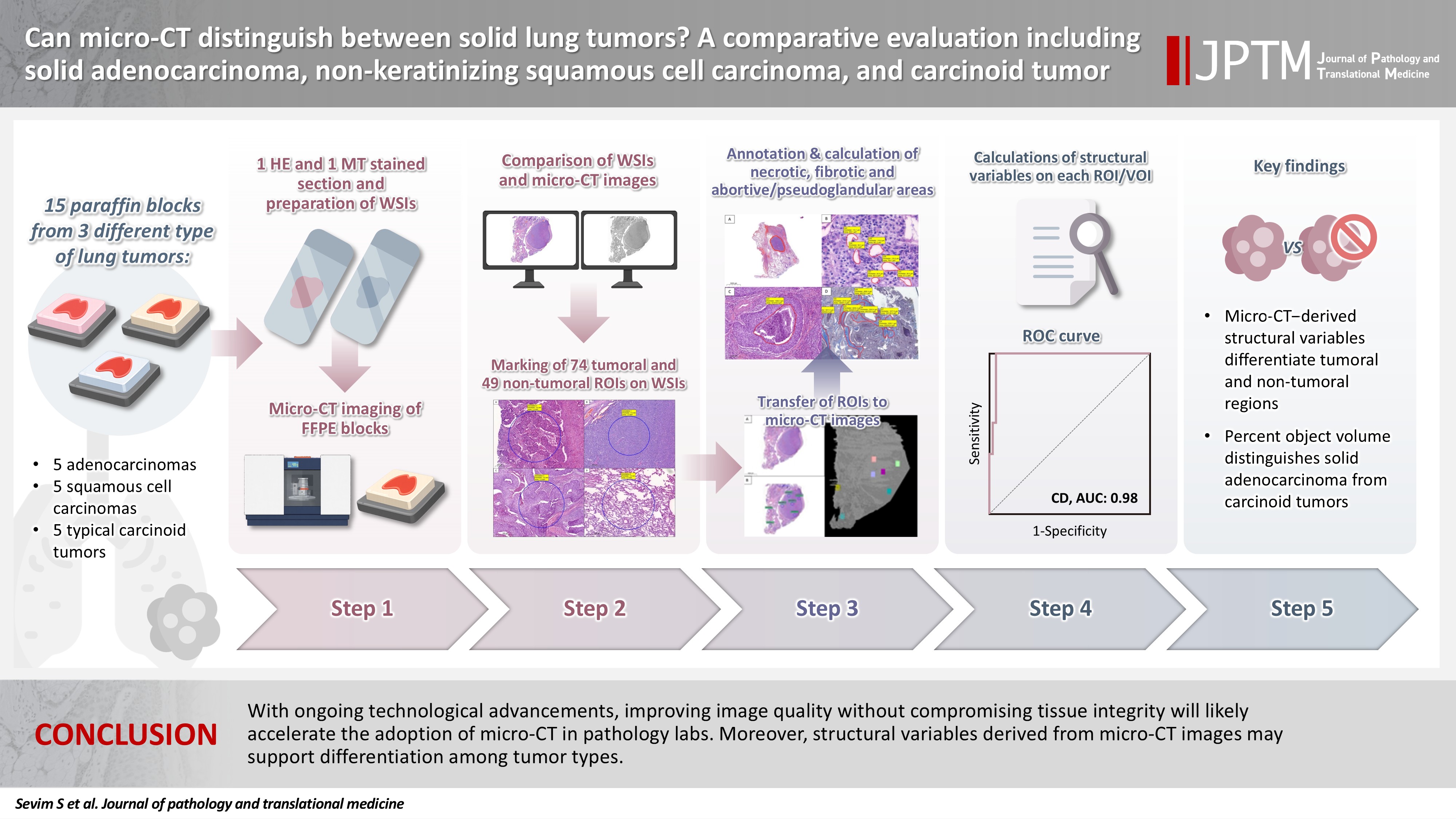
- Can micro-CT distinguish between solid lung tumors? A comparative evaluation including solid adenocarcinoma, non-keratinizing squamous cell carcinoma, and carcinoid tumor
- Selim Sevim, Serpil Dizbay Sak, Kaan Orhan, Arda Buyuksungur, Duru Karasoy, Hilal Ozakinci, Ayten Kayi Cangir
- J Pathol Transl Med. 2026;60(2):231-245. Published online March 10, 2026
- DOI: https://doi.org/10.4132/jptm.2025.12.16
- 354 View
- 40 Download
-
 Abstract
 PDF
PDF Supplementary Material
 - Background
Some pulmonary carcinomas display a solid pattern, and immunohistochemistry is commonly used for tumor differentiation. Micro–computed tomography (micro-CT), with its ability to produce detailed three-dimensional images using small voxel sizes, may offer additional insights. This study investigates whether three solid tumor types, solid adenocarcinoma (sAC), non-keratinizing squamous cell carcinoma, and carcinoid tumor (CaT), can be differentiated using micro-CT. Methods: Fifteen paraffin blocks, five for each type, were scanned with micro-CT (Skyscan 1275, Bruker). These images were compared to whole slide images (WSIs) of the same tumors. Consequently, tumoral (n = 74) and non-tumoral (n = 49) regions of interest (tumor ROIs [tROIs] and non-tumor ROIs [ntROIs]) were selected on the micro-CT images and evaluated in terms of certain structural variables (percent object volume, structure model index, structure thickness, structure linear density, connectivity, connectivity density, open porosity, closed porosity) to investigate whether tumors can be differentiated from normal parenchyma and from each other. Results: Although detailed images comparable to WSIs could not be obtained, it was considered an important advantage to be able to examine the entire depth of the paraffin blocks. tROIs and ntROIs could be distinguished based on all variables (p < .001). Additionally, sAC showed a notable difference from CaT in “percent object volume” (p = .011). Conclusions: With ongoing technological advancements, improving image quality without compromising tissue integrity will likely accelerate the adoption of micro-CT in pathology labs. Moreover, structural variables derived from micro-CT images may support differentiation among tumor types.
Case Report
- Fine Needle Aspiration Cytology of Small Cell Carcinoma of the Parotid Gland: A Case Report .
- Chan Kwon Jung, Eun Sun Jung, Youn Soo Lee, Sun Moo Kim, Byung Kee Kim
- J Pathol Transl Med. 1999;10(2):163-167.
- 2,038 View
- 23 Download
-
Abstract
PDF
- Primary small cell carcinoma of the salivary gland is a rare neoplasm that accounts for approximately 1.8% of all primary major salivary gland malignancies. Because of its rarity, it is difficult to diagnose small cell carcinoma of the parotid gland by fine needle aspiration cytology(FNAC). We experienced a case of primary small cell carcinoma of the parotid gland in a 72-year-old woman who presented with two palpable masses of the left infraauricular and ocular regions of two to three month's duration, respectively. Aspirate smears from the left infraauricular area were highly cellular on necrotic and lymphocytic background and showed individually dispersed cells or three-dimensional clusters of small cells. The tumor cells were round to oval with a very high nucleocytoplasmic ratio. Nuclei were about two times the size of lymphocytes and had uniformly dispersed but hyperchromatic to pyknotic chromatin. Nucleoli were occasionally visible but were generally inconspicuous. Numerous mitotic figures were detected. The clusters of these small tumor cells exhibited angular nuclear molding, irregular nuclear outlines, and occasionally rosette like arrangement. The tumor was confirmed by histology and immunohistochemistry.
Original Article
- Primary Undifferentiated Carcinoma of the Endometrium with Small Cell and Trophoblastic Differentiation.
- Chul Hwan Kim, Seoung Hye Park, In Sun Kim, Seung Yong Paik
- Korean J Pathol. 1990;24(1):58-64.
- 2,184 View
- 19 Download
-
Abstract
PDF
- This report describes a very rare case of primary undifferentiated carcinoma of the endometrium with small cell and trophoblastic differentiation. The patient was 54-year-old woman with complaints of vaginal bleeding and palpable lower abdominal mass. The light microscopic findings revealed predominantly small cells with round nuclei, spindle cells, and large cells with hyperchromatic bizarre nuclei. Foci of syncytiotrophoblastic giant cells are scattered, especially in the hemorrhagic areas. Immunohistochemical stainging for neuron specific enolase and beta-hCG showed positive reactions to small cells and syncytiotrophoblastic giant cells, respectively. Argentaffin and argyrophil stains, however, showed negative reactions to small cells. The histogenesis of small cell undifferentiated carcinoma of the endometrium remains unclear; however, it may arise from epithelial precursors instead of neuroendocrine cells, and syncytiotrophoblastic cells may be differentiated or dedifferentiated from the undifferentiated carcinoma cells.
Case Reports
- Carcinoid Tumor of the Uterine Cercix: A light and electron microscopic study of two cases.
- Moon Hyang Park, Jung Dal Lee, Yoon Young Hwang
- Korean J Pathol. 1990;24(1):70-76.
- 2,044 View
- 12 Download
-
Abstract
PDF
- Two cases of carcinoid tumor of the uterine cervix were reported with emphasis on the histologic, cytologic, histochemical and electron microscopic appearance of tumor cells. Based on the light microscopic findings, one case was a well differentiated carcinoid with acinus formation, and the other was a poorly differentiated anaplastic type, being composed of small cells similar to those of oat cell carcinoma of the lung. Both tumors demonstrated scattered argyrophilic cells on Grimelius stain, and contained neurosecretory granules on electron microscopy. They were in stages II b and IV, respectively, at the time of presentation. The latter patient was treated with vinblastin, platinol and bleomycin, but died in 9 months after the initial diagnosis. The former was lost to follow-up study. Importance of distinction between this highly malignant tumor and other varieties of cervical cancer was emphasized.
- Cytologic Findings of Primary Small Cell Carcinoma of the Urinary Bladder: A case report.
- Mi Seon Kwon, Geung Hwan Ahn, Jin Haeng Chung, Seung Sook Lee, Jae Soo Koh
- J Pathol Transl Med. 2001;12(2):121-126.
- 2,295 View
- 26 Download
-
Abstract
PDF
- Primary small cell carcinoma of the urinary bladder is a rare malignant tumor. A more rapidly fatal course may be seen in advanced stages of small cell carcinoma as compared to similar stages of urothelial carcinoma. It is very important to recognize this distinct form of bladder cancer by urinary cytology. The differential diagnosis of small cell carcinoma of the urinary bladder includes metastatic small cell carcinoma, urothelial carcinoma, and primary or secondary malignant lymphoma. This article highlights the urinary cytologic diagnosis of a case of primary small cell carcinoma. A 59-year-old male presented with gross hematuria for five months. Urinary cytology showed high cellularity consisting of tiny monotonous tumor cells in the necrotic background. The tumor cells occurred predominantly singly, but a few in clusters. The cytoplasm was so scanty that only a very narrow rim of it was seen. The nuclei were oval or round and had finely stippled chromatin. Rarely, the nuclei contain visible nucleoli. Frequently cell molding was noted in clusters. Many single cells demonstrated nuclear pyknosis or karyorrhexis. The histologic findings of transurethral resection and partial cystectomy specimen were those of small cell carcinoma. Cytologic distinction may be very difficult but careful attention to clinical features and cellualr details can classify these neoplasms correctly.
Original Article
- Immunohistochemical Study on the Expression of p53 and Bcl-2 Proteins in Non-Small Cell Lung Carcinomas.
- Ok Ju Lee, Do Youn Park, Kang Suek Suh
- Korean J Pathol. 1997;31(9):823-831.
- 2,044 View
- 17 Download
-
Abstract
PDF
- To address the possible prognostic value of p53 and Bcl-2 proteins in non-small cell lung carcinomas (NSCLCs), the authors studied 43 cases of NSCLCs diagnosed between the years 1990 to 1995 at Pusan National University Hospital. The patients were treated either by pneumonectomy or lobectomy of the lung. The expression of p53 and Bcl-2 proteins was semiquantitatively analyzed in paraffin sections by immunohistochemical method and correlated with clinicopathologic prognostic parameters of NSCLCs. Overexpression of the p53 protein was found in 31 cases (72.1%) of the 43 NSCLCs. Overexpression of the p53 protein was significantly correlated with the decreasing degree of histologic differentiation, increasing tumor stage, and cigarette smoking. Bcl-2 expression was found in 19 cases (44.2%) of the 43 NSCLCs. Increased expression of the Bcl-2 protein was significantly correlated only with decreasing tumor stage. An inverse relationship was found between p53 and Bcl-2 proteins, but it was not statistically significant. Thus p53 and Bcl-2 proteins, as demonstrated immunohistochemically in routine paraffin sections, could be of value in prediction of the aggressiveness and prognosis of NSCLCs, in agreement with the central role of p53 and Bcl-2 proteins in the evolution of NSCLCs associated with cigarette smoking.
Case Report
- Cytodiagnosis of Primary Small Cell Carcinoma of the Urinary Bladder: A Case Report.
- Hye Sun Kim, Aee Ree Kim, Chul Hwan Kim, Yang Seok Chae, Nam Hee Won
- J Pathol Transl Med. 1994;5(2):167-171.
- 1,879 View
- 15 Download
-
Abstract
PDF
- Samll cell carcinoma of the urinary bladder is a rare tumor which occurs in about 0.48% of all bladder tumors. We report cytologic features of small cell carcinoma of the urinary bladder in a 66-year-old man who had painless total gross hematuria, which was confirmed by partial cystectomy. In urine cytology, abundant tumor cells appeared in scattered and clustered forms in a bloody background. The tumor cells were small and uniform in size with a high nuclear/cytoplasmic ratio. The nuclei of the tumor cells were hyperchromatic, characteristically molded and showed inconspicuous nucleoli. The cytoplasms were scanty and plae blue.
Original Articles
- Differential Diagnosis between Small Cell Carcinoma and Adenocarcinoma of Lung in Fine Needle Aspiration Cytology.
- Young Hee Choi, Jae Soo Koh, Sunhoo Park, Min Suk Kim, Soo Youn Cho, Jung Soon Kim, Hwa Jung Ha, Seung Sook Lee
- J Pathol Transl Med. 2006;17(2):120-125.
- 2,873 View
- 71 Download
-
Abstract
PDF
- Distinguishing small cell carcinoma from other lung malignancies is of great clinico-therapeutic significance. Small cell carcinoma is an aggressive tumor with a tendency to metastasize early. Survival time if untreated is low but this tumor is highly responsive to chemotherapy. We have occasionally experienced difficulties in differentiation between adenocarcinoma and small cell carcinoma of the lung in fine needle aspiration cytology (FNAC). The aim of this study was to investigate the possibility of distinguishing small cell carcinoma from adenocarcinoma of the lung in FNAC. We evaluated cytomorphological features of FNAC specimens from 62 small cell carcinomas and 57 adenocarcinomas from the lung that were confirmed by biopsy and/or immunohistochemistry on cell block. Cytomorphological details of the two tumors were compared. Nuclear smearing and nearly absent cytoplasm were the most distinct findings in small cell carcinoma compared to adenocarcinoma (p<0.05). Necrotic background, architecture and chromatin pattern, nuclear molding and nucleoli were significantly different (p<0.05). Nuclear size, nuclear membrane nature and nuclear size variation however were not helpful in distinguishing the two tumors. Combining several features described above, small cell carcinoma can be properly differentiated from adenocarcinoma on FNAC. FNAC is proposed as a diagnostic tool of small cell carcinoma of the lung in the case of inaccessibility to biopsy, and so may allow the proper therapeutic strategies to be determined in such cases.
- Fine Needle Aspiration Cytology of Metastatic Cell Carcinoma of Lymph Nodes: Comparison to Non-Hodgkin's Lymphoma on 5 Cases.
- Yeon Mee Kim, Hye Je Cho, Ill Hyang Ko
- J Pathol Transl Med. 1996;7(1):44-50.
- 4,020 View
- 93 Download
-
Abstract
PDF
- Small cell carcinoma of the lung is characterized by cells with finely stippled chromatin and scanty cytoplasm as well as a particularly aggressive clinical course and favorable response to the chemotherapy. Recently percutaneous fine needle aspiration(FNA) biopsy has become both widely established and highly respected for the diagnosis of lung cancer. However metastatic small cell carcinoma of lymph node should be cytologically differentiated from the small round cell tumor of particular sites, especially malignant lymphoma, because small cell carcinoma of classic oat cell type may simulate small cell non-Hodgkin's lymphoma. We report five cases of metastatic small cell carcinoma of intermediate cell type diagnosed by FNA of the enlarged lymph nodes of the neck and axilla. The cytologic smears contained diffuse small neoplastic cells larger than lymphocytes with dense, pyknotic nuclei and extremely scanty cytoplasm. Apparently viable large tumor cells have vesicular nuclei with granular, sometimes very coarse chromatin. The characteristic cytologic features of small cell carcinoma as compared to malignant lymphoma were as follows.: 1) small cells with dense pyknotic nuclei are evenly distributed in the background of apparently viable larger tumor cells, admixed with mature lymphocytes and phagocytic macrophages. 2) small loose aggregates of cells with nuclear molding are indicative of small cell carcinoma rather than non-Hodgkin's lymphoma. 3) the cytoplasmic and nuclear fragments of tumor necrosis are more dominant in the smears of small cell carcinoma. 4) nuclear membrane and nucleoli are generally indistinct in small cell carcinoma due to condensation of chromatin.
Case Reports
- Voided Urine Cytology of Small Cell Carcinoma of the Urinary Bladder: A Case Report.
- Won Ae Lee, Seung Ha Lee
- J Pathol Transl Med. 2007;18(2):153-156.
- 1,939 View
- 15 Download
-
Abstract
PDF
- Primary small cell carcinoma of the urinary bladder is an extremely rare but important entity. We experienced a case of small cell carcinoma of the urinary bladder diagnosed by urine cytology. A 59-year-old man presented with gross hematuria and dysuria, and a calcified mass was detected at the left ureterovesical junction by cystoscopy. Abdominal ultrasonography revealed focal wall thickening at the left lateral side of the urinary bladder, and urine cytology findings were of an inflammatory background and atypical small round cells with minute hyperchromatic or pyknotic nuclei, scant cytoplasm, and rare nucleoli. In addition, atypical cells were scattered in an isolated single cell pattern or in small loose clusters with prominent nuclear molding. Subsequent histological and immunohistochemical examinations confirmed a diagnosis of small cell carcinoma.
- Carcinosarcoma of the Esophagus: A report of case.
- Sug Hyung Lee, Won Sang Park, Young Jin Choi, An Hee Lee, Sun Moo Kim
- Korean J Pathol. 1992;26(2):191-196.
- 1,968 View
- 18 Download
-
Abstract
PDF
- Carcinosarcoma of the esophagus is a rare neoplasm composed of both carcinoma and spindle sarcomatous area. Usually the carcinoma component is a squamous cell carcinoma but rarely adenocarcinoma or undifferentiated carcinoma is found. The histogenesis of the sarcomatous component is still unknown. A case of ulcerated polypoid lesion with a stalk in esophagus was reported. Microscopically it was composed of spindle shaped cells interminled with squamous cell carcinoma and small cell carcinoma nests. No distinct transition between spindle shaped cells and carcinoma are was observed. Immunoreactivity to cytokeratin was observed in both carcinomatous and spindle cell component, but electron microscopic examination failed to demonstrated desmosome or tonofilaments in spindle cells. Undifferentiated small cell nests were reactive to neuron specific enolase and contained membrane bounded secretory granule in electron microscopy.
- Small Cell Carcinoma of the Ovary: A case report.
- Young Bae Kim, Sook Hee Hong, Kyu Rae Kim
- Korean J Pathol. 1992;26(4):399-404.
- 2,170 View
- 14 Download
-
Abstract
PDF
- Small cell carcinoma of the ovary is rare malignancy occurring in women under 40 years of age(average, 23 years), which is associated with hypercalcemia in two thirds of cases. Its histogenesis is uncertain, but the possibilities of common epithelial, neuroendocrine, sex cord stromal and germ cell origin are suggested. All reported cases were proved to have rapid fatal course despite various therapy and 5 years suvival rate was only 10%. We report one case of a 20-year old woman with primary small cell carcinoma of the left ovary. The ovary was markedly enlarged and completely replaced by a mass, measuring 21x16x8 cm. Microscopic examination revealed dimorphic population of small and large malignant cell producing immature follicle-like structure which is characteristic of small cell carcinoma of the ovary. These pathological findings were similar to those of granulosa cell tumor, which is required to make differential diagnosis from small cell carcinoma. Immunohistochemical stains for cytokeratin and vimentin were positive, but those for S-100 protein and NSE were negative. One month after the initial operation, the tumor has recurred and the second and the second palliative operation followed by 3 cycles of chemotherapy was done. The patient showed disseminated metastasis at present time.
Original Articles
- Small Cell Carcinoma of the Uterine Cervix.
- Jinwon Seo, Jong Sun Choi, Geunghwan Ahn
- Korean J Pathol. 2003;37(6):373-378.
- 2,131 View
- 17 Download
-
Abstract
PDF
- BACKGROUND
Small cell carcinoma of the uterine cervix is a rare and aggressive neoplasm, for which there have been few diagnostic markers.
METHODS
Eleven cases of small cell carcinoma of the uterine cervix were retrieved from pathology files. Immunohistochemical stains were performed for two epithelial markers (cytokeratin [AE1/AE3] and epithelial membrane antigen [EMA]) and four neuroendocrine markers (neuron-specific enolase [NSE], CD56, chromogranin, and synaptophysin).
RESULTS
Of the nine cases followed up, two with initial distant metastasis died within one year. All seven remaining cases were diagnosed at stage Ib, and showed no evidence of recurrence. Nine cases were positive for one or more epithelial markers. Two cases showed positivity for epithelial markers in less than 10% of their tumor cells. The immunoreactivity for neuroendocrine markers showed variable results; four cases were reactive for chromogranin, four were positive for synaptophysin, and seven were reactive for CD56. All cases were positive for NSE.
CONCLUSIONS
A diagnostic panel of chromogranin, synaptophysin, and CD56 rather than a single marker would be useful for the diagnosis of small-cell carcinoma of the uterine cervix.
- The Clinicopathological Characteristics of Gastrointestinal Neuroendocrine Tumors; An Analysis of 65 Cases.
- Hyunjoo Lee, Jungwoo Choi, Jung Suk An, Hyunchul Kim, Bong Kyung Shin, Aeree Kim, Hankyeom Kim, Insun Kim
- Korean J Pathol. 2007;41(3):149-157.
- 2,355 View
- 27 Download
-
Abstract
PDF
- Background
: This study was designed to investigate gastrointestinal neuroendocrine tumors with an emphasis on their clinicopathological characteristics.
Methods
: Sixty-five cases were reviewed and classified as typical carcinoid (TC), atypical carcinoid (AC), large cell neuroendocrine carcinoma (LCNEC) and small cell carcinoma (SmCC). We performed immunohistochemistry to characterize the expression of the immunoreactivity for synaptophysin, chromogranin, gastrin, somatostatin, thyroid transcription factor-1, p53 and Ki-67.
Results
: Most commonly, the tumors were located in the rectum (54%), followed by the stomach (23%) and colon (9%). Histologically, the tumors were classified as 49 TCs, 4 ACs, 6 LCNECs and 6 SmCCs. Most tumors were stained positive for synaptophysin and/or chromogranin. Four LCNECs and one SmCC were p53-positive. The carcinoids revealed a low level (<5%) of reactivity for Ki-67, while > or =30% of the cells showed reactivity for Ki-67 in the majority of LCNECs and SmCCs. Six patients with metastatic carcinoids were older than those patients without metastasis (64 vs 48 years, respectively, p=0.004). Furthermore, the size of tumors was larger for the patients with metastatic carcinoids than for the patients with nonmetastatic carcinoids (2.3 vs 0.5 cm, respectively, p=0.005).
Conclusion
: Old age, large tumor size and muscle invasion are associated with high grade neuroendocrine tumor and lymph node metastasis for those patients with carcinoids.
 First
First Prev
Prev


