 E-submission
Articles
- Page Path
- HOME > J Pathol Transl Med > Volume 51(3); 2017 > Article
-
Brief Case Report
Human Papillomavirus Infection–Associated Adenoid Cystic Carcinoma of the Hard Palate - Arthur Minwoo Chung, Dong Il Sun1, Eun Sun Jung, Youn Soo Lee
-
Journal of Pathology and Translational Medicine 2017;51(3):329-331.
DOI: https://doi.org/10.4132/jptm.2016.07.07
Published online: December 5, 2016
Department of Hospital Pathology, College of Medicine, The Catholic University of Korea, Seoul, Korea
1Department of Otorhinolaryngology, College of Medicine, The Catholic University of Korea, Seoul, Korea
- Corresponding Author Arthur Minwoo Chung, MD Department of Hospital Pathology, College of Medicine, The Catholic University of Korea, 222 Banpo-daero, Seocho-gu, Seoul 06591, Korea Tel: +82-2-2258-1613, Fax: +82-2-2258-1628, E-mail: minu0731@gmail.com
• Received: April 5, 2016 • Revised: June 10, 2016 • Accepted: July 7, 2016
© 2017 The Korean Society of Pathologists/The Korean Society for Cytopathology
This is an Open Access article distributed under the terms of the Creative Commons Attribution Non-Commercial License (http://creativecommons.org/licenses/by-nc/4.0) which permits unrestricted non-commercial use, distribution, and reproduction in any medium, provided the original work is properly cited.
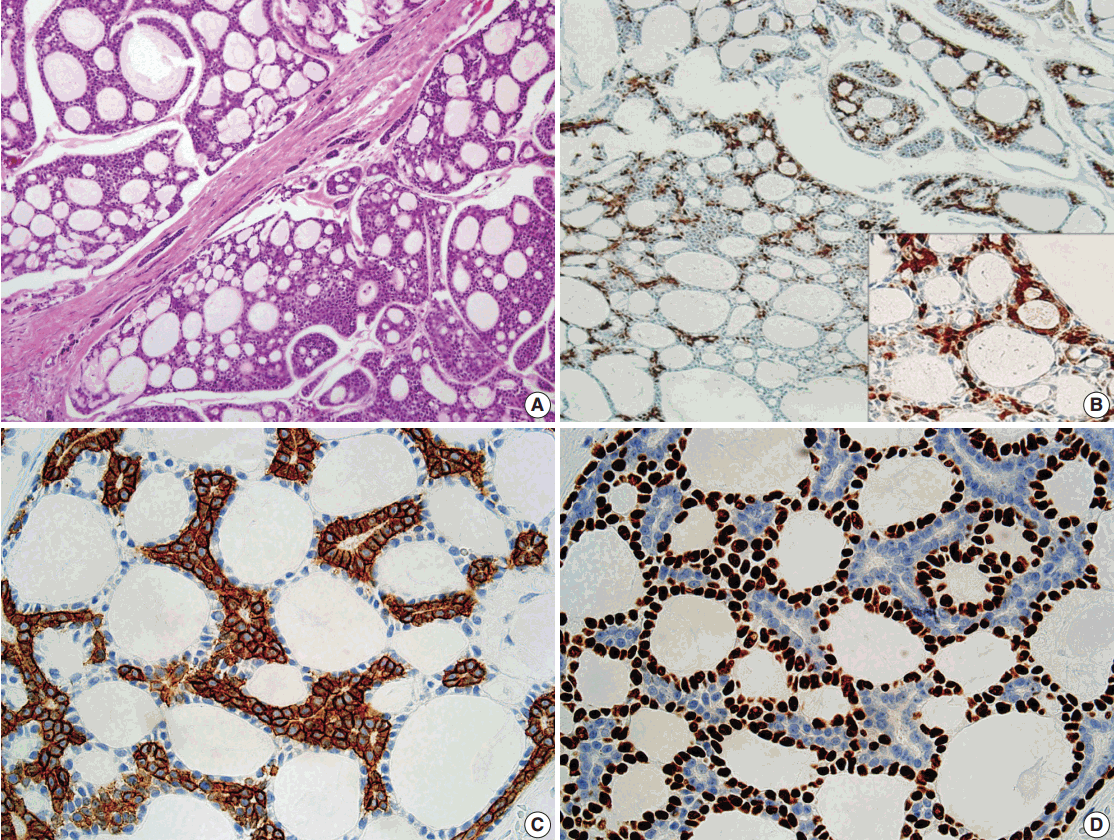
- A 55-year-old male patient presented with a 3.9-cm mass in the right hard palate, which appeared 3 months ago and had increased in size over time. The patient was otherwise healthy without a remarkable medical history. He was a nonsmoker and did not have a history of alcohol or other substance use disorders. Magnetic resonance imaging revealed a lobular contoured mass in the right hard palate, extending to the retromolar trigone and involving the pterygoid muscle and plates. The patient underwent a right inferior maxillectomy. The cut surface of the resected specimen showed a whitish gray and firm mass with irregular margins, measuring 3.9 cm in diameter (Fig. 1). The mass was involving the maxillary bone. Microscopically, the lesion showed poorly circumscribed infiltrates of tumor cells forming a cribriform pattern (Fig. 2A). There was no squamous dysplasia of the overlying surface epithelium. Small to medium bland cells with scant cytoplasm surrounded pseudocysts, and mitotic figures were rarely seen. Perineural invasion was commonly seen, as was venous invasion. Approximately 75% of tumor cells were positive for p16 (RTU, Roche, Branford, CT, USA) (Fig. 2B) on immunohistochemistry. The ductal cells were positive for CD117 (1:400, DAKO, Glostrup, Denmark) (Fig. 2C), whereas the myoepithelial cells were strongly positive for p63 (1:200, DAKO) (Fig. 2D). HPV detection and genotyping were performed using the BMT HPV 9G DNA kit provided by Biometrix Technology Inc. (Chuncheon, Korea). DNA was isolated from the tissue using the DNA extraction kit, and amplification was performed with a polymerase chain reaction mixture consisting of extracted target DNA, the primer set, and polymerase chain reaction premix. Hybridization was performed using an HPV DNA chip at room temperature for 30 minutes. HPV DNA hybridization was detected using the DNA Chip Scanner (ScanArray Gx, Perkin-Elmer Life and Analytical Science, Boston, MA, USA). In this case, the tumor tissue DNA was positive for HPV type 16. The resection margins were free of tumor. On the basis of these findings, the histopathological diagnosis was HPV-associated ACC. After discussion with the head and neck multidisciplinary team, the patient underwent postoperative radiation therapy. No recurrence was evident at 6-month follow-up.
CASE REPORT
- HPV has been shown to be etiologically related to the development of 20%–25% of head and neck carcinomas. It has a strong predilection for the oropharynx, as up to 80% of nonkeratinizing oropharyngeal squamous cell carcinomas are related to HPV infection [3]. The likelihood of detecting HPV outside of the oropharynx is very low, especially in the nasal cavity [4]. Using an HPV DNA chip, we report the first case of ACC of the hard palate associated with HPV infection.
- Although p16 protein overexpression can be detected in ACC of the head and neck, the staining pattern of p16 is mostly focal (less than 75% of cells are stained), and the presence of co-existing HPV infection is rare [2,5]. Even though p16 protein overexpression was observed in our case, we used an HPV DNA chip for confirmation.
- The most important differential diagnosis to consider is HPV-related carcinoma with adenoid cystic-like features due to its morphologic and immunohistochemical features. Its morphologic resemblance to ACC is based on architectural patterns, a dual population of cells, and immunohistochemical expression of myoepithelial markers. However, HPV-related carcinoma with adenoid cystic-like features has a predominantly solid growth pattern, and it is associated with squamous dysplasia of the overlying surface epithelium [1]. Furthermore, HPV 33 is the most common high-risk HPV type in HPV-related carcinoma; however, HPV type 16 was detected in our case.
- Because of their overlapping morphological features, basaloid squamous cell carcinoma must be distinguished from ACC. Unlike basaloid squamous cell carcinoma, ACC usually lacks squamous differentiation. The most important differentiating characteristic of ACC is the presence of a myoepithelial cell component. In our case, the tumor has this dual population.
- In summary, we present an HPV-associated ACC of the hard palate. HPV-associated ACC is extremely rare, and only two cases of HPV-related ACC arising in the nasal cavity have been reported. Here, we report the first documented case of ACC in the hard palate with the presence of p16 protein expression and co-existent HPV type 16 infection.
DISCUSSION
Fig. 1.On cut sections, an ill-defined, whitish gray mass (white circle), measuring 3.9 cm in diameter, involves the maxillary bone.
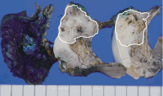
Fig. 2.Microscopic findings of the adenoid cystic carcinoma of the hard palate. (A) Adenoid cystic carcinoma with a cribriform pattern. (B) p16 protein expression is seen in 75% of cells. The ductal cells are positive for CD117 (C), and the myoepithelial cells are positive for p63 (D).
- 1. Hwang SJ, Ok S, Lee HM, Lee E, Park IH. Human papillomavirus-related carcinoma with adenoid cystic-like features of the inferior turbinate: a case report. Auris Nasus Larynx 2015; 42: 53-5. ArticlePubMed
- 2. Boland JM, McPhail ED, Garcia JJ, Lewis JE, Schembri-Wismayer DJ. Detection of human papilloma virus and p16 expression in high-grade adenoid cystic carcinoma of the head and neck. Mod Pathol 2012; 25: 529-36. ArticlePubMedPDF
- 3. Bishop JA, Guo TW, Smith DF, et al. Human papillomavirus-related carcinomas of the sinonasal tract. Am J Surg Pathol 2013; 37: 185-92. ArticlePubMedPMC
- 4. Bishop JA, Ogawa T, Stelow EB, et al. Human papillomavirus-related carcinoma with adenoid cystic-like features: a peculiar variant of head and neck cancer restricted to the sinonasal tract. Am J Surg Pathol 2013; 37: 836-44. PubMedPMC
- 5. Maruya S, Kurotaki H, Shimoyama N, Kaimori M, Shinkawa H, Yagihashi S. Expression of p16 protein and hypermethylation status of its promoter gene in adenoid cystic carcinoma of the head and neck. ORL J Otorhinolaryngol Relat Spec 2003; 65: 26-32. ArticlePubMedPDF
REFERENCES
Figure & Data
References
Citations
Citations to this article as recorded by 
- Adenoid Cystic Carcinoma of the Skull Base: Response to Radiation Therapy and Outcomes in a Retrospective Case Series
Shekhar K. Gadkaree, Anuraag S. Parikh, Alejandro I. Rodarte, Ashton Lehmann, Stacey T. Gray, Derrick T. Lin
Journal of Neurological Surgery Part B: Skull Base.2020; 81(05): 505. CrossRef
 PubReader
 ePub Link
-
 Cite this Article
- Cite this Article
-
- Close
- Download Citation
- Close
- Figure
-


Human Papillomavirus Infection–Associated Adenoid Cystic Carcinoma of the Hard Palate


Fig. 1. On cut sections, an ill-defined, whitish gray mass (white circle), measuring 3.9 cm in diameter, involves the maxillary bone.
Fig. 2. Microscopic findings of the adenoid cystic carcinoma of the hard palate. (A) Adenoid cystic carcinoma with a cribriform pattern. (B) p16 protein expression is seen in 75% of cells. The ductal cells are positive for CD117 (C), and the myoepithelial cells are positive for p63 (D).
Fig. 1.
Fig. 2.
Human Papillomavirus Infection–Associated Adenoid Cystic Carcinoma of the Hard Palate

