Articles
- Page Path
- HOME > J Pathol Transl Med > Volume 52(1); 2018 > Article
-
Brief Case Report
Hyalinizing Cholecystitis and Associated Carcinoma: A Case Report - Youngjin Kang, Yang-Seok Chae, Chul Hwan Kim, Youngseok Lee, Dong-Sik Kim1, Young-Dong Yu1, Joo Young Kim
-
Journal of Pathology and Translational Medicine 2018;52(1):64-66.
DOI: https://doi.org/10.4132/jptm.2016.11.04
Published online: April 4, 2017
Department of Pathology, Korea University Anam Hospital, Korea University College of Medicine, Seoul, Korea
1Department of Surgery, Korea University Anam Hospital, Korea University College of Medicine, Seoul, Korea
- Corresponding Author Joo Young Kim, MD, PhD Department of Pathology, Korea University Anam Hospital, Korea University College of Medicine, 73 Inchon-ro, Seongbuk-gu, Seoul 02841, Korea Tel: +82-2-920-6268, Fax: +82-2-920-6576, E-mail: lepetit80@hanmail.net
• Received: September 2, 2016 • Revised: October 31, 2016 • Accepted: November 3, 2016
© 2018 The Korean Society of Pathologists/The Korean Society for Cytopathology
This is an Open Access article distributed under the terms of the Creative Commons Attribution Non-Commercial License (http://creativecommons.org/licenses/by-nc/4.0) which permits unrestricted non-commercial use, distribution, and reproduction in any medium, provided the original work is properly cited.
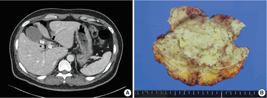
- A 54-year-old man visited the hospital due to fatigue, pruritus, and diarrhea. Laboratory findings revealed elevations in aspartate aminotransferase (98 IU/L; reference range, 3 to 45 IU/L), alanine transaminase (232 IU/L; reference range, 3 to 45 IU/L), alkaline phosphatase (320 IU/L; reference range, 30 to 120 IU/L), and gamma-glutamyl transpeptidase (619 IU/L; reference range, 9 to 64 IU/L). Biliary computed tomography scan and magnetic resonance cholangiography revealed enhancing wall thickening of the cystic duct and common hepatic duct and multiple calcified gallstones as well as sludge in the gallbladder with mild wall thickening (Fig. 1A). Radiologically, carcinoma of the primary bile duct or cystic duct and associated chronic calculous cholecystitis was suspected, and bile duct resection with cholecystectomy was performed.
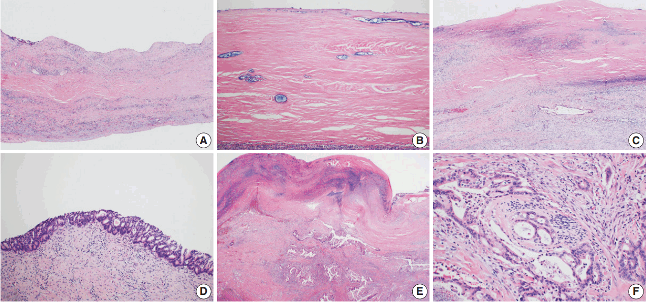
- An 8.0 × 5.5 × 5.5 cm gallbladder was submitted for intraoperative frozen diagnosis. Grossly, the gallbladder wall was diffusely fibrotic, and the mucosal surface was covered by yellowish necrotic material with a gallstone (2.0 × 1.2 × 1.0 cm). There were no mass-like lesions (Fig. 1B). Two random frozen sections were examined microscopically, and an inflammatory lesion without malignancy was suggested. After the operation, we also received the segmentally resected common hepatic duct (1.7 cm in length, 0.6 cm in diameter) and the attached cystic duct (1.7 cm in length, 0.4 cm in diameter). The bile duct wall was diffusely thickened; however, the mucosa of the ducts was smooth and did not form mass-like lesions. The entire permanent sections of the gallbladder and bile ducts were examined microscopically. The gallbladder wall was nearly completely replaced by dense lamellated eosinophilic hyaline material, with multifocal neutrophilic and lymphoplasmacytic infiltration. Focal intramural calcifications were identified (Fig. 2A, B). Multifocal invasive glands with irregular borders were sparsely scattered in the hyalinized gallbladder wall in a longitudinal arrangement (Fig. 2C). The mucosal surface of the gallbladder showed extensive denudation of the epithelium, but some foci of high-grade biliary intraepithelial neoplasia were noted (Fig. 2D). There were focal areas composed of clusters of invasive glands in the gallbladder wall (Fig. 2E). The invasive glands were composed of stratified depolarized atypical cells with enlarged hyperchromatic nuclei containing coarse chromatin and occasionally prominent nucleoli. Some glands also showed perineural invasion (Fig. 2F). Diffuse involvement of the walls of the common hepatic duct and cystic duct by adenocarcinoma was observed (Fig. 3A, B); however, the mucosa was relatively spared, with no intraepithelial lesions (Fig. 3C). Considering all of these findings, we made the diagnosis of HC and associated adenocarcinoma, with direct invasion of the cystic duct and common hepatic duct.
- This study was approved by the Institutional Review Board of Korea University Anam Hospital (ED17246), and informed consent was waived.
CASE REPORT
- HC is a recently described, distinctive entity, comprising 1.6% of cholecystectomy specimens [1]. HC is characterized by diffuse and dense hyalinization of the gallbladder wall with complete effacement of normal histologic components. Focal to extensive calcification may also be seen. HC is more often associated with carcinoma; the frequency of carcinoma was 15% compared to 4% of all cholecystectomy specimens in a South American cohort [1]. However, to identify the carcinoma component is diagnostically challenging because it does not form distinct mass lesions or induce significant thickening of the gallbladder wall [1]. Furthermore, the malignant glands are not easily found due to the paucity and sparse distribution of tumor glands within an abundant hyalinizing sclerotic background. In our case, there were no mass-like lesions, and the entire gallbladder wall was diffusely fibrotic with no significant wall thickening. The carcinoma component was not found in two randomly submitted frozen sections; however, it was identified after extensive sampling of permanent sections. Therefore, careful examination with extensive or entire sampling of the gallbladder is crucial in HC to identify associated carcinoma.
- Preoperative diagnosis of HC-related carcinoma is also highly challenging. Patel et al. [1] reported that about 70% of malignancies had been diagnosed as benign preoperatively. This might be because HC-associated carcinomas typically do not form a mass or result in thickening of the gallbladder. Subsequently, surgical resection can be delayed until progression of the malignancy. In our case, the malignancy was identified radiologically after the tumor directly extended to the extrahepatic bile duct, and the preoperative diagnosis was primary bile duct cancer with chronic calculous cholecystitis. The final pathologic stage was T3 (direct invasion of the extrahepatic bile ducts). The late discovery of carcinoma is associated with advanced stage at diagnosis and poor prognosis of HC-related carcinomas. The median survival for HC-associated carcinoma is shorter than that of usual carcinomas [1].
- In conclusion, HC is a rare variant of chronic cholecystitis and is often associated with carcinoma with aggressive behavior. Suspicion of the possibility of HC and identification of HC as an unusual variant of chronic cholecystitis are important in gross examination of cholecystectomy specimens. After the identification of HC, extensive sampling and meticulous microscopic examination are essential to determine the possibility of associated carcinoma. In addition, close follow-up is recommended because carcinoma associated with HC can have more aggressive behavior than typical gallbladder cancer.
DISCUSSION
Fig. 1.Computed tomography and gross findings of hyalinizing cholecystitis and associated carcinoma. (A) Biliary computed tomography scan reveals subtle enhancement of wall thickening at the confluence of the cystic duct and the common hepatic duct (arrow). There is mild wall thickening of the gallbladder with multiple calcified gallstones in the neck portion (arrowhead). (B) Grossly, the gallbladder wall shows diffuse fibrosis and is covered by yellowish necrotic materials, without mass-like lesions.


Fig. 2.Microscopic findings of hyalinizing cholecystitis and associated carcinoma. (A, B) The gallbladder wall is replaced by dense lamellated eosinophilic hyaline material. Inflammatory cells are also seen. (C) A few invasive glands are longitudinally arranged in the hyalinized gallbladder wall with a denuded epithelium. (D) Multifocal carcinoma in situ lesions are found on the surface. (E) Focal clusters of invasive glands are identified in the hyalinized wall. (F) Invasive glands have irregular borders and cytologic atypia with perineural invasion.


Fig. 3.Microscopic findings of the common hepatic and cystic ducts. (A, B) Invasive glands infiltrate the common hepatic and cystic ducts beneath the surface epithelium. (C) The surface epithelium is relatively spared, with no intraepithelial lesion.


- 1. Patel S, Roa JC, Tapia O, et al. Hyalinizing cholecystitis and associated carcinomas: clinicopathologic analysis of a distinctive variant of cholecystitis with porcelain-like features and accompanying diagnostically challenging carcinomas. Am J Surg Pathol 2011; 35: 1104-13. PubMed
- 2. Kane RA, Jacobs R, Katz J, Costello P. Porcelain gallbladder: ultrasound and CT appearance. Radiology 1984; 152: 137-41. ArticlePubMed
- 3. Ochsner SF, Carrera GM. Calcification of the gallbladder (“porcelain gallbladder”). Am J Roentgenol Radium Ther Nucl Med 1963; 89: 847-53. PubMed
- 4. Weiner PL, Lawson TL. The radiology corner: porcelain gallbladder. Am J Gastroenterol 1975; 64: 224-7. PubMed
- 5. Shimizu M, Miura J, Tanaka T, Itoh H, Saitoh Y. Porcelain gallbladder: relation between its type by ultrasound and incidence of cancer. J Clin Gastroenterol 1989; 11: 471-6. PubMed
- 6. Stephen AE, Berger DL. Carcinoma in the porcelain gallbladder: a relationship revisited. Surgery 2001; 129: 699-703. ArticlePubMed
- 7. Towfigh S, McFadden DW, Cortina GR, et al. Porcelain gallbladder is not associated with gallbladder carcinoma. Am Surg 2001; 67: 7-10. ArticlePubMedPDF
- 8. Gupta RK, Patton KT. Hyalinizing cholecystitis with features of immunoglobulin G4-related disease-coincidence or an unrecognized association? A case report. Hum Pathol 2015; 46: 625-8. ArticlePubMed
REFERENCES
Figure & Data
References
Citations
Citations to this article as recorded by 

- Unexpected intraoperative finding of a hyalinizing cholecystitis in a patient with gallbladder calculi
Klaudia Gjinoska, Andrej Nikolovski, Emil Stoicovski, Zan Mitrev
Journal of Surgical Case Reports.2024;[Epub] CrossRef - 18F-FDG PET/CT in Hyalinized Cholecystitis
Esra Arslan, Aytül Hande Yardimci, Enver Yarikkaya, Göksel Alçin, Tevfik Fikret Çermik
Clinical Nuclear Medicine.2021; 46(4): e228. CrossRef - Gallbladder Carcinoma and Its Differential Diagnosis at MRI: What Radiologists Should Know
Camila Lopes Vendrami, Michael J. Magnetta, Pardeep K. Mittal, Courtney C. Moreno, Frank H. Miller
RadioGraphics.2021; 41(1): 78. CrossRef - A simple method for diagnosing gallbladder malignant tumors with subserosa invasion by endoscopic ultrasonography
Mitsuru Sugimoto, Hiroki Irie, Mika Takasumi, Minami Hashimoto, Yuka Oka, Tadayuki Takagi, Rei Suzuki, Naoki Konno, Hiroyuki Asama, Yuki Sato, Jun Nakamura, Tsunetaka Kato, Ryoichiro Kobashi, Yuko Hashimoto, Shigeru Marubashi, Takuto Hikichi, Hiromasa Ohi
BMC Cancer.2021;[Epub] CrossRef - KRONİK KOLESİSTİTİN NADİR VE MALİGNİTE POTANSİYELİ YÜKSEK OLAN ALT TİPİ; HYALİNİZE KOLESİSTİT: OLGU SUNUMU
Leymune PARLAK, Bahar MEMİŞ
Balıkesir Medical Journal.2019; 3(3): 129. CrossRef
 PubReader
 ePub Link
-
 Cite this Article
- Cite this Article
-
- Close
- Download Citation
- Close
- Figure
Hyalinizing Cholecystitis and Associated Carcinoma: A Case Report


Fig. 1. Computed tomography and gross findings of hyalinizing cholecystitis and associated carcinoma. (A) Biliary computed tomography scan reveals subtle enhancement of wall thickening at the confluence of the cystic duct and the common hepatic duct (arrow). There is mild wall thickening of the gallbladder with multiple calcified gallstones in the neck portion (arrowhead). (B) Grossly, the gallbladder wall shows diffuse fibrosis and is covered by yellowish necrotic materials, without mass-like lesions.
Fig. 2. Microscopic findings of hyalinizing cholecystitis and associated carcinoma. (A, B) The gallbladder wall is replaced by dense lamellated eosinophilic hyaline material. Inflammatory cells are also seen. (C) A few invasive glands are longitudinally arranged in the hyalinized gallbladder wall with a denuded epithelium. (D) Multifocal carcinoma in situ lesions are found on the surface. (E) Focal clusters of invasive glands are identified in the hyalinized wall. (F) Invasive glands have irregular borders and cytologic atypia with perineural invasion.
Fig. 3. Microscopic findings of the common hepatic and cystic ducts. (A, B) Invasive glands infiltrate the common hepatic and cystic ducts beneath the surface epithelium. (C) The surface epithelium is relatively spared, with no intraepithelial lesion.
Fig. 1.
Fig. 2.
Fig. 3.
Hyalinizing Cholecystitis and Associated Carcinoma: A Case Report

 E-submission


