E-submission
Articles
- Page Path
- HOME > J Pathol Transl Med > Forthcoming articles > Article
-
Case Study
Gastric amphicrine carcinoma in the stomach: an unexpected presentation of MUTYH-associated polyposis -
Seungkyun Lee1
 , Byung-Hoon Min2, Young-Gon Kim3, Soomin Ahn1, Kyoung-Mee Kim1
 -
DOI: https://doi.org/10.4132/jptm.2026.06.24
Published online: September 7, 2026
1Department of Pathology and Translational Genomics, Samsung Medical Center, Sungkyunkwan University School of Medicine, Seoul, Korea
2Department of Medicine, Samsung Medical Center, Sungkyunkwan University School of Medicine, Seoul, Korea
3Department of Laboratory Medicine and Genetics, Samsung Medical Center, Sungkyunkwan University School of Medicine, Seoul, Korea
- Corresponding Author: Kyoung-Mee Kim, MD, PhD Department of Pathology and Translational Genomics, Samsung Medical Center, Sungkyunkwan University School of Medicine, 81 Irwon-ro, Gangnam-gu, Seoul 06351, Korea Tel: +82-2-3410-2768, Fax: +82-2-3410-0025, E-mail: kkmkys@skku.edu
© The Korean Society of Pathologists/The Korean Society for Cytopathology
This is an Open Access article distributed under the terms of the Creative Commons Attribution Non-Commercial License (https://creativecommons.org/licenses/by-nc/4.0) which permits unrestricted non-commercial use, distribution, and reproduction in any medium, provided the original work is properly cited.
- 797 Views
- 12 Download


Abstract
- Amphicrine carcinomas of the stomach, defined by dual exocrine and neuroendocrine differentiation within the same neoplastic cell, are exceedingly rare. MUTYH-associated polyposis (MAP) is an autosomal recessive polyposis syndrome characterized by multiple colorectal adenomas and variable upper gastrointestinal involvement; however, amphicrine carcinomas have not been previously documented in this setting. We report a gastric amphicrine carcinoma arising in the background of extensive fundic gland polyposis in a patient with MAP. Endoscopy revealed a 3.5-cm flat elevated lesion in the gastric fundus amid extensive fundic gland polyposis. Histologically, the tumor consisted of a single population of cells exhibiting combined glandular and neuroendocrine differentiation without zonal or biphasic architecture, and many of these cells demonstrated true amphicrine morphology. Immunohistochemistry confirmed co-expression of cytokeratin and the neuroendocrine markers chromogranin A and synaptophysin in the same cell population. Germline targeted next-generation sequencing identified biallelic MUTYH variants in trans (c.733C>T, p.Arg245Cys [likely pathogenic]; c.842C>T, p.Ala281Val [variant of uncertain significance]), supporting a diagnosis of MAP. To our knowledge, this is the first reported case of a gastric amphicrine carcinoma in a MAP patient, expanding the spectrum of MAP-associated upper gastrointestinal neoplasia and underscoring the importance of vigilant endoscopic surveillance in hereditary polyposis syndromes.
- Amphicrine carcinomas are rare epithelial neoplasms characterized by simultaneous exocrine and neuroendocrine differentiation within individual tumor cells [1]. This distinguishes them from mixed neuroendocrine–non-neuroendocrine neoplasms (MiNEN), in which the two components are morphologically and immunophenotypically distinct [2].
- MUTYH-associated polyposis (MAP) results from biallelic pathogenic variants in MUTYH, a base excision repair gene [3,4]. MAP typically manifests with tens to hundreds of colorectal adenomas and an increased risk of colorectal carcinoma [4]. Upper gastrointestinal lesions—including duodenal adenomas and fundic gland polyps—are recognized, but gastric carcinoma is uncommon [5], and amphicrine carcinomas have not been previously reported in this context.
- Herein, we describe the first documented case of a gastric amphicrine carcinoma arising in a patient with MAP. This case broadens the known phenotypic range of MAP.
INTRODUCTION
- Clinical features
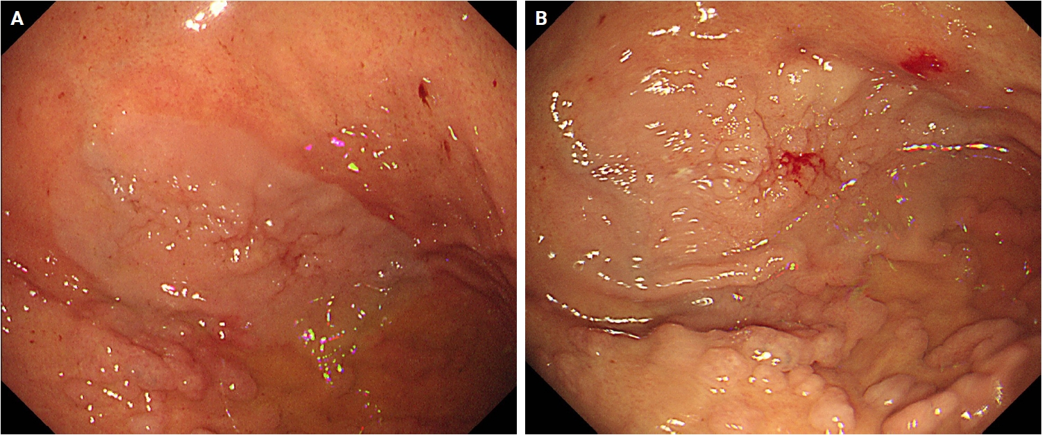
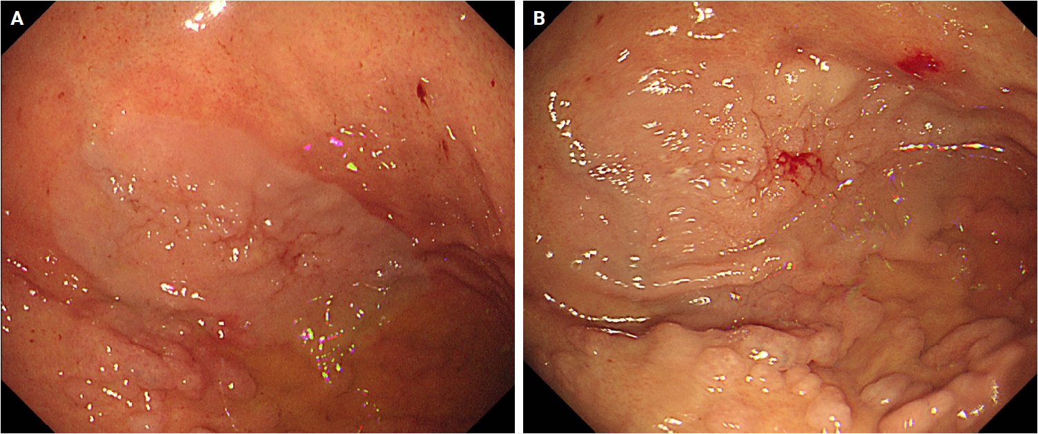
- A patient undergoing surveillance endoscopy for extensive gastrointestinal polyposis was evaluated. Colonoscopy revealed approximately 100 colorectal adenomas. Upper endoscopy demonstrated extensive gastric polyposis, predominantly fundic gland polyps numbering around 100 (Fig. 1B). A 3.5-cm whitish flat elevated lesion was identified on the anterior wall of the gastric fundus (Fig. 1A), and endoscopic biopsy was performed.
- Pathological findings
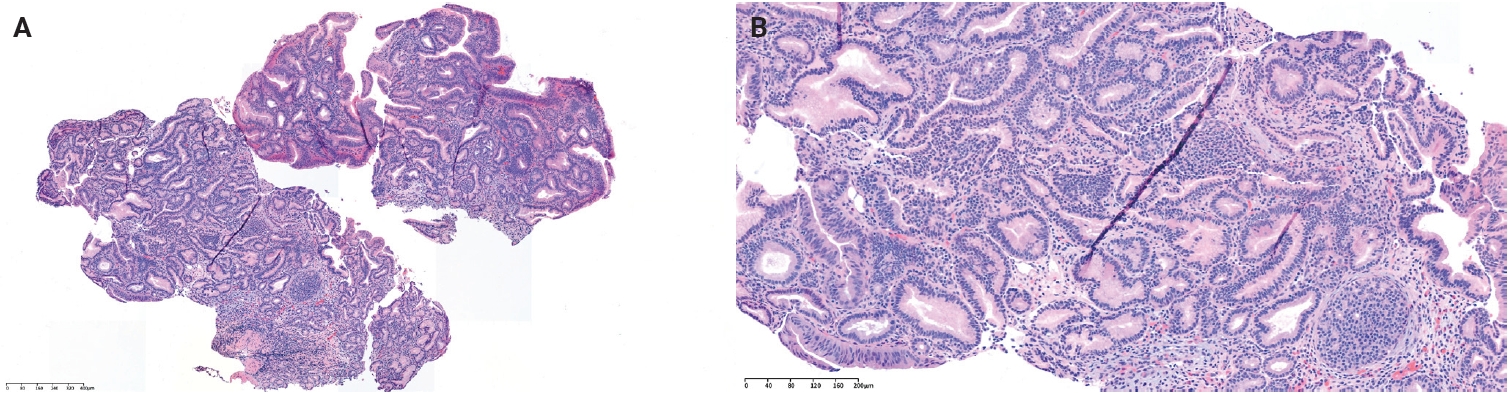
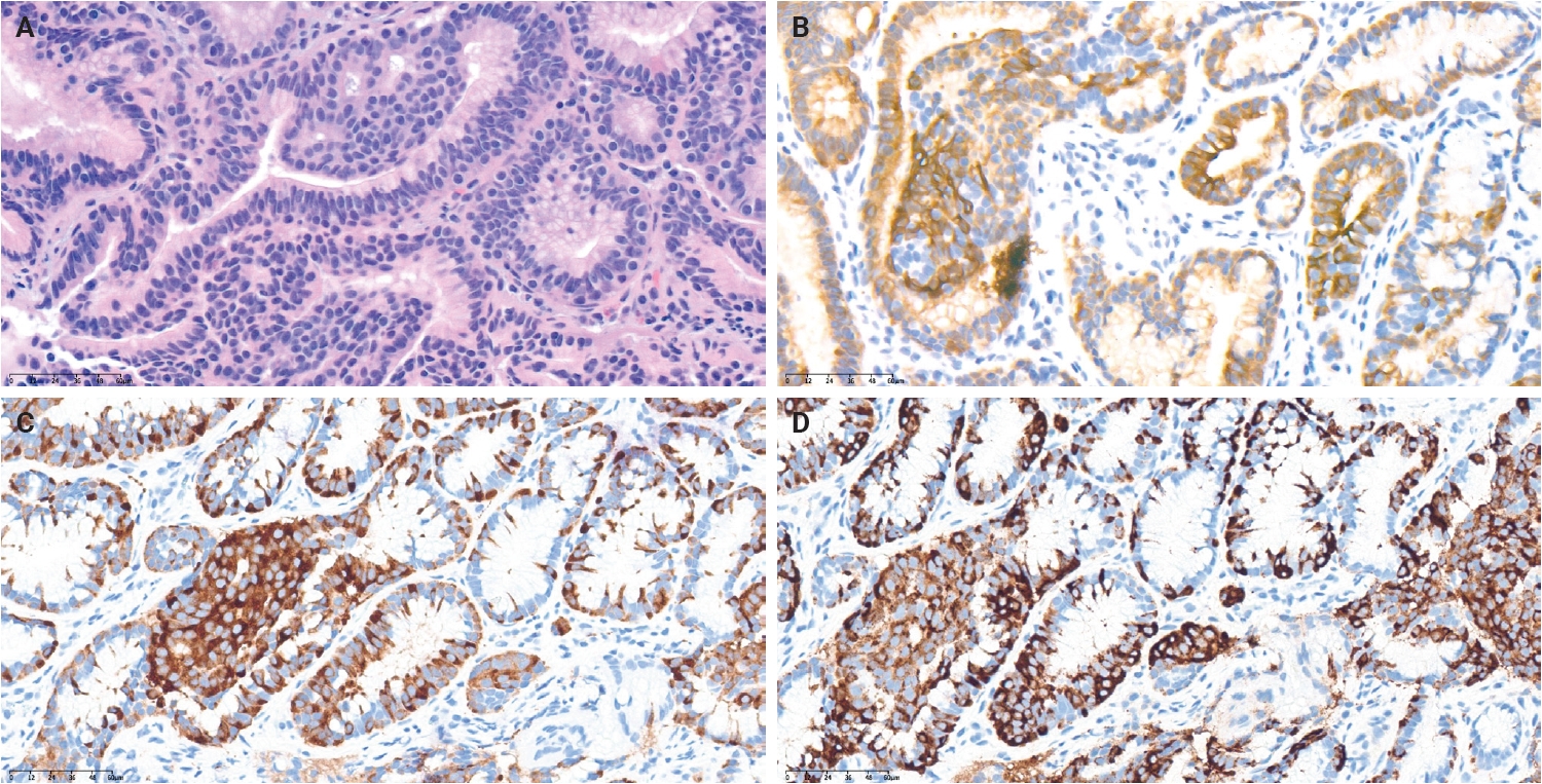
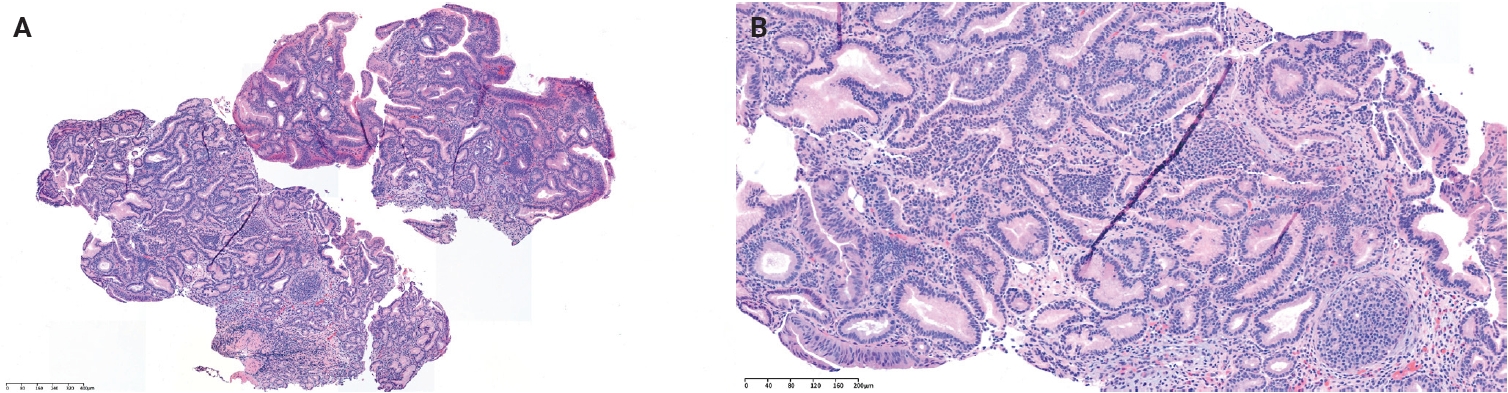
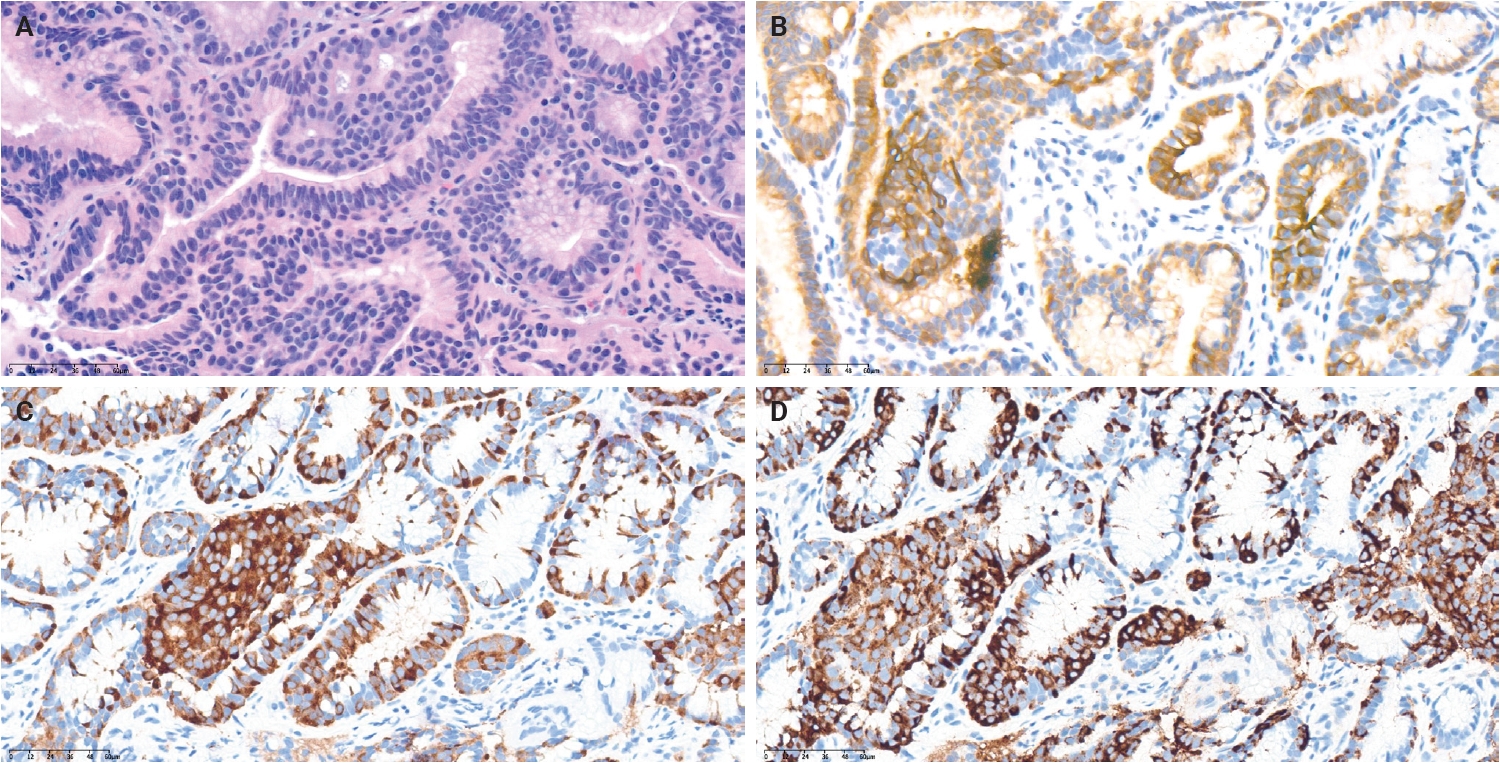
- The lesion arose in a background of extensive fundic gland polyposis. Microscopic examination revealed a carcinoma composed of a single, relatively uniform population of cells arranged in tubular, cord-like, and trabecular patterns, without a zonal or biphasic separation into distinct glandular and neuroendocrine areas (Fig. 2A). Individual tumor cells concurrently exhibited glandular differentiation, including lumen formation, and neuroendocrine cytologic features, such as round nuclei, finely granular (“salt-and-pepper”) chromatin, and eosinophilic cytoplasm, indicating amphicrine differentiation at the single-cell level (Figs. 2B, 3A). On immunohistochemistry, the tumor cells showed diffuse cytokeratin 7 positivity (Fig. 3B) together with strong, diffuse co-expression of synaptophysin (Fig. 3C) and chromogranin A (Fig. 3D) in the same cell population. The Ki-67 proliferation index was 2%.
- Genetic analysis
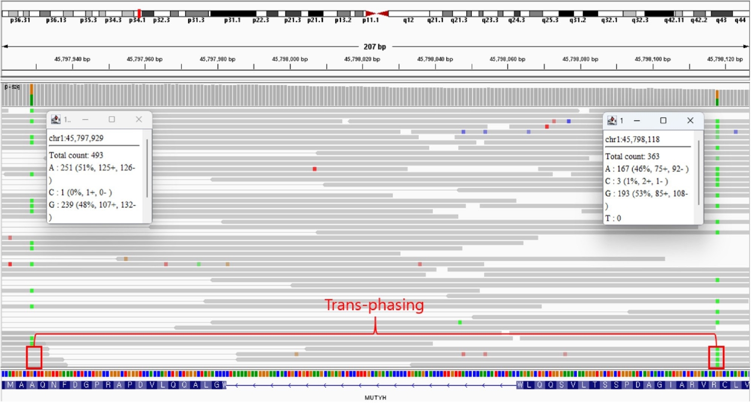
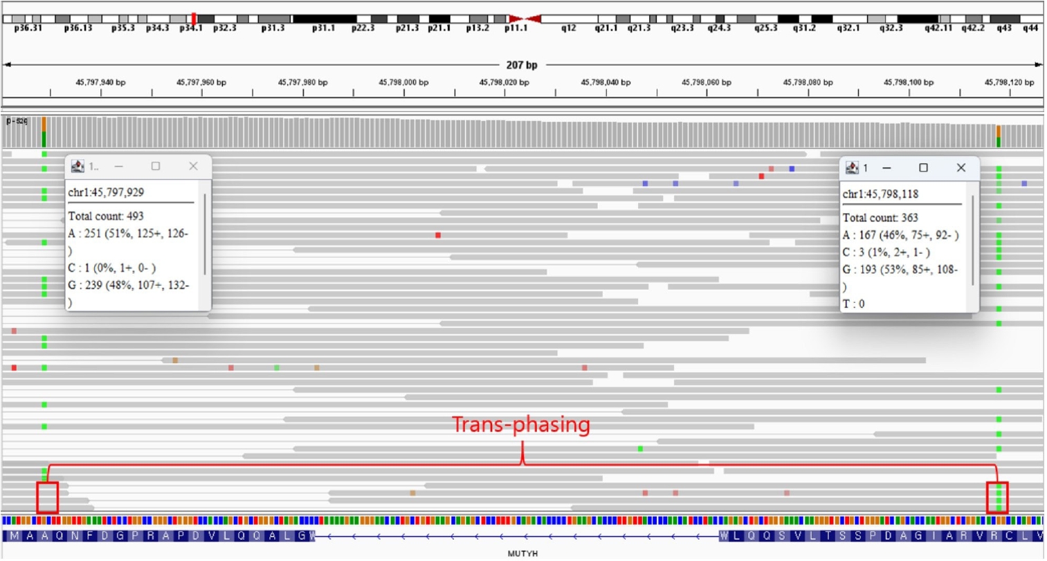
- Germline targeted next-generation sequencing (NGS) was performed using genomic DNA isolated from peripheral-blood leukocytes, establishing the constitutional (germline) nature of the variants. Using a hybridization capture-based enrichment method (NextSeq-G1V v2.0) on the Illumina NextSeq platform (reference genome hg19/GRCh37), two heterozygous MUTYH variants were identified, with allele fractions consistent with germline heterozygosity. The first was a likely pathogenic variant in NM_001128425.1 (MUTYH) exon 10, c.733C>T (p.Arg245Cys); the second was a variant of uncertain significance in exon 11, c.842C>T (p.Ala281Val). Trans-phasing analysis of the NGS data confirmed that the two variants reside on different alleles, establishing biallelic (compound heterozygous) MUTYH involvement (Fig. 4). Together with the patient’s polyposis phenotype (approximately 100 colorectal adenomas), these germline findings support the diagnosis of MAP; the uncertain significance of the p.Ala281Val variant is acknowledged. The patient’s recovery was uneventful, and follow-up is ongoing.
CASE REPORT
- This case represents the first reported gastric amphicrine carcinoma in a patient with MAP. Amphicrine carcinomas are rare and biologically distinct from MiNEN, as they exhibit hybrid differentiation at the single-cell level [1,2]. Their pathogenesis remains poorly understood. MAP results from defective base-excision repair, leading to oxidative DNA damage accumulation [3,4]. Whether biallelic MUTYH inactivation contributes mechanistically to amphicrine differentiation is unknown; based on a single case, this co-occurrence may be incidental, and no causal relationship can be inferred. The germline nature of the biallelic MUTYH variants was established by sequencing peripheral-blood leukocyte DNA. The c.733C>T (p.Arg245Cys) allele has been reported as pathogenic in MAP, whereas p.Ala281Val currently remains a variant of uncertain significance; their presence in trans, together with the patient’s marked polyposis phenotype, is consistent with a diagnosis of MUTYH-associated polyposis, although the uncertain significance of the second variant is acknowledged as a limitation.
- As seen in other hereditary cancer syndromes such as the fibroblastic reticular cell tumor case associated with Lynch syndrome, where “whole-exome sequencing revealed 631 putative somatic mutations” and uncovered an unexpected tumor association, this case highlights how molecular testing can reveal novel tumor phenotypes linked to germline predisposition [6].
- Given the rarity of gastric carcinoma in MAP and the absence of prior reports of amphicrine carcinomas, this case expands the recognized spectrum of MAP-associated upper gastrointestinal neoplasia. It also underscores the importance of meticulous endoscopic surveillance in hereditary polyposis syndromes.
- To our knowledge, this is the first reported case of a gastric amphicrine carcinoma arising in a patient with MUTYH-associated polyposis. This case broadens the phenotypic spectrum of MAP and highlights the value of integrating histopathology, immunophenotyping, and germline testing in the evaluation of unusual gastric neoplasms.
DISCUSSION
Ethics Statement
All procedures performed in the current study were approved by the Institutional Review Board (IRB) of the Samsung Medical Center (IRB No. 2026-04-028) in accordance with the 1964 Helsinki Declaration and its later amendments. Formal written informed consent was not required with a waiver by the appropriate IRB.
Availability of Data and Material
The datasets generated or analyzed during the study are available from the corresponding author on reasonable request.
Code Availability
Not applicable.
Author Contributions
Conceptualization: KMK. Investigation: SL. Visualization: SL, BHM, YGK. Writing—original draft: SL. Writing—review & editing: SL, BHM, YGK, SA, KMK. Approval of final manuscript: all authors.
Conflicts of Interest
The authors declare that they have no potential conflicts of interest.
Funding Statement
No funding to declare.
- 1. Jesinghaus M, Schmitt MP, Foersch S, Konukiewitz B. MiNEN, amphicrine carcinomas, and conventional carcinomas with neuroendocrine differentiation: diagnostic criteria, open questions, and future perspectives. Virchows Arch 2026; 488: 33-43. ArticlePubMedPDF
- 2. La Rosa S, Sessa F, Uccella S. Mixed neuroendocrine-nonneuroendocrine neoplasms (MiNENs): unifying the concept of a heterogeneous group of neoplasms. Endocr Pathol 2016; 27: 284-311. ArticlePubMedPDF
- 3. Sieber OM, Lipton L, Crabtree M, et al. Multiple colorectal adenomas, classic adenomatous polyposis, and germ-line mutations in MYH. N Engl J Med 2003; 348: 791-9. ArticlePubMed
- 4. Nielsen M, Morreau H, Vasen HF, Hes FJ. MUTYH-associated polyposis (MAP). Crit Rev Oncol Hematol 2011; 79: 1-16. ArticlePubMed
- 5. Vogt S, Jones N, Christian D, et al. Expanded extracolonic tumor spectrum in MUTYH-associated polyposis. Gastroenterology 2009; 137: 1976-85. ArticlePubMed
- 6. Lopez LV, Marker DF, Bailey N, et al. Soft tissue fibroblastic reticular cell tumor with whole-exome sequencing findings: an unexpected presentation of Lynch syndrome. AJSP Rev Rep 2019; 24: 288-91. Article
REFERENCES
Figure & Data
References
Citations

 PubReader
 ePub Link
-
 Cite this Article
- Cite this Article
-
- Close
- Download Citation
- Close
 XML Download
- Figure
-