E-submission

Articles
- Page Path
- HOME > J Pathol Transl Med > Forthcoming articles > Article
-
Case Study
Silicone lymphadenopathy of the axilla: a potential for misdiagnosis as metastatic carcinoma on fine-needle aspiration cytology -
Nah Ihm Kim1
 , Hyo-Jae Lee2, Ji Shin Lee1
-
DOI: https://doi.org/10.4132/jptm.2026.09.08
Published online: November 3, 2026
1Department of Pathology, Chonnam National University Medical School, Gwangju, Korea
2Department of Radiology, Chonnam National University Medical School, Gwangju, Korea
- Corresponding Author: Ji Shin Lee, MD Department of Pathology, Chonnam National University Hwasun Hospital, 322 Seoyang-ro, Hwasun-eup, Hwasun 58128, Korea Tel: +82-61-379-7072 Fax: +82-61-379-7079 E-mail: jshinlee@hanmail.net
© The Korean Society of Pathologists/The Korean Society for Cytopathology
This is an Open Access article distributed under the terms of the Creative Commons Attribution Non-Commercial License (https://creativecommons.org/licenses/by-nc/4.0) which permits unrestricted non-commercial use, distribution, and reproduction in any medium, provided the original work is properly cited.
- 52 Views
- 1 Download


Abstract
- Silicone lymphadenopathy is an uncommon but well-recognized complication of silicone implant placement resulting from migration of silicone to regional lymph nodes. In patients with prior breast implantation, silicone lymphadenopathy most commonly involves the axillary lymph nodes. Because axillary lymphadenopathy may reflect malignant disease, including metastatic carcinoma, thorough evaluation to exclude neoplasia is essential. Fine-needle aspiration cytology (FNAC) serves as a valuable initial diagnostic modality in the assessment of axillary lymphadenopathy; however, silicone-induced foreign body reactions within lymph nodes may be misinterpreted as metastatic malignancy when silicone lymphadenopathy is not included in the initial differential diagnosis. Herein, a case of axillary silicone lymphadenopathy initially misdiagnosed as metastatic carcinoma on FNAC is presented. This case highlights the importance of recognizing the distinctive cytomorphologic features of silicone lymphadenopathy, which may be sufficiently characteristic to prompt consideration of prior breast implant history during cytopathologic evaluation.
- Silicone lymphadenopathy is characterized by enlargement of regional lymph nodes secondary to silicone deposition following lymphatic migration of silicone material [1]. Silicone implants have been widely utilized for breast augmentation and post-mastectomy reconstruction [2]. Silicone lymphadenopathy associated with breast implants most commonly involves the axillary lymph nodes [2-11].
- Axillary lymphadenopathy may arise from both benign and malignant etiologies. Fine-needle aspiration cytology (FNAC) is a valuable initial diagnostic modality for excluding neoplastic causes of axillary lymphadenopathy [12]. However, only a limited number of case reports have described the cytologic features of silicone lymphadenopathy in the diagnostic setting [3,4,8]. The characteristic cytologic findings of silicone lymphadenopathy include granulomatous inflammation, numerous histiocytes, and multinucleated giant cells within a lymphoid background. Multiple variably sized vacuoles consistent with silicone are typically identified within the cytoplasm of histiocytes and multinucleated giant cells. Nevertheless, when silicone lymphadenopathy is not considered in the initial differential diagnosis, particularly in the absence of a documented history of silicone breast implants, the condition may be difficult to recognize and may be erroneously interpreted as malignant lymphadenopathy.
- Herein, we report a case of axillary silicone lymphadenopathy initially misdiagnosed as metastatic carcinoma on FNAC. Although a history of prior silicone implantation is not always available to the cytopathologist, the cytologic appearance of silicone lymphadenopathy may be sufficiently characteristic to prompt consideration of this clinical history.
INTRODUCTION
- A 45-year-old woman presented to an outside institution with a palpable mass in the right axillary region. The mass had been initially detected 1 year earlier and had recently increased in size. FNAC was performed at an outside institution and subsequently interpreted at a specialized diagnostic center, yielding a diagnosis of metastatic carcinoma with atypical clear cells containing cytoplasmic vacuoles. She was subsequently referred to our rapid-access oncology clinic for evaluation of metastatic carcinoma of unknown primary origin.
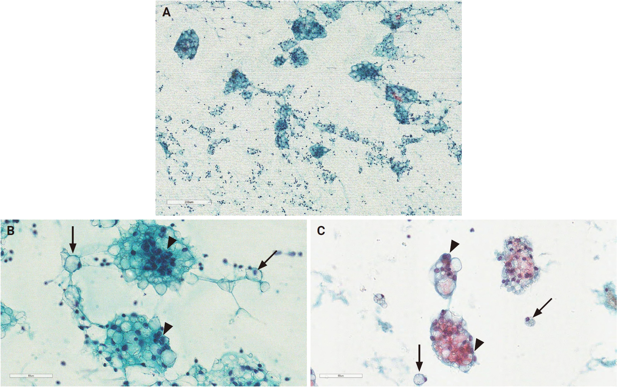
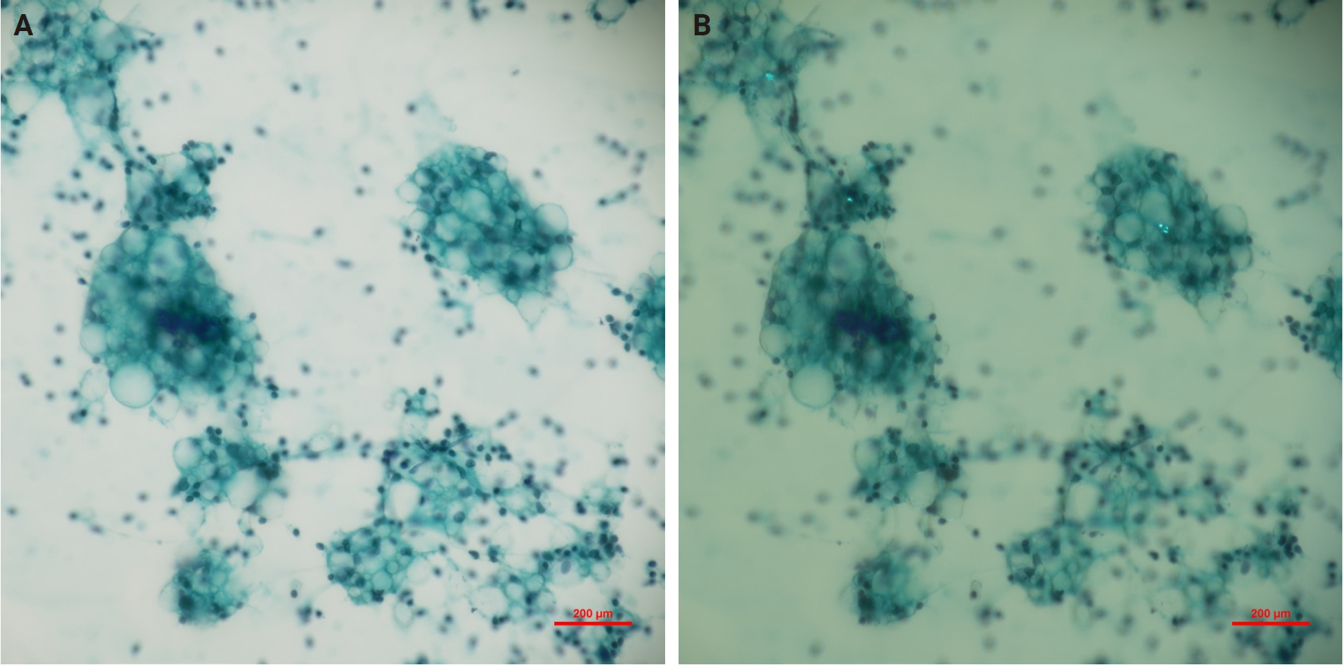
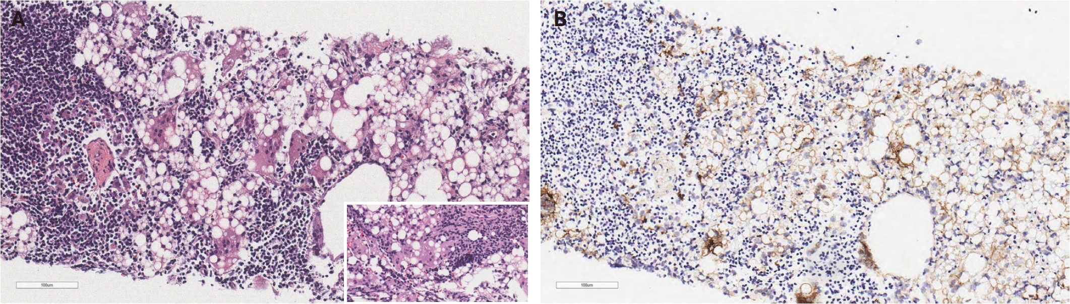
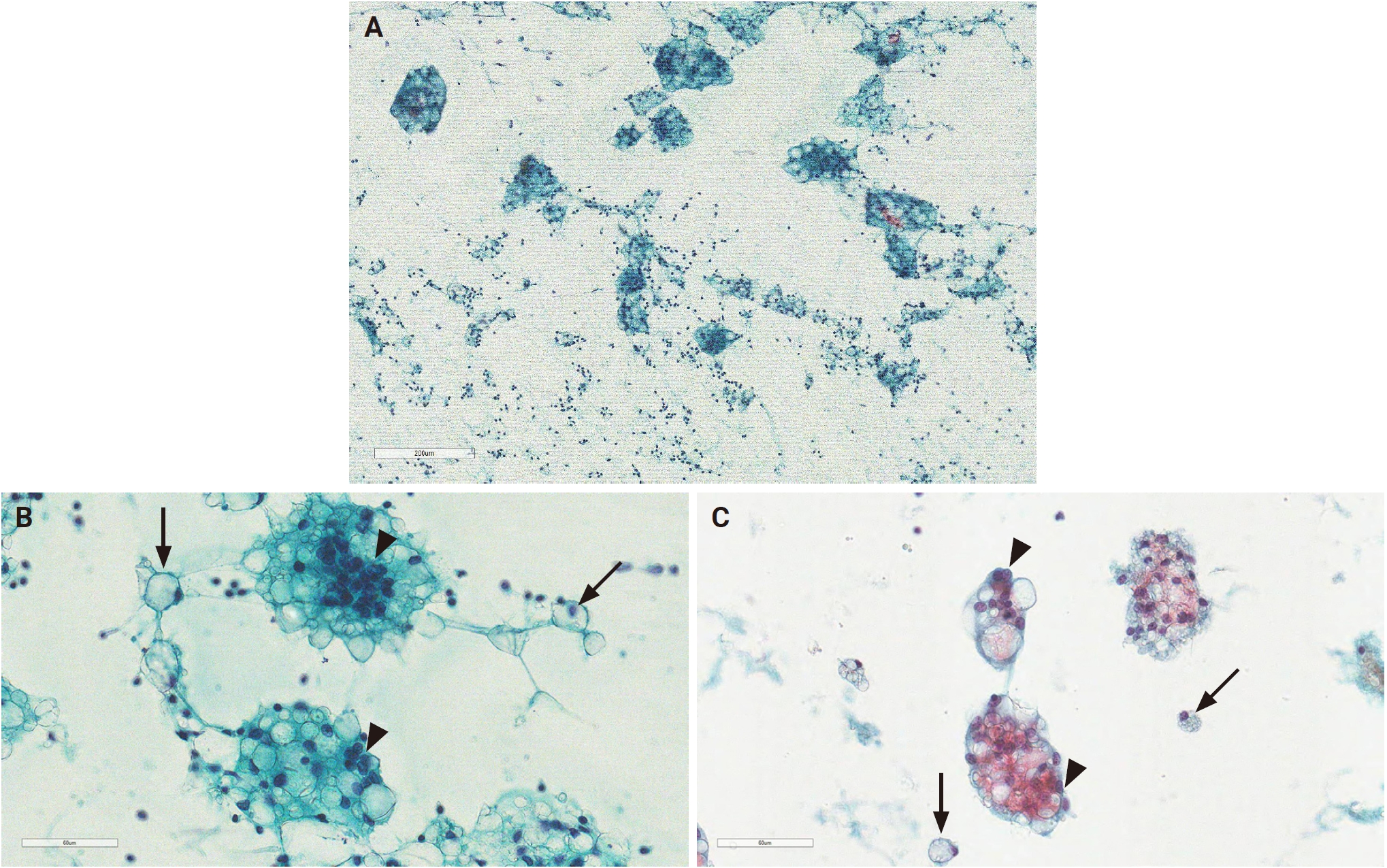
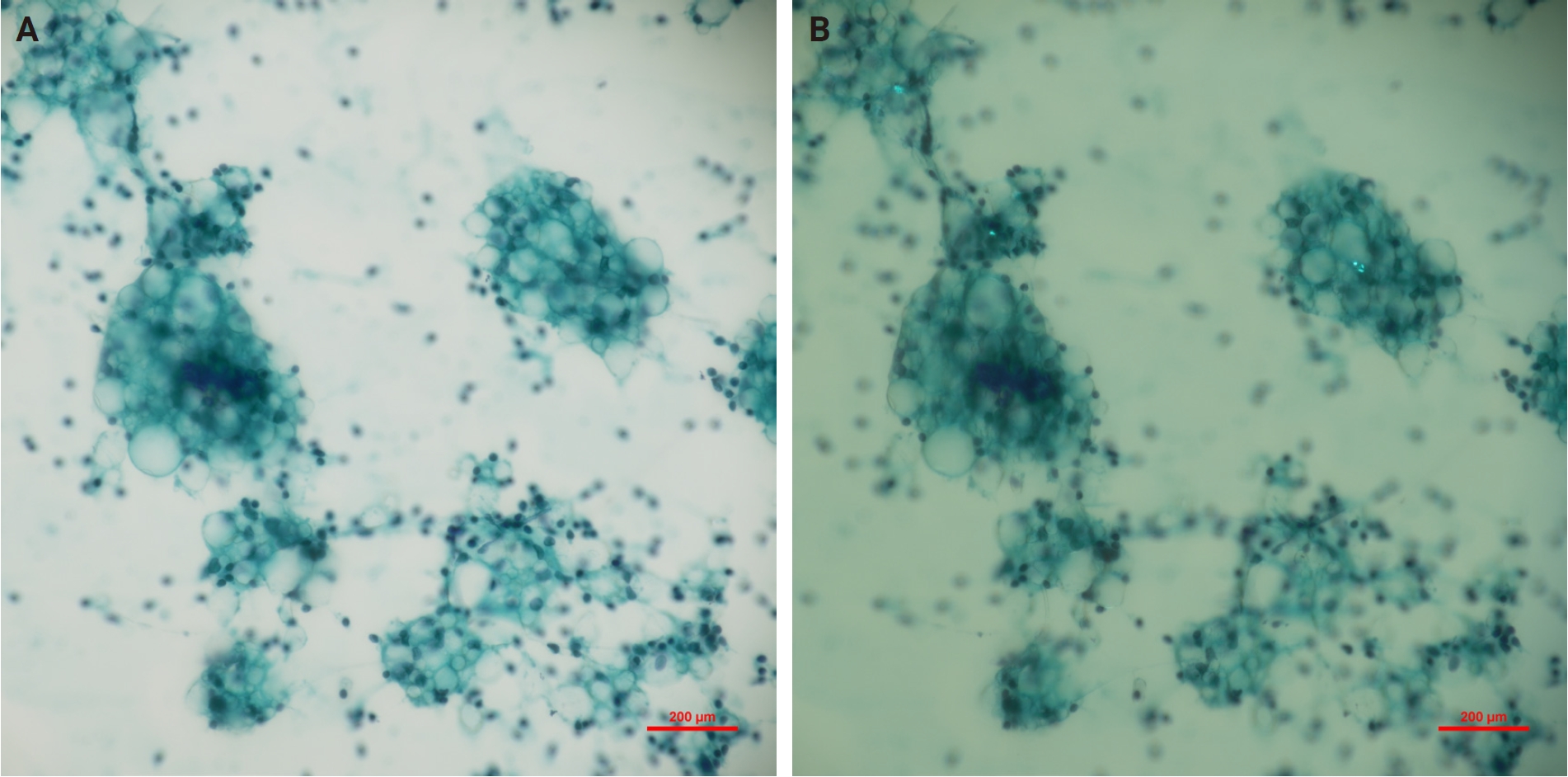
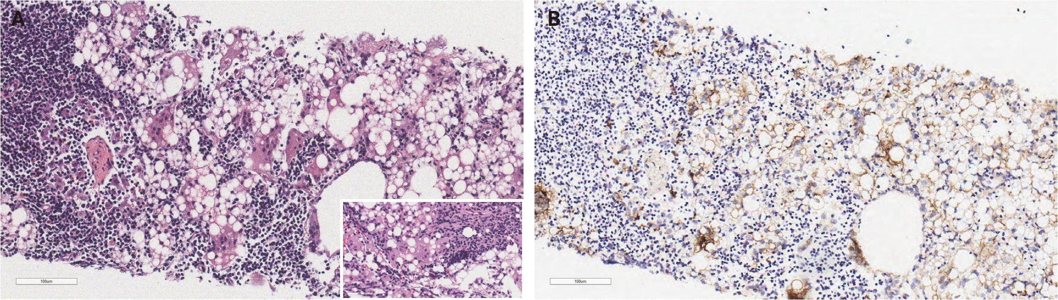
- Ultrasound-guided FNAC and core-needle biopsy of the right axillary lymph node were performed at the initial visit. Cytologic examination revealed several cellular clusters and scattered single cells within a lymphocytic background (Fig. 1A). The cells exhibited round nuclei with inconspicuous nucleoli and finely distributed chromatin. Cytoplasmic microvacuoles compressed the nuclei, imparting a signet ring cell-like or lipoblast-like appearance (Fig. 1B). These vacuoles appeared either empty or contained refractile, colorless material. Similar vacuoles were also observed in the extracellular space. Multinucleated giant cells containing vacuoles of varying sizes (ranging from 5 to 50 µm) were frequently identified. Notably, no birefringent material was observed under polarized microscopy (Fig. 2A, B). There was no evidence of necrosis, cytologic atypia, or mitotic activity. These cytologic findings were suspicious of silicone lymphadenopathy. A retrospective review of the FNAC slides from the specialized diagnostic center revealed cytomorphological features fundamentally similar to those observed at our institution. The specimen exhibited cells with signet ring cell features, characterized by intracytoplasmic vacuoles that eccentrically displaced the nuclei, mimicking metastatic signet ring cell carcinoma (Fig. 1C). However, the specimen lacked a mucinous, bloody, or necrotic background, and neither distinct irregular nuclear membranes nor prominent nucleoli were observed. Furthermore, multinucleated giant cells containing numerous, variably sized vacuoles were observed, along with scattered extracellular vacuoles. Core biopsy demonstrated lymph node tissue containing epithelioid cells with large vacuoles, scattered lymphocytes, and multinucleated giant cells. Asteroid bodies were also identified within the cytoplasm of giant cells (Fig. 3A). The histiocytic nature of the epithelioid cells and multinucleated giant cells was confirmed by CD68 positivity (Fig. 3B). The empty spaces were interpreted as vacuoles containing silicone-type material. These histologic findings were diagnostic of silicone lymphadenopathy. Upon further inquiry, the patient confirmed a history of bilateral cosmetic breast augmentation with silicone prostheses 10 years earlier.
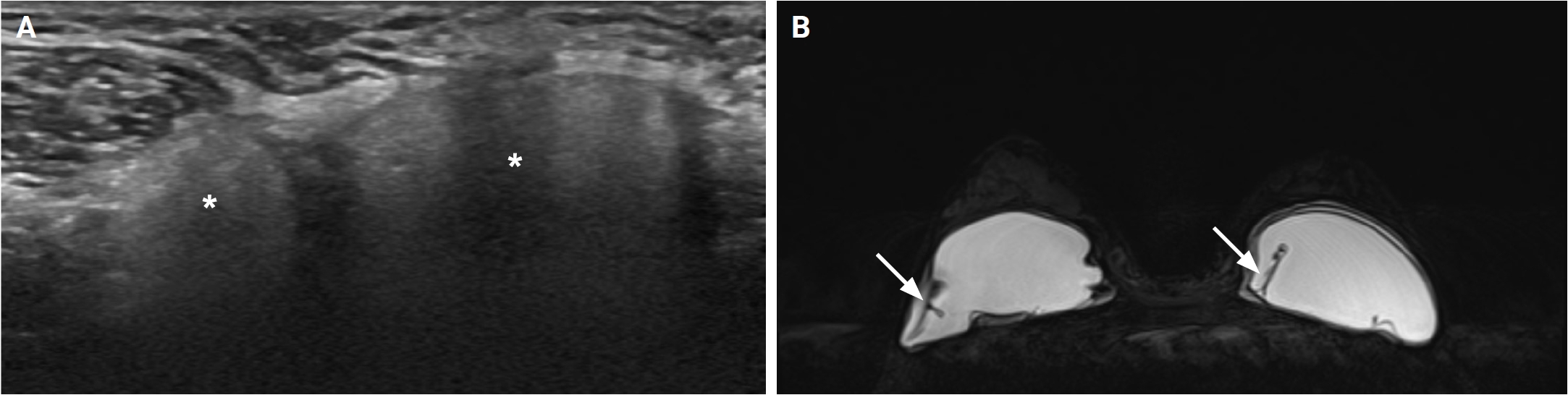
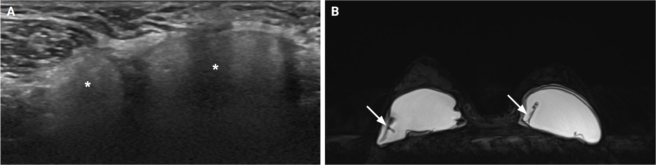
- Subsequent ultrasound and magnetic resonance imaging (MRI) evaluation of the breasts and axillae demonstrated bilateral intracapsular rupture of the silicone implants and silicone lymphadenopathy (Fig. 4A, B). Additional computed tomography of the chest, abdomen, and pelvis revealed no pathologic lesions, with no evidence of malignancy in the internal organs or bilateral breast parenchyma.
- The patient was discharged and referred back to the outside institution for further evaluation of leakage and rupture of the silicone breast prostheses.
CASE REPORT
- Silicone lymphadenopathy is an uncommon entity that typically presents as painless or painful nodal enlargement [2]. FNAC is an accurate initial diagnostic technique that may aid in the evaluation of lymphadenopathy [12]. Only a limited number of reports have described the cytologic findings of silicone lymphadenopathy, with the most characteristic features consisting of a reactive lymphoid background containing numerous multinucleated giant cells and histiocytes with multiple variably sized vacuoles consistent with silicone [3,4,8]. In the appropriate clinical context, these cytologic findings may be sufficiently characteristic to support a diagnosis of silicone lymphadenopathy. Santos-Briz et al. [4] described the FNAC findings of silicone lymphadenopathy and suggested that FNAC is an accurate method for diagnosing silicone lymphadenopathy in patients with breast implants presenting with an axillary mass. Tabatowski et al. [3] were the first to describe the cytologic findings of silicone lymphadenopathy in a patient with a breast implant. Although these authors considered the cytologic findings diagnostic in the appropriate clinical setting, they recommended lymph node excision to exclude a concomitant malignant neoplasm.
- However, definitive diagnosis of silicone lymphadenopathy by FNAC alone may be challenging in the absence of a known history of silicone breast implantation. In lymph node FNAC specimens demonstrating epithelioid cells and multinucleated giant cells, the differential diagnosis includes other granulomatous processes, including lipogranuloma. When considering the diagnosis of lipogranuloma, it is important to note that most patients have a clinical history related to lipid deposition (e.g., injection, surgery, or trauma) [13]. Although silicone lymphadenopathy also presents with chronic granulomatous inflammation, it is pathologically distinct. The hallmark of silicone lymphadenopathy is the presence of refractile, non-birefringent, colorless material, which can be identified both within the cytoplasm of histiocytes and multinucleated giant cells and as extracellular droplets [4,7,14]. In contrast, lipogranuloma is characterized by lipid vacuoles that typically lack these refractile particles [13]. Cytoplasmic microvacuoles may compress the nuclei of epithelioid cells, resulting in a signet ring cell–like morphology. This appearance represents a potential diagnostic pitfall, as it can mimic metastatic carcinoma, such as breast lobular carcinoma or signet ring cell adenocarcinoma [6,9]. Owing to the restricted access to the patient’s clinical history at the specialized diagnostic center, the clear cells containing vacuoles on FNAC were initially misidentified as metastatic carcinoma rather than silicone lymphadenopathy. However, malignant signet ring cells typically exhibit small and numerous cytoplasmic vacuoles, along with enlarged, irregular nuclei and prominent nucleoli [15]. Notably, they lack the foreign body reaction and the intracytoplasmic, variably sized, refractile, non-birefringent particles that are characteristic of silicone lymphadenopathy [4,7,14]. In silicone lymphadenopathy, cytoplasmic microvacuoles may compress the nuclei, thereby imparting a lipoblast-like appearance and necessitating a differential diagnosis from liposarcoma. However, a true lipoblast is cytologically characterized by an atypical, hyperchromatic nucleus that is distinctly scalloped by multiple, sharply defined lipid vacuoles within the cytoplasm [16]. Furthermore, liposarcoma typically exhibits other malignant features such as round or spindle-shaped cells, significant cytonuclear atypia, mitotic figures, a myxoid stroma, and characteristic arborizing vascular structures, which were absent in the present case. The patient had undergone bilateral cosmetic silicone breast implantation 10 years earlier. Subsequent ultrasonography of the bilateral axillae revealed the pathognomonic “snowstorm” artifact, confirming silicone lymphadenopathy. Concurrently, breast MRI demonstrated the collapsed layers of the elastomer shell folded within the implant lumen (e.g., teardrop signs and subcapsular lines), providing evidence of bilateral intracapsular rupture. Comprehensive radiologic evaluation revealed no malignant tumors in the internal organs, including the breasts. The final diagnosis was axillary silicone lymphadenopathy.
- Once the diagnosis of silicone lymphadenopathy has been established, no additional treatment is generally required [2]. Surgical excision should be reserved for symptomatic patients. In the present case, the patient was referred back to the local clinic at the outside institution for further evaluation of leakage and rupture of the silicone breast implants.
- Although axillary silicone lymphadenopathy is uncommon, it should be considered in the differential diagnosis of axillary lymphadenopathy. The cytologic findings of silicone lymphadenopathy may be diagnostic in the appropriate clinical setting. Awareness of the distinctive cytomorphologic features of silicone lymphadenopathy is essential for cytopathologists to avoid diagnostic misinterpretation and facilitate appropriate patient management.
DISCUSSION
Ethics Statement
This report was approved by the Institutional Review Board (IRB) of Chonnam National University Hwasun Hospital (No. CNUHH-2026-075, May 13, 2026) and complied with the principles of the 1964 Declaration of Helsinki and its later amendments. The requirement for written informed consent was waived by the IRB of Chonnam National University Hwasun Hospital.
Availability of Data and Material
The datasets generated or analyzed during the study are available from the corresponding author upon reasonable request.
Code Availability
Not applicable.
Author Contributions
Conceptualization: NIK, JSL. Investigation: NIK. Methodology: NIK, HJL. Project administration: NIK. Resources: NIK, JSL. Supervision: JSL. Visualization: NIK, HJL. Writing—original draft preparation: NIK, HJL. Writing—review & editing: NIK, JSL. Approval of final manuscript: all authors.
Conflicts of Interest
The authors declare that they have no potential conflicts of interest.
Funding Statement
No funding to declare.
- 1. Christie AJ, Weinberger KA, Dietrich M. Silicone lymphadenopathy and synovitis. Complications of silicone elastomer finger joint prostheses. JAMA 1977; 237: 1463-4. ArticlePubMed
- 2. Zambacos GJ, Molnar C, Mandrekas AD. Silicone lymphadenopathy after breast augmentation: case reports, review of the literature, and current thoughts. Aesthetic Plast Surg 2013; 37: 278-89. ArticlePubMedPDF
- 3. Tabatowski K, Elson CE, Johnston WW. Silicone lymphadenopathy in a patient with a mammary prosthesis: fine needle aspiration cytology, histology and analytical electron microscopy. Acta Cytol 1990; 34: 10-4. PubMed
- 4. Santos-Briz A Jr, Lopez-Rios F, Santos-Briz A, De Agustin PP. Granulomatous reaction to silicone in axillary lymph nodes: a case report with cytologic findings. Acta Cytol 1999; 43: 1163-5. ArticlePubMed
- 5. Dragoumis DM, Assimaki AS, Vrizas TI, Tsiftsoglou AP. Axillary silicone lymphadenopathy secondary to augmentation mammaplasty. Indian J Plast Surg 2010; 43: 206-9. ArticlePubMedPMC
- 6. Omakobia E, Porter G, Armstrong S, Denton K. Silicone lymphadenopathy: an unexpected cause of neck lumps. J Laryngol Otol 2012; 126: 970-3. ArticlePubMed
- 7. Gundeslioglu AO, Hakverdi S, Erdem O, Ozen EC, Inan I, Emlik D. Axillary lipogranuloma mimicking carcinoma metastasis after silicone breast implant rupture: a case report. J Plast Reconstr Aesthet Surg 2013; 66: e72-5. ArticlePubMed
- 8. Malzone MG, Campanile AC, Gioioso A, et al. Silicone lymphadenopathy: presentation of a further case containing asteroid bodies on fine-needle cytology sample. Diagn Cytopathol 2015; 43: 57-9. ArticlePubMed
- 9. Lee Y, Song SE, Yoon ES, Bae JW, Jung SP. Extensive silicone lymphadenopathy after breast implant insertion mimicking malignant lymphadenopathy. Ann Surg Treat Res 2017; 93: 331-5. ArticlePubMedPMCPDF
- 10. Klang E, Yosepovich A, Krosser A, et al. Detection of pathologically proven silicone lymphadenopathy: ultrasonography versus magnetic resonance imaging. J Ultrasound Med 2018; 37: 969-75. ArticlePubMedPDF
- 11. Salemis NS. Axillary silicone lymphadenopathy: an important differential in patients with post-mastectomy reconstruction with silicone gel implants. Breast J 2020; 26: 1821-2. ArticlePubMedPDF
- 12. Jaffer S, Zakowski M. Fine-needle aspiration biopsy of axillary lymph nodes. Diagn Cytopathol 2002; 26: 69-74. ArticlePubMed
- 13. Fundakowski CE, Chapman JR, Sargi Z. Lipogranuloma of the head and neck. Int J Case Rep Med 2013; 2013: 410335.Article
- 14. Jimenez-Heffernan JA, Rosell A, Vazquez-Espinosa E, Idoate-Ortueta C, Solis-Garcia M, Pacios E. Cytologic features of a pleural effusion after silicone breast implant rupture. Virchows Arch 2023; 482: 1065-8. ArticlePubMedPDF
- 15. Zhu H, Khattab R, Ondrejka SL, Reynolds JP. Signet-ring cells in pleural and peritoneal effusions identified on Wright stains: a diagnostic pitfall. Cytojournal 2022; 19: 12.ArticlePubMedPMC
- 16. Klijanienko J, Caillaud JM, Lagace R. Fine-needle aspiration in liposarcoma: cytohistologic correlative study including well-differentiated, myxoid, and pleomorphic variants. Diagn Cytopathol 2004; 30: 307-12. ArticlePubMed
REFERENCES
Figure & Data
References
Citations

 PubReader
 ePub Link
-
 Cite this Article
- Cite this Article
-
- Close
- Download Citation
- Close
 XML Download
- Figure
-