- KRAS Mutation Test in Korean Patients with Colorectal Carcinomas: A Methodological Comparison between Sanger Sequencing and a Real-Time PCR-Based Assay
-
Sung Hak Lee, Arthur Minwoo Chung, Ahwon Lee, Woo Jin Oh, Yeong Jin Choi, Youn-Soo Lee, Eun Sun Jung
-
J Pathol Transl Med. 2017;51(1):24-31. Published online December 25, 2016
-
DOI: https://doi.org/10.4132/jptm.2016.10.03
-
-
10,803
View
-
168
Download
-
5
Web of Science
-
5
Crossref
-
 Abstract Abstract
 PDF PDF Supplementary Material Supplementary Material
- Background
Mutations in the KRAS gene have been identified in approximately 50% of colorectal cancers (CRCs). KRAS mutations are well established biomarkers in anti–epidermal growth factor receptor therapy. Therefore, assessment of KRAS mutations is needed in CRC patients to ensure appropriate treatment.
Methods
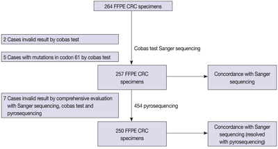
We compared the analytical performance of the cobas test to Sanger sequencing in 264 CRC cases. In addition, discordant specimens were evaluated by 454 pyrosequencing.
Results
KRAS mutations for codons 12/13 were detected in 43.2% of cases (114/264) by Sanger sequencing. Of 257 evaluable specimens for comparison, KRAS mutations were detected in 112 cases (43.6%) by Sanger sequencing and 118 cases (45.9%) by the cobas test. Concordance between the cobas test and Sanger sequencing for each lot was 93.8% positive percent agreement (PPA) and 91.0% negative percent agreement (NPA) for codons 12/13. Results from the cobas test and Sanger sequencing were discordant for 20 cases (7.8%). Twenty discrepant cases were subsequently subjected to 454 pyrosequencing. After comprehensive analysis of the results from combined Sanger sequencing–454 pyrosequencing and the cobas test, PPA was 97.5% and NPA was 100%.
Conclusions
The cobas test is an accurate and sensitive test for detecting KRAS-activating mutations and has analytical power equivalent to Sanger sequencing. Prescreening using the cobas test with subsequent application of Sanger sequencing is the best strategy for routine detection of KRAS mutations in CRC.
-
Citations
Citations to this article as recorded by  - Single-center study on clinicopathological and typical molecular pathologic features of metastatic brain tumor
Su Hwa Kim, Young Suk Lee, Sung Hak Lee, Yeoun Eun Sung, Ahwon Lee, Jun Kang, Jae-Sung Park, Sin Soo Jeun, Youn Soo Lee
Journal of Pathology and Translational Medicine.2023; 57(4): 217. CrossRef - Assessment of KRAS and NRAS status in metastatic colorectal cancer: Experience of the National Institute of Oncology in Rabat Morocco
Chaimaa Mounjid, Hajar El Agouri, Youssef Mahdi, Abdelilah Laraqui, En-nacer Chtati, Soumaya Ech-charif, Mouna Khmou, Youssef Bakri, Amine Souadka, Basma El Khannoussi
Annals of Cancer Research and Therapy.2022; 30(2): 80. CrossRef - The current understanding on the impact of KRAS on colorectal cancer
Mingjing Meng, Keying Zhong, Ting Jiang, Zhongqiu Liu, Hiu Yee Kwan, Tao Su
Biomedicine & Pharmacotherapy.2021; 140: 111717. CrossRef - Droplet digital PCR revealed high concordance between primary tumors and lymph node metastases in multiplex screening of KRAS mutations in colorectal cancer
Barbora Vanova, Michal Kalman, Karin Jasek, Ivana Kasubova, Tatiana Burjanivova, Anna Farkasova, Peter Kruzliak, Dietrich Busselberg, Lukas Plank, Zora Lasabova
Clinical and Experimental Medicine.2019; 19(2): 219. CrossRef - CRISPR Technology for Breast Cancer: Diagnostics, Modeling, and Therapy
Rachel L. Mintz, Madeleine A. Gao, Kahmun Lo, Yeh‐Hsing Lao, Mingqiang Li, Kam W. Leong
Advanced Biosystems.2018;[Epub] CrossRef
- Mesenchymal Stromal Cells Promote Tumor Progression in Fibrosarcoma and Gastric Cancer Cells
-
Byunghoo Song, Bokyung Kim, Se-Ha Choi, Kyo Young Song, Yang-Guk Chung, Youn-Soo Lee, Gyeongsin Park
-
Korean J Pathol. 2014;48(3):217-224. Published online June 26, 2014
-
DOI: https://doi.org/10.4132/KoreanJPathol.2014.48.3.217
-
-
8,419
View
-
54
Download
-
8
Crossref
-
Abstract
PDF
- Background
Extensive evidence has accumulated regarding the role of mesenchymal stromal cells (MSCs) in tumor progression, but the exact effects and mechanisms underlying this role remain unclear. We investigated the effects of MSC-associated tumor progression in MSC-sarcoma models and a gastric cancer metastatic model. MethodsWe conducted an in vitro growth kinetics assay and an in vivo tumor progression assay for sarcoma cells and gastric cancer cells in the presence or absence of MSCs. ResultsMSC-cocultured human fibrosarcoma cells (HT1080) showed accelerated growth compared with HT1080 alone (79- vs 37-fold change, p<.050). For HT1080, human MSC-coinjected tumors showed significantly greater and highly infiltrative growth compared to those of HT1080 alone (p=.035). For mouse fibrosarcoma cells (WEHI164), mouse MSC-coinjected tumors had greater volume than those of WEHI164 alone (p=.141). For rat sarcoma cells (RR1022), rat MSC-coinjected tumors exhibited greater volume and infiltrative growth than those of RR1022 alone (p=.050). For human gastric cancer cells (5FU), tumors of 5FU alone were compact, nodular in shape, and expansile with good demarcation and no definite lung metastatic nodules, whereas tumors grown in the presence of human MSCs showed highly desmoplastic and infiltrative growth and multiple lung metastasis. ConclusionsWe observed morphological evidence for MSC-associated tumor progression of fibrosarcomas and gastric cancer cells.
-
Citations
Citations to this article as recorded by - Transition between canonical to non-canonical Wnt signaling during interactions between mesenchymal stem cells and osteosarcomas
Asulin Masha, Ghedalia-Peled Noa Ben, Erez Ifat Cohen, Ventura Yvonne, Vago Razi
Open Journal of Orthopedics and Rheumatology.2020; : 037. CrossRef - Mesenchymal stem-cell therapy for perianal fistulas in Crohn’s disease: a systematic review and meta-analysis
F. Cheng, Z. Huang, Z. Li
Techniques in Coloproctology.2019; 23(7): 613. CrossRef - Human mesenchymal stromal cells do not promote recurrence of soft tissue sarcomas in mouse xenografts after radiation and surgery
PAOLA A. FILOMENO, KYUNG-PHIL KIM, NARA YOON, IRAN RASHEDI, VICTOR DAYAN, RITA A. KANDEL, XING-HUA WANG, TANIA C. FELIZARDO, ELLIOT BERINSTEIN, SALOMEH JELVEH, ANDREA FILOMENO, JEFFREY A. MEDIN, PETER C. FERGUSON, ARMAND KEATING
Cytotherapy.2018; 20(8): 1001. CrossRef - Review article: mesenchymal stromal cell therapy for inflammatory bowel diseases
C. Grégoire, C. Lechanteur, A. Briquet, É. Baudoux, F. Baron, E. Louis, Y. Beguin
Alimentary Pharmacology & Therapeutics.2017; 45(2): 205. CrossRef - Effect of hGC-MSCs from human gastric cancer tissue on cell proliferation, invasion and epithelial-mesenchymal transition in tumor tissue of gastric cancer tumor-bearing mice
Lin Song, Xin Zhou, Hong-Jun Jia, Mei Du, Jin-Ling Zhang, Liang Li
Asian Pacific Journal of Tropical Medicine.2016; 9(8): 796. CrossRef - BMP9 inhibits the growth and migration of lung adenocarcinoma A549 cells in a bone marrow stromal cell-derived microenvironment through the MAPK/ERK and NF-κB pathways
JING WANG, YAGUANG WENG, MINGHAO ZHANG, YA LI, MENGTIAN FAN, YANGLIU GUO, YANTING SUN, WANG LI, QIONG SHI
Oncology Reports.2016; 36(1): 410. CrossRef - Comparative proteomic analysis of fibrosarcoma and skin fibroblast cell lines
Ogunc Meral, Hamdi Uysal
Tumor Biology.2015; 36(2): 561. CrossRef - Involvement of Wnt/β-catenin signaling in the mesenchymal stem cells promote metastatic growth and chemoresistance of cholangiocarcinoma
Weiwei Wang, Wei Zhong, Jiahui Yuan, Congcong Yan, Shaoping Hu, Yinping Tong, Yubin Mao, Tianhui Hu, Bing Zhang, Gang Song
Oncotarget.2015; 6(39): 42276. CrossRef
|
 E-submission